[Preprint]. 2024 Apr 9:2024.03.21.586084.
doi: 10.1101/2024.03.21.586084.

Endogenous CD28 drives CAR T cell responses in multiple myeloma

Affiliations

Endogenous CD28 drives CAR T cell responses in multiple myeloma

Mackenzie M Lieberman et al. bioRxiv. .

Abstract

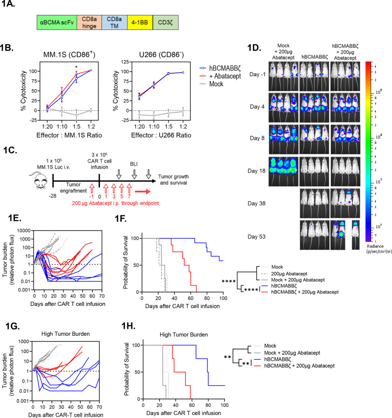
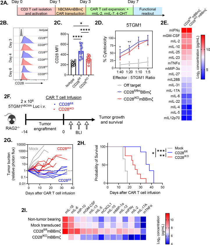
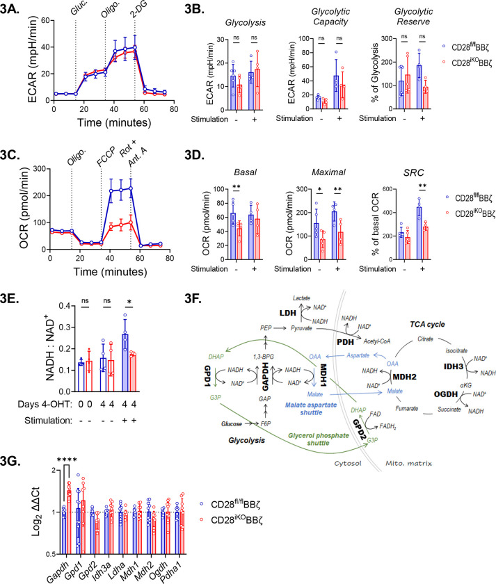
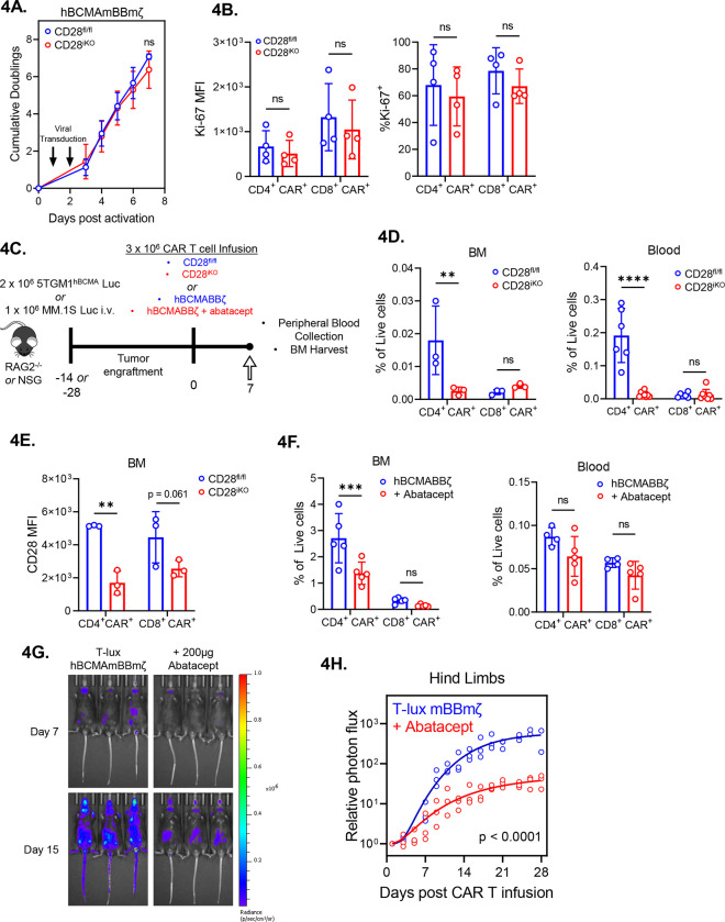
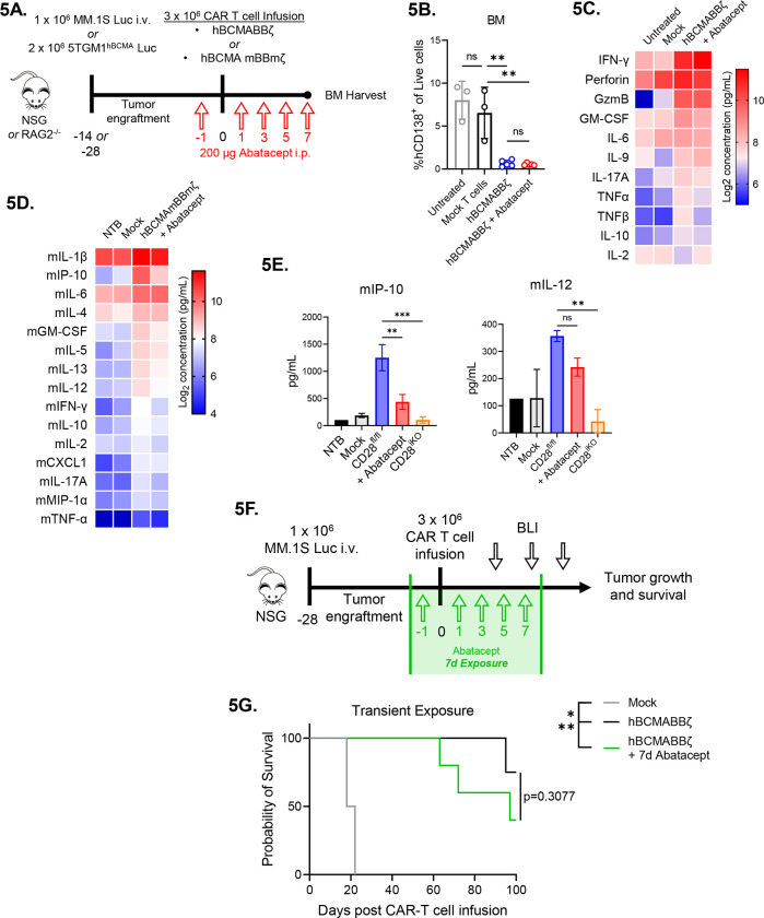
Recent FDA approvals of chimeric antigen receptor (CAR) T cell therapy for multiple myeloma (MM) have reshaped the therapeutic landscape for this incurable cancer. In pivotal clinical trials B cell maturation antigen (BCMA) targeted, 4-1BB co-stimulated (BBζ) CAR T cells dramatically outperformed standard-of-care chemotherapy, yet most patients experienced MM relapse within two years of therapy, underscoring the need to improve CAR T cell efficacy in MM. We set out to determine if inhibition of MM bone marrow microenvironment (BME) survival signaling could increase sensitivity to CAR T cells. In contrast to expectations, blocking the CD28 MM survival signal with abatacept (CTLA4-Ig) accelerated disease relapse following CAR T therapy in preclinical models, potentially due to blocking CD28 signaling in CAR T cells. Knockout studies confirmed that endogenous CD28 expressed on BBζ CAR T cells drove in vivo anti-MM activity. Mechanistically, CD28 reprogrammed mitochondrial metabolism to maintain redox balance and CAR T cell proliferation in the MM BME. Transient CD28 inhibition with abatacept restrained rapid BBζ CAR T cell expansion and limited inflammatory cytokines in the MM BME without significantly affecting long-term survival of treated mice. Overall, data directly demonstrate a need for CD28 signaling for sustained in vivo function of CAR T cells and indicate that transient CD28 blockade could reduce cytokine release and associated toxicities.

Keywords: CAR T cells; CD28; co-stimulation; metabolism; multiple myeloma; tumor microenvironment.


Conflict of interest statement

Conflicts of Interest R.J.B. has licensed intellectual property to and collects royalties from Bristol Myers Squibb (BMS), Caribou, and Sanofi. R.J.B. received research funding from BMS. R.J.B. is a consultant to BMS, Atara Biotherapeutics Inc, and Triumvira. R.J.B is a member of the scientific advisory board for Triumvira.

Figures

Figure 1:
Figure 1:. Endogenous CD28 blockade impairs hBCMABBζ CAR T cell anti-myeloma efficacy.
(A) Schematic of second-generation retroviral CAR construct used to generate BCMA targeted human CAR T cells. (B) CAR T cell cytotoxic activity during a 24-hr. co-culture with luciferase-tagged MM.1S (left) or U266 (right) myeloma cells ± abatacept. Data are shown as mean ± SD and representative of hBCMABBζ CAR T cells generated from 3 healthy donors. *p<0.05 by two-way analysis of variance (ANOVA) with Tukey’s multiple comparison test. (C) Diagram of experimental setup used to evaluate hBCMABBζ CAR T + abatacept therapy in a human MM xenograft model. NSG mice were intravenously inoculated with 1 × 10⁶ MM.1S-luc myeloma cells on day −28 and treated with CAR T cells on day 0. Mice received 200 μg abatacept 3x/week beginning the day before infusion and continuing through endpoint. Tumor burden was monitored by IVIS bioluminescent imaging (BLI) 2x/week. (D) Representative bioluminescent images of MM.1S bearing mice on specified days following CAR T cell infusion. (E) Tumor burden expressed as relative photon flux measured by BLI from MM.1S-luc bearing mice treated with hBCMABBζ CAR T or control T cells ± abatacept (200 μg, 3x/week). Each line represents an individual mouse (n = 7 mice per CAR T cell treated group). (F) Kaplan – Meier analysis of survival of hBCMABBζ CAR T or control T cells ± abatacept (200 μg, 3x/week) treated MM.1S-luc bearing mice (n = 8 – 12 mice per CAR T cell treated group). Median survival of hBCMABBζ CAR T treated mice was >100 days post CAR T cell infusion vs. 55 days for hBCMABBζ CAR T + abatacept treated mice. ****p<0.0001 by log-rank Mantel-Cox test. (G) Tumor burden expressed as relative photon flux measured by BLI from MM.1S high tumor burden mice (inoculated on day −35) treated with hBCMABBζ CAR T or control T cells ± abatacept (200 μg, 3x/week). Each line represents an individual mouse (n = 4 mice per CAR T cell treated group). (H) Kaplan – Meier analysis of survival of hBCMABBζ CAR T or control T cells ± abatacept (200 μg, 3x/week) treated MM.1S-luc high tumor burden mice (n = 4 mice per group). Median survival of hBCMABBζ CAR T treated mice was 80 days vs. 45 days for hBCMABBζ CAR T + abatacept treated mice. ***p<0.0001 by log-rank Mantel-Cox test.
Figure 2:
Figure 2:. CD28iKO hBCMAmBBmζ CAR T cells are functionally impaired in vivo.
(A) Schematic depicting the process of manufacturing mouse CD28 knockout (CD28iKO) hBCMAmBBmζ CAR T cell. (B) Surface CD28 protein expression on CD28fl/fl versus CD28iKO mouse T cells during CAR T cell manufacture. (C) Median fluorescent intensity (MFI) of CD28 measured by flow cytometry at the conclusion of CD28fl/fl versus CD28iKO mouse CAR T manufacture. Data shown as mean ± SD from 10+ independent experiments. *p<0.05, ****p<0.0001 by one-way ANOVA. (D) CD28fl/fl versus CD28iKO hBCMAmBBmζ CAR T cell cytotoxic activity during 24 hr. co-culture with luciferase-tagged 5TGM1hBCMA mouse myeloma cells. Cell viability was assessed by luciferase assay. Data are shown as mean ± SD and representative of at least 3 independent experiments. *p<0.05, **p<0.01 by two-way ANOVA with Tukey’s multiple comparison test. (E) Heatmap representation of culture supernatant mouse cytokine concentrations measured by multiplexed Luminex assays at the conclusion of a 24-hr. co-culture of hBCMAmBBmζ CD28fl/fl or CD28iKO CAR T cells with 5TGM1hBCMA myeloma cells. Log2 transformed cytokine concentrations represent the mean of 4 independent experiments. (F) Diagram of experimental setup used to evaluate CD28fl/fl versus CD28iKO hBCMAmBBmζ CAR T cell therapy in a mouse MM xenograft model. RAG2−/− mice were inoculated intravenously with 2 × 10⁶ 5TGM1hBCMA-luc cells on day −14 and treated with CD28fl/fl or CD28iKO CAR T cells on day 0. Tumor burden was monitored by IVIS bioluminescent imaging (BLI) 2x/week through endpoint. (G) Tumor burden expressed as relative photon flux measured by BLI from 5TGM1hBCMA-luc bearing mice treated with CD28fl/fl or CD28iKO hBCMAmBBmζ CAR T or control T cells. Each line represents an individual mouse (n = 6 mice per CAR T cell treated group). (H) Kaplan – Meier analysis of survival of CD28fl/fl or CD28iKO hBCMAmBBmζ CAR T cell or control T cell treated 5TGM1hBCMA-luc bearing mice (n = 6 mice per CAR T cell treated group). Median survival of CD28fl/fl hBCMAmBBmζ CAR T treated mice was 38 days post-CAR T cell infusion vs. 24 days for CD28iKO hBCMAmBBmζ CAR T treated mice. *p<0.05, **p<0.01, ***p<0.001 by log-rank Mantel-Cox test. (I) Heatmap representation of cytokine levels in the MM BME 7 days following infusion of CD28fl/fl or CD28iKO hBCMAmBBmζ CAR T cells into 5TGM1hBCMA-luc bearing mice. Bilateral hind limbs were harvested, and BM was flushed into 15 μL PBS for multiplexed cytokine analysis. Log2 transformed cytokine concentrations represent the mean of 3 mice per group.
Figure 3:
Figure 3:. Perturbation of oxidative metabolism and redox homeostasis in stimulated CD28iKO hBCMAmBBmζ CAR T cells.
(A) Representative Seahorse Glycolysis Stress Test performed 24 hr. after stimulation of CD28fl/fl or CD28iKO hBCMAmBBmζ CAR T cells with 5TGM1hBCMA myeloma cells. Connected data points represent mean extracellular acidification rate (ECAR) ± SD of four technical replicates at indicated time points during a representative experiment that was repeated at least 3 times. Dashes indicate the timing of glucose, oligomycin (Oligo) and 2-deoxyglucose (2-DG) injection. (B) Quantified rates of glycolysis (left), glycolytic capacity (middle), and glycolytic reserve (left) in CD28fl/fl or CD28iKO hBCMAmBBmζ CAR T cells ± 24-hr. stimulation with 5TGM1hBCMA myeloma cells. Bars represent mean ± SD, dots represent independent experiments. ns = not significant by one-way ANOVA. (C) Representative Seahorse Mito Stress Test performed 24 hr. after stimulation of CD28fl/fl or CD28iKO hBCMAmBBmζ CAR T cells with 5TGM1hBCMA myeloma cells. Connected data points represent mean oxygen consumption rate (OCR) ± SD of four technical replicates at indicated time points during a representative experiment that was repeated at least 3 times. Dashes indicate the timing of oligomycin (Oligo), FCCP, and rotenone (Rot) + antimycin A (Ant. A) injection. (D) Quantified basal OCR (right), uncoupled maximal respiration (middle), and spare respiratory capacity (SRC) expressed as percent of basal OCR (left) in CD28fl/fl or CD28iKO hBCMAmBBmζ CAR T cells ± 24 hr. stimulation with 5TGM1hBCMA myeloma cells. Bars represent mean ± SD, dots represent independent experiments. *p<0.05, **p<0.01 by one-way ANOVA. (E) Ratio of NADH to NAD+ ratios in CD28fl/fl or CD28iKO hBCMAmBBmζ CAR T cells prior to 4-OHT mediated CD28 deletion or ± 24 hr. stimulation with 5TGM1hBCMA myeloma cells. Bars represent mean ± SD from 3 independent experiments. *p<0.05 by paired Student’s t test. (F) Diagrams depicting enzymes of central carbon metabolism that interconvert NADH and NAD+. (G) Relative expression of mRNAs coding enzymes depicted in (F) in CD28iKO versus CD28fl/fl hBCMAmBBmζ CAR T cells following 24 hr. stimulation with 5TGM1hBCMA myeloma cells. Bars represent mean log2 transformed ΔΔCt values ± SD, dots technical replicates pooled from 3 independent experiments. Tbp and Actb were used as endogenous controls. ****p<0.0001 by two-way ANOVA.
Figure 4:
Figure 4:. Diminished in vivo expansion of 4-BB co-stimulated CD28iKO CAR T cells.
(A) Expansion of CD28fl/fl versus CD28iKO hBMCAmBBmζ CAR T cells over the course of manufacturing. ns = not significant by two-way ANOVA. (B) Expression of the proliferation marker Ki-67 in CD28fl/fl versus CD28iKO hBCMAmBBmζ CAR T cells following 24 hr. stimulation with 5TGM1 target cells as assessed by flow cytometric analysis. ns = not significant by one-way ANOVA. (C) Diagram of experimental setup. 5TGM1hBCMA bearing RAG2−/− mice were treated with CD28fl/fl or CD28iKO hBCMAmBBmζ CAR T cells (4D and 4E) or MM.1S bearing NSG mice were treated with hBCMABBζ CAR T cells ± 200μg abatacept on days −1, 1, 3, 5, 7 (4F) and euthanized 7 days post adoptive transfer for blood collection and hind limb BM harvest. (D) CAR T cell frequency assessed by flow cytometry in bone marrow (BM, left) and peripheral blood (right) one week after adoptive transfer of CD28fl/fl or CD28iKO hBCMAmBBmζ CAR T cells into 5TGM1hBCMA myeloma bearing mice. Bars represent mean ± SD, dots indicate individual mice (n = 3–6 mice per group). **p<0.01, ****p<0.0001 by two-way ANOVA with Tukey’s multiple comparison test. (E) CD28 surface protein expression on BM-infiltrating CD28fl/fl or CD28iKO hBCMAmBBmζ CAR T cells one week after adoptive transfer into 5TGM1hBCMA myeloma bearing mice. (F) CAR T cell frequency assessed by flow cytometry in bone marrow (BM, left) and peripheral blood (right) one week after adoptive transfer of hBCMABBζ CAR T cells into MM.1S myeloma bearing mice ± abatacept. CAR T population identified by surface staining and analyzed by flow cytometry. Bars represent mean ± SD, dots indicate individual mice (n = 4–5 mice per group). ***p<0.001 by two-way ANOVA with Tukey’s multiple comparison test. (G) Representative IVIS bioluminescence images of T-lux luciferase expressing hBCMAmBBmζ CAR T cells ± abatacept (200μg, 3x/week) on day 7 or day 15 after infusion. (H) Quantification of photon flux by IVIS imaging within the hind limb region of interest (ROI) over a four-week period following T-lux hBCMAmBBmζ CAR T cell infusion ± abatacept (200μg, 3x/week) into 5TGM1hBCMA myeloma bearing mice. Dots represent individual mice (n = 3 mice per group), lines are best-fit sigmoidal curves, and significance was determined by mixed effects modeling.
Figure 5:
Figure 5:. Transient CD28 blockade limits inflammatory cytokines in the MM BME without affecting survival of hBCMAmBBmζ CAR T cell treated mice.
(A) Diagram of experimental setup. MM.1S bearing NSG mice were treated with hBCMABBζ CAR T cells ± 200μg abatacept on days −1, 1, 3, 5, 7 (5B and 5C) or 5TGM1hBCMA bearing RAG2−/− mice were treated with CD28fl/fl or CD28iKO hBCMAmBBmζ CAR T cells (5D and 5E) and euthanized 7 days post adoptive transfer for hind limb BM harvest. (B) Myeloma burden assessed by flow cytometry for human CD138+ cells in the bone marrow of MM.1S bearing mice treated as described in 5A. Bars represent mean ± SD, dots indicate individual mice. **p<0.01 by one-way ANOVA, ns = not significant. (C) Heatmap representation of human cytokine levels in the MM BME 7 days after treatment of MM.1S bearing mice with hBCMABBζ CAR T cells ± abatacept. Bilateral hind limbs were harvested, and BM was flushed into 15 μL PBS for multiplexed cytokine analysis. Log2 transformed cytokine concentrations represent the mean of 3–6 mice per group. (D) Heatmap representation of murine cytokine levels in the MM BME 7 days after treatment of 5TGM1hBCMA bearing mice with hBCMAmBBmζ CAR T cells ± abatacept. Bilateral hind limbs were harvested, and BM was flushed into 15 μL PBS for multiplexed cytokine analysis. Log2 transformed cytokine concentrations represent the mean of 2–3 mice per group. (E) Bar graphs showing concentrations of murine IP-10 (left) and IL-12 (right) in the MM BME 7 days after treatment of 5TGM1hBCMA bearing mice with CD28fl/fl hBCMAmBBmζ CAR T cells ± abatacept versus CD28iKO hBCMAmBBmζ CAR T cells. Bars represent mean ± SD of 2–3 biological replicates. **p<0.01, ***p<0.001 by one-way ANOVA. (F) Diagram of experimental setup. MM.1S bearing NSG mice were treated with hBCMABBζ CAR T cells ± 200μg abatacept on days −1, 1, 3, 5, 7 and tumor burden was monitored by bioluminescent imaging 2x/week until endpoint. (G) Kaplan – Meier analysis of survival of hBCMABBζ CAR T ± transient abatacept or mock transduced T cell treated MM.1S-luc bearing mice (n = 4–5 mice per CAR T cell treated group). Median survival of hBCMABBζ CAR T treated mice was >100 days post CAR T cell infusion vs. 97 days for hBCMABBζ CAR T + transient abatacept treated mice. Statistical significance was determined by log-rank Mantel-Cox test.

References

    1. Cappell K. M. & Kochenderfer J. N. Long-term outcomes following CAR T cell therapy: what we know so far. Nat Rev Clin Oncol 20, 359–371, doi:10.1038/s41571-023-00754-1 (2023). - DOI - PMC - PubMed
    1. Cappell K. M. et al. Long-Term Follow-Up of Anti-CD19 Chimeric Antigen Receptor T-Cell Therapy. J Clin Oncol 38, 3805–3815, doi:10.1200/jco.20.01467 (2020). - DOI - PMC - PubMed
    1. Kochenderfer J. N. et al. B-cell depletion and remissions of malignancy along with cytokine-associated toxicity in a clinical trial of anti-CD19 chimeric-antigen-receptor-transduced T cells. Blood 119, 2709–2720, doi:10.1182/blood-2011-10-384388 (2012). - DOI - PMC - PubMed
    1. Kochenderfer J. N. et al. Eradication of B-lineage cells and regression of lymphoma in a patient treated with autologous T cells genetically engineered to recognize CD19. Blood 116, 4099–4102, doi:10.1182/blood-2010-04-281931 (2010). - DOI - PMC - PubMed
    1. Kalos M. et al. T cells with chimeric antigen receptors have potent antitumor effects and can establish memory in patients with advanced leukemia. Sci Transl Med 3, 95ra73, doi:10.1126/scitranslmed.3002842 (2011). - DOI - PMC - PubMed

Publication types