

Tandem Mass Tag-Based Proteomic Analysis of Normal and Degenerated Human Intervertebral Discs
- PMID: 38563035
- PMCID: PMC10982071
- DOI: 10.2147/JPR.S449044
Tandem Mass Tag-Based Proteomic Analysis of Normal and Degenerated Human Intervertebral Discs
Abstract
Background: Intervertebral disc degeneration (IVDD) is the main cause of low back pain (LBP), but the specific regulatory factors, pathways and specific molecular mechanisms remain unclear.
Methods: We identified and quantitatively analyzed Pfirrmann Grade II (n=3) and Pfirrmann Grade IV (n=3) pulposus samples via MRI. The differential abundance of proteins in the samples was determined and quantitatively analyzed by relative and absolute quantitative analysis of the isotope marker levels combined with the liquid chromatography-tandem mass spectrometry (LC‒MSMS/MS).
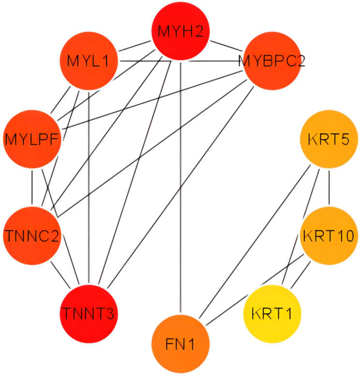
Results: A total of 70 proteins (30 significantly increased proteins (> 1.2-fold change) and 40 significantly decreased proteins (< 0.8-fold change)) showed different levels among the groups. Kyoto Encyclopedia of Genes and Genomes and Gene Ontology (GO) enrichment analyses and Western blot analysis showed that CYCS, RAC1, and PSMD14 may play important roles in IVDD and that Epstein‒Barr virus infection, viral myocarditis, colorectal cancer, nonalcoholic fatty liver disease (NAFLD) and amyotrophic lateral sclerosis (ALS) are the main pathways involved in IVDD.
Conclusion: CYCS, RAC1 and PSMD14 may play important roles in IVDD, and Epstein‒Barr virus infection, viral myocarditis, colorectal cancer, NAFLD and ALS may be the main pathways involved in IVDD.
Keywords: LC‒MS/MS; TMT; low back pain; nucleus pulposus; proteomics.
© 2024 Fu et al.
Conflict of interest statement
The authors declare that they have no competing interests in this work.
Figures


Similar articles
-
Differential proteomic analysis of fetal and geriatric lumbar nucleus pulposus: immunoinflammation and age-related intervertebral disc degeneration.BMC Musculoskelet Disord. 2020 Jun 2;21(1):339. doi: 10.1186/s12891-020-03329-8. BMC Musculoskelet Disord. 2020. PMID: 32487144 Free PMC article.
-
Cartilage endplate stem cells inhibit intervertebral disc degeneration by releasing exosomes to nucleus pulposus cells to activate Akt/autophagy.Stem Cells. 2021 Apr;39(4):467-481. doi: 10.1002/stem.3322. Epub 2021 Jan 18. Stem Cells. 2021. PMID: 33459443 Free PMC article.
-
The Upregulation of COX2 in Human Degenerated Nucleus Pulposus: The Association of Inflammation with Intervertebral Disc Degeneration.Mediators Inflamm. 2021 Oct 18;2021:2933199. doi: 10.1155/2021/2933199. eCollection 2021. Mediators Inflamm. 2021. PMID: 34707460 Free PMC article.
-
Biomaterials delivery strategies to repair degenerated intervertebral discs by regulating the inflammatory microenvironment.Front Immunol. 2023 Jan 23;14:1051606. doi: 10.3389/fimmu.2023.1051606. eCollection 2023. Front Immunol. 2023. PMID: 36756124 Free PMC article. Review.
-
The pathological mechanisms of circRNAs in mediating intervertebral disc degeneration.Noncoding RNA Res. 2023 Sep 18;8(4):633-640. doi: 10.1016/j.ncrna.2023.09.004. eCollection 2023 Dec. Noncoding RNA Res. 2023. PMID: 37780894 Free PMC article. Review.
References
LinkOut - more resources
Full Text Sources
Research Materials
Miscellaneous
