Extracellular Microvesicles Modified with Arginine-Rich Peptides for Active Macropinocytosis Induction and Delivery of Therapeutic Molecules
- PMID: 38563247
- PMCID: PMC11011658
- DOI: 10.1021/acsami.3c14592
Extracellular Microvesicles Modified with Arginine-Rich Peptides for Active Macropinocytosis Induction and Delivery of Therapeutic Molecules
Abstract
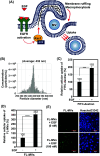
Extracellular vesicles (EVs), including exosomes and microvesicles (MVs), transfer bioactive molecules from donor to recipient cells in various pathophysiological settings, thereby mediating intercellular communication. Despite their significant roles in extracellular signaling, the cellular uptake mechanisms of different EV subpopulations remain unknown. In particular, plasma membrane-derived MVs are larger vesicles (100 nm to 1 μm in diameter) and may serve as efficient molecular delivery systems due to their large capacity; however, because of size limitations, receptor-mediated endocytosis is considered an inefficient means for cellular MV uptake. This study demonstrated that macropinocytosis (lamellipodia formation and plasma membrane ruffling, causing the engulfment of large fluid volumes outside cells) can enhance cellular MV uptake. We developed experimental techniques to induce macropinocytosis-mediated MV uptake by modifying MV membranes with arginine-rich cell-penetrating peptides for the intracellular delivery of therapeutic molecules.
Keywords: arginine-rich cell-penetrating peptides; endocytosis; exosomes; extracellular vesicles; macropinocytosis; microvesicles; peptide modification.
Conflict of interest statement
The authors declare no competing financial interest.
Figures


Similar articles
-
Arginine-rich cell-penetrating peptide-modified extracellular vesicles for active macropinocytosis induction and efficient intracellular delivery.Sci Rep. 2017 May 16;7(1):1991. doi: 10.1038/s41598-017-02014-6. Sci Rep. 2017. PMID: 28512335 Free PMC article.
-
Vectorization of biomacromolecules into cells using extracellular vesicles with enhanced internalization induced by macropinocytosis.Sci Rep. 2016 Oct 17;6:34937. doi: 10.1038/srep34937. Sci Rep. 2016. PMID: 27748399 Free PMC article.
-
Effects of Lyophilization of Arginine-rich Cell-penetrating Peptide-modified Extracellular Vesicles on Intracellular Delivery.Anticancer Res. 2019 Dec;39(12):6701-6709. doi: 10.21873/anticanres.13885. Anticancer Res. 2019. PMID: 31810935
-
A review of the regulatory mechanisms of extracellular vesicles-mediated intercellular communication.Cell Commun Signal. 2023 Apr 13;21(1):77. doi: 10.1186/s12964-023-01103-6. Cell Commun Signal. 2023. PMID: 37055761 Free PMC article. Review.
-
Extracellular vesicles biogenesis and uptake concepts: A comprehensive guide to studying host-pathogen communication.Mol Microbiol. 2024 Nov;122(5):613-629. doi: 10.1111/mmi.15168. Epub 2023 Sep 27. Mol Microbiol. 2024. PMID: 37758682 Review.
Cited by
-
Mechanisms of extracellular vesicle uptake and implications for the design of cancer therapeutics.J Extracell Biol. 2024 Oct 30;3(11):e70017. doi: 10.1002/jex2.70017. eCollection 2024 Nov. J Extracell Biol. 2024. PMID: 39483807 Free PMC article. Review.
-
The Different Cellular Entry Routes for Drug Delivery Using Cell Penetrating Peptides.Biol Cell. 2025 Jun;117(6):e70012. doi: 10.1111/boc.70012. Biol Cell. 2025. PMID: 40490965 Free PMC article. Review.
-
Survival strategies of cancer cells: the role of macropinocytosis in nutrient acquisition, metabolic reprogramming, and therapeutic targeting.Autophagy. 2025 Apr;21(4):693-718. doi: 10.1080/15548627.2025.2452149. Epub 2025 Jan 26. Autophagy. 2025. PMID: 39817564 Free PMC article. Review.
References
-
- Lázaro-Ibáñez E.; Sanz-Garcia A.; Visakorpi T.; Escobedo-Lucea C.; Siljander P.; Ayuso-Sacido A.; Yliperttula M. Different gDNA content in the subpopulations of prostate cancer extracellular vesicles: apoptotic bodies, microvesicles, and exosomes. Prostate 2014, 74 (14), 1379–1390. 10.1002/pros.22853. - DOI - PMC - PubMed
MeSH terms
Substances
LinkOut - more resources
Full Text Sources

