

Extracranial Vascular Anomalies Driven by RAS/MAPK Variants: Spectrum and Genotype-Phenotype Correlations
- PMID: 38563363
- PMCID: PMC11262533
- DOI: 10.1161/JAHA.123.033287
Extracranial Vascular Anomalies Driven by RAS/MAPK Variants: Spectrum and Genotype-Phenotype Correlations
Abstract
Background: We aimed to correlate alterations in the rat sarcoma virus (RAS)/mitogen-activated protein kinase pathway in vascular anomalies to the clinical phenotype for improved patient and treatment stratification.
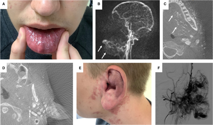
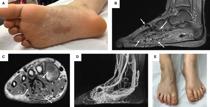
Methods and results: This retrospective multicenter cohort study included 29 patients with extracranial vascular anomalies containing mosaic pathogenic variants (PVs) in genes of the RAS/mitogen-activated protein kinase pathway. Tissue samples were collected during invasive treatment or clinically indicated biopsies. PVs were detected by the targeted sequencing of panels of genes known to be associated with vascular anomalies, performed using DNA from affected tissue. Subgroup analyses were performed according to the affected genes with regard to phenotypic characteristics in a descriptive manner. Twenty-five vascular malformations, 3 vascular tumors, and 1 patient with both a vascular malformation and vascular tumor presented the following distribution of PVs in genes: Kirsten rat sarcoma viral oncogene (n=10), neuroblastoma ras viral oncogene homolog (n=1), Harvey rat sarcoma viral oncogene homolog (n=5), V-Raf murine sarcoma viral oncogene homolog B (n=8), and mitogen-activated protein kinase kinase 1 (n=5). Patients with RAS PVs had advanced disease stages according to the Schobinger classification (stage 3-4: RAS, 9/13 versus non-RAS, 3/11) and more frequent progression after treatment (RAS, 10/13 versus non-RAS, 2/11). Lesions with Kirsten rat sarcoma viral oncogene PVs infiltrated more tissue layers compared with the other PVs including other RAS PVs (multiple tissue layers: Kirsten rat sarcoma viral oncogene, 8/10 versus other PVs, 6/19).
Conclusions: This comparison of patients with various PVs in genes of the RAS/MAPK pathway provides potential associations with certain morphological and clinical phenotypes. RAS variants were associated with more aggressive phenotypes, generating preliminary data and hypothesis for future larger studies.
Keywords: RAS/MAPK pathway; clinical characteristics; mosaicism; pathogenic variants; vascular anomalies.
Figures

References
-
- Wassef M, Borsik M, Cerceau P, Faucon B, Laurian C, Le Clerc N, Lemarchand‐Venencie F, Massoni C, Salvan D, Bisdorff‐Bresson A. Classification des tumeurs et malformations vasculaires. Apport de la classification ISSVA 2014/2018. Article in French. Ann Pathol. 2021;41:58–70. doi: 10.1016/j.annpat.2020.11.004 - DOI - PubMed
-
- Schmidt VF, Masthoff M, Goldann C, Deniz S, Öcal O, Häberle B, Köhler M, Seidensticker M, Ricke J, Wohlgemuth WA, et al. Percutaneous sclerotherapy of venous malformations of the hand: a multicenter analysis. Cardiovasc Intervent Radiol. 2021;44:1543–1550. doi: 10.1007/s00270-021-02926-x - DOI - PMC - PubMed
-
- Schmidt VF, Masthoff M, Goldann C, Ehrl D, Deniz S, Öcal O, Seidensticker M, Ricke J, Köhler M, Brill R, et al. Image‐guided embolization of arteriovenous malformations of the hand using ethylene‐vinyl alcohol copolymer. Diagn Interv Radiol. 2022;28:486–494. doi: 10.5152/dir.2022.21644 - DOI - PMC - PubMed
Publication types
MeSH terms
Substances
LinkOut - more resources
Full Text Sources
Research Materials
Miscellaneous
