. 2024 Apr 16;13(8):e032734.
doi: 10.1161/JAHA.123.032734. Epub 2024 Apr 2.

Long-Term Clinical-Pathologic Results of Enzyme Replacement Therapy in Prehypertrophic Fabry Disease Cardiomyopathy

Affiliations

Long-Term Clinical-Pathologic Results of Enzyme Replacement Therapy in Prehypertrophic Fabry Disease Cardiomyopathy

Andrea Frustaci et al. J Am Heart Assoc. .

Abstract

Background: The limited ability of enzyme replacement therapy (ERT) in removing globotriaosylceramide from cardiomyocytes is recognized for advanced Fabry disease cardiomyopathy (FDCM). Prehypertrophic FDCM is believed to be cured or stabilized by ERT. However, no pathologic confirmation is available. We report here on the long-term clinical-pathologic impact of ERT on prehypertrophic FDCM.

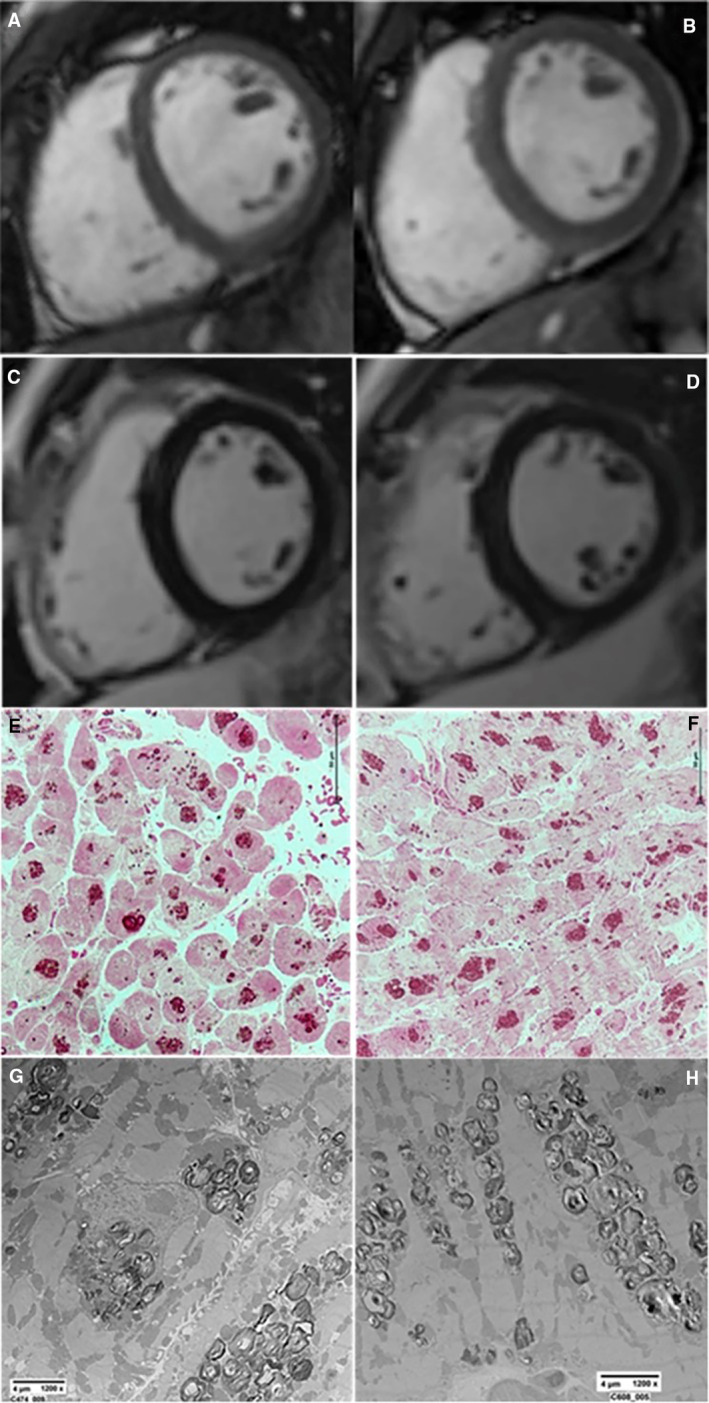
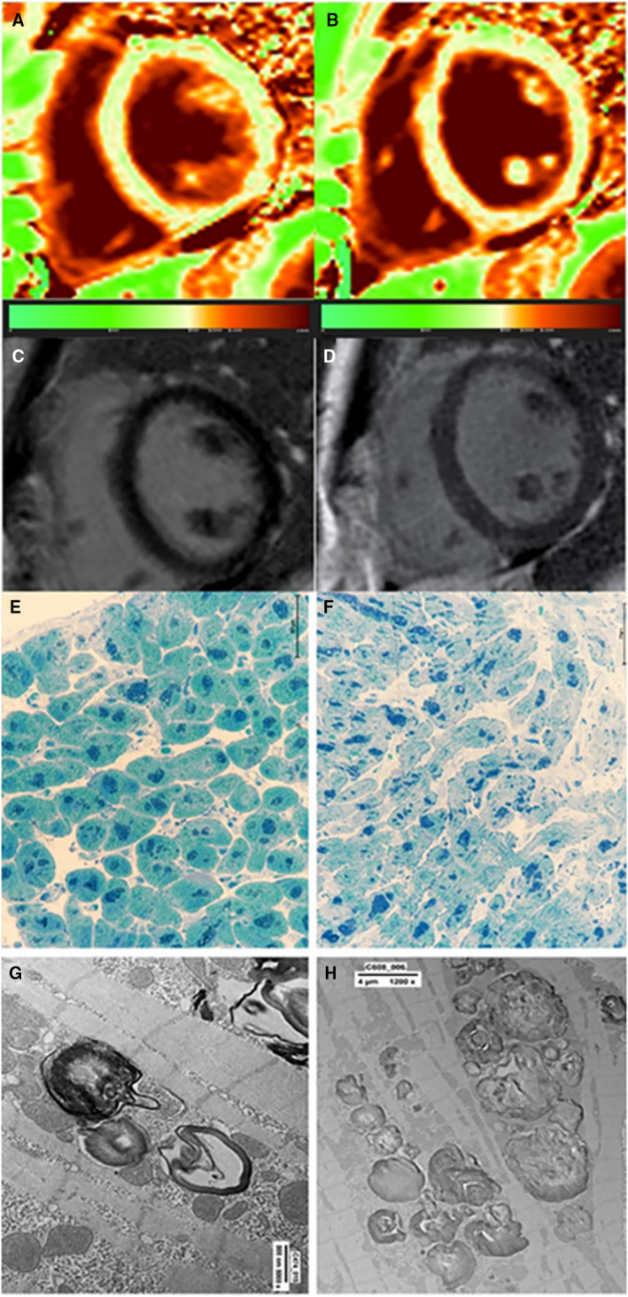
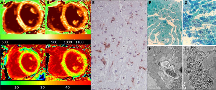
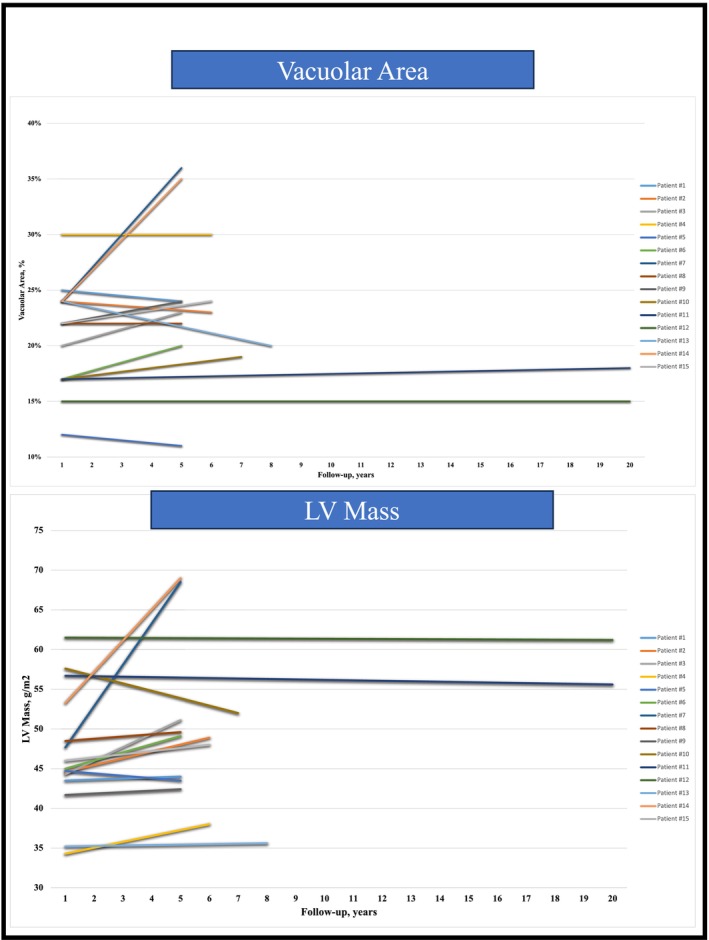
Methods and results: Fifteen patients with Fabry disease with left ventricular maximal wall thickness ≤10.5 mm at cardiac magnetic resonance required endomyocardial biopsy because of angina and ventricular arrhythmias. Endomyocardial biopsy showed coronary small-vessel disease in the angina cohort, and vacuoles in smooth muscle cells and cardiomyocytes ≈20% of the cell surface containing myelin bodies at electron microscopy. Patients received α-agalsidase in 8 cases, and β-agalsidase in 7 cases. Both groups experienced symptom improvement except 1 patients treated with α-agalsidase and 1 treated with β-agalsidase. After ERT administration ranging from 4 to 20 years, all patients had control cardiac magnetic resonance and left ventricular endomyocardial biopsy because of persistence of symptoms or patient inquiry on disease resolution. In 13 asymptomatic patients with FDCM, left ventricular maximal wall thickness and left ventricular mass, cardiomyocyte diameter, vacuole surface/cell surface ratio, and vessels remained unchanged or minimally increased (left ventricular mass increased by <2%) even after 20 years of observation, and storage material was still present at electron microscopy. In 2 symptomatic patients, FDCM progressed, with larger and more engulfed by globotriaosylceramide myocytes being associated with myocardial virus-negative lymphocytic inflammation.

Conclusions: ERT stabilizes storage deposits and myocyte dimensions in 87% of patients with prehypertrophic FDCM. Globotriaosylceramide is never completely removed even after long-term treatment. Immune-mediated myocardial inflammation can overlap, limiting ERT activity.

Keywords: Fabry disease cardiomyopathy; enzyme replacement therapy; globotrioasylceramide; mannose‐6‐phosphate receptors.


Figures

Figure 1
Figure 1. Patient 11 (24‐year‐old man) with genetic and biopsy‐proven diagnosis of FDCM before and after 20 years of ERT.
CineMR (A and B) and LGE (C and D) images, performed at baseline (A and C) and 20 years after ERT (B and D) show normal LV wall thickness (8 and 10 mm) and absence of fibrotic areas.  Semithin section of Epon‐embedded EMB samples, stained with basic fuchsin, showing mildly hypertrophied cardiomyocytes containing storage material that result unmodified after long‐term ERT administration at baseline (E) and 20 years after ERT (F). Ultrastructural evidence of storage material consisting of myelin bodies at baseline (G) that remained unchanged after ERT (H). CineMR indicates cine magnetic resonance; EMB, endomyocardial biopsy; ERT, enzyme replacement therapy; FDCM, Fabry disease cardiomyopathy; and LGE, late gadolinium enhancement.
Figure 2
Figure 2. Clinical–pathologic results of 48 months ERT in a 27‐year‐old man (patient 6) affected by prehypertrophic FDCM.
CMR acquired on midventricular short axis view at baseline (A and C) and after treatment (B and D). The analysis of native T1 maps (A and B) show a diffuse reduction of the myocardial T1 value and a slight increase at follow‐up (nT1: 920 vs 949.5 milliseconds). Myocardial ECV and T2 values (maps not shown in the figure) were within normal range in both exams. LGE images at baseline (C) and after treatment (D) show that neither ventricular hypertrophy nor areas of fibrosis occurred over the observation interval. Semithin sections showing diffuse intracytoplasmic accumulation of dark blue deposits at baseline (E) that mildly reduce after treatment (F). Ultrastructural detection of myelin bodies at baseline (G) that are still visible after ERT (H). CMR indicates cardiac magnetic resonance; ECV, extracellular volume; FDCM, Fabry disease cardiomyopathy; and LGE, late gadolinium enhancement.
Figure 3
Figure 3. CMR images (A through D) with histologic (E through G) and ultrastructural (H and I) documentation of ERT results in a 52‐year‐old (patient 7) woman with prehypertrophic FDCM.
CMR images are acquired at baseline (A and C) and after 60 months ERT (B and D). Native T1 (A and B) and ECV (C and D) maps show progressive increase of myocardial mass (LV mass/BSA from 37.7 to 68.5 g/m2), myocardial nT1 and ECV values (nT1 from 946 to 968 milliseconds; ECV from 30% to 34% at follow‐up), reflecting occurrence of interstitial fibrosis. E, Section of left ventricular endomyocardial biopsy stained with immunohistochemistry for CD45Ro show hypertrophied cardiomyocytes containing large vacuoles with T lymphocytes associated with necrosis of adjacent cells, denoting over overlapping myocarditis. F and G, Diffuse intracytoplasmic accumulation of small dark blue deposits that at TEM can be identified as typical glycolipid bodies at baseline (F). After 5 years of ERT the amount of dark blue deposit is drastically increased in number and size (G). Ultrastructural details of glycolipid bodies at baseline (H) and after 5 years of ERT (I) denoting increase of storage material. BSA indicates body surface area; CMR, cardiac magnetic resonance; ERT, enzyme replacement therapy; FDCM, Fabry disease cardiomyopathy; LV, left ventricular; and TEM, transmission electronic microscope.
Figure 4
Figure 4. Vacuolar area (%) and LV mass changes during follow‐up of 15 patients with prehypertrophic FDCM treated with long‐term ERT.
Data show stabilization of vacuolar area and LV mass in 13 patients, whereas overlapping myocarditis indicates disease progression in 2 patients. ERT indicates enzyme replacement therapy; FDCM, Fabry disease cardiomyopathy; and LV, left ventricular.

Similar articles

Cited by

References

    1. Desnick RJ, Brady R, Barranger J, Collins AJ, Germain DP, Goldman M, Grabowski G, Packman S, Wilcox WR. Fabry disease, an under‐recognized multisystemic disorder: expert recommendations for diagnosis, management, and enzyme replacement therapy. Ann Intern Med. 2003;138:338–346. - PubMed
    1. Desnick RJ. Fabry disease (α‐galactosidase A deficiency). Genetic Diseases of the Kidney. Elsevier; 2009:597–616.
    1. Germain DP. Fabry disease. Orphanet J Rare Dis. 2010;5:30. doi: 10.1186/1750-1172-5-30 - DOI - PMC - PubMed
    1. Thurberg BL, Fallon JT, Mitchell R, Aretz T, Gordon RE, O'Callaghan MW. Cardiac microvascular pathology in Fabry disease evaluation of endomyocardial biopsies before and after enzyme replacement therapy. Circulation. 2009;119:2561–2567. doi: 10.1161/CIRCULATIONAHA.108.841494 - DOI - PubMed
    1. Najafian B, Svarstad E, Bostad L, Gubler MC, Tøndel C, Whitley C, Mauer M. Progressive podocyte injury and glo‐botriaosylceramide (GB‐3) accumulation in young patients with Fabry disease. Kidney Int. 2011;79:663–670. doi: 10.1038/ki.2010.484 - DOI - PMC - PubMed

MeSH terms