. 2024 May 14;98(5):e0017724.
doi: 10.1128/jvi.00177-24. Epub 2024 Apr 2.

Regulation of ubiquitination and antiviral activity of Cactin by deubiquitinase Usp14 in Drosophila

Affiliations

Regulation of ubiquitination and antiviral activity of Cactin by deubiquitinase Usp14 in Drosophila

Qiqi Rong et al. J Virol. .

Abstract

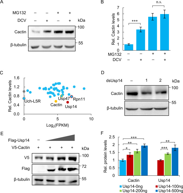
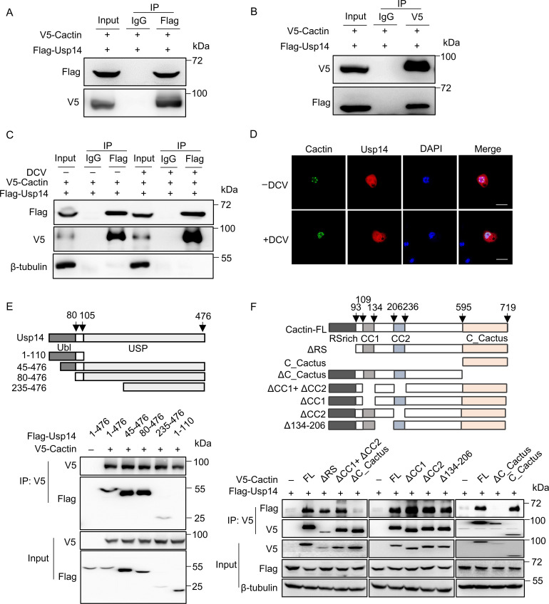
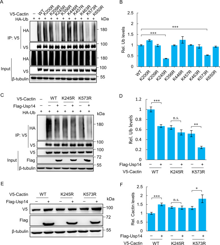
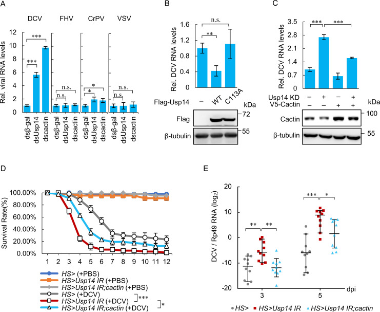
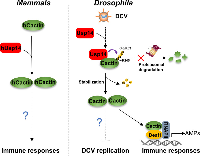
Cactin, a highly conserved protein, plays a crucial role in various physiological processes in eukaryotes, including innate immunity. Recently, the function of Cactin in the innate immunity of Drosophila has been explored, revealing that Cactin regulates a non-canonical signaling pathway associated with the Toll and Imd pathways via the Cactin-Deaf1 axis. In addition, Cactin exhibits specific antiviral activity against the Drosophila C virus (DCV) in Drosophila, with an unknown mechanism. During DCV infection, it has been confirmed that the protein level and antiviral activity of Cactin are regulated by ubiquitination. However, the precise ubiquitination and deubiquitination mechanisms of Cactin in Drosophila remain unexplored. In this study, we identified ubiquitin-specific protease 14 (Usp14) as a major deubiquitinase for Cactin through comprehensive deubiquitinase screening. Our results demonstrate that Usp14 interacts with the C_Cactus domain of Cactin via its USP domain. Usp14 efficiently removes K48- and K63-linked polyubiquitin chains from Cactin, thereby preventing its degradation through the ubiquitin-proteasome pathway. Usp14 significantly inhibits DCV replication in Drosophila cells by stabilizing Cactin. Moreover, Usp14-deficient fruit flies exhibit increased susceptibility to DCV infection compared to wild-type flies. Collectively, our findings reveal the regulation of ubiquitination and antiviral activity of Cactin by the deubiquitinase Usp14, providing valuable insights into the modulation of Cactin-mediated antiviral activity in Drosophila.IMPORTANCEViral infections pose a severe threat to human health, marked by high pathogenicity and mortality rates. Innate antiviral pathways, such as Toll, Imd, and JAK-STAT, are generally conserved across insects and mammals. Recently, the multi-functionality of Cactin in innate immunity has been identified in Drosophila. In addition to regulating a non-canonical signaling pathway through the Cactin-Deaf1 axis, Cactin exhibits specialized antiviral activity against the Drosophila C virus (DCV) with an unknown mechanism. A previous study emphasized the significance of the Cactin level, regulated by the ubiquitin-proteasome pathway, in modulating antiviral signaling. However, the regulatory mechanisms governing Cactin remain unexplored. In this study, we demonstrate that Usp14 stabilizes Cactin by preventing its ubiquitination and subsequent degradation. Furthermore, Usp14 plays a crucial role in regulating the antiviral function mediated by Cactin. Therefore, our findings elucidate the regulatory mechanism of Cactin in Drosophila, offering a potential target for the prevention and treatment of viral infections.

Keywords: Cactin; Drosophila; antiviral immunity; deubiquitinase; ubiquitination.


Conflict of interest statement

The authors declare no conflict of interest.

Figures

Fig 1
Fig 1
Deubiquitinase Usp14 upregulates Cactin protein levels. (A) S2 cells were infected with DCV and treated with either MG132 (50 µM) or DMSO (control) for 4 hours. Protein extracts were subjected to immunoblotting using an anti-Cactin antibody. (B) Quantification of the blots from (A) using ImageJ software to calculate the Cactin to β-tubulin ratio. (C) S2 cells were treated with dsRNA targeting various DUB genes or β-gal (control), and protein extracts were subjected to immunoblotting using an anti-Cactin antibody. Relative gray values of Cactin were analyzed using ImageJ software. The horizontal axis represents log2(FPKM) of DUBs in the RNA-seq data, with dots representing the average gray value of Cactin from three replicates. The negative control (β-gal) is shown as a gray dot, and the four target genes are labeled as colored dots. (D) S2 cells were treated with two independent Usp14 dsRNAs or β-gal (control), and protein extracts were subjected to immunoblotting using an anti-Cactin antibody. (E) S2 cells were co-transfected with pMT-V5-Cactin and increasing amounts of pMT-Flag-Usp14. Protein extracts were subjected to immunoblotting using anti-V5 and anti-Flag antibodies. (F) Quantitative analysis of the western blot bands from (E) using ImageJ software to show the ratios of V5-Cactin and Flag-Usp14 to β-tubulin. Data are presented as mean ± standard deviation (SD) from three independent experiments (B, C, F). Statistical analysis was performed using Student’s t-test (B) or one-way ANOVA (F). Significance levels: *P < 0.05; **P < 0.01; ***P < 0.001; n.s., not significant. Representative results from triplicate experiments are shown (A, D, E).
Fig 2
Fig 2
Usp14 forms physical interactions with Cactin. (A) Co-expression of Flag-tagged Usp14 and V5-tagged Cactin in S2 cells followed by immunoprecipitation with anti-Flag and anti-IgG antibodies. Immunoblotting was performed using anti-V5 and anti-Flag antibodies. (B) Co-expression of Flag-tagged Usp14 and V5-tagged Cactin in S2 cells followed by immunoprecipitation with anti-V5 and anti-IgG antibodies. Immunoblotting was performed using anti-V5 and anti-Flag antibodies. (C) Co-expression of Flag-Usp14 and V5-Cactin in S2 cells with or without DCV infection for 24 hours. Cell lysates were immunoprecipitated using anti-Flag or control IgG antibodies, followed by immunoblotting with anti-V5 and anti-Flag antibodies. (D) Co-expression of Cactin-eGFP and Usp14-mCherry in S2 cells with or without DCV infection for 24 hours. Immunofluorescence was performed, where red fluorescence represents Usp14, green fluorescence represents Cactin, and blue fluorescence indicates nuclear staining with DAPI. Scale bar: 10 µm. (E) Co-expression of V5-Cactin with wild-type Usp14 or indicated Usp14 mutants in S2 cells. Cell lysates were immunoprecipitated with anti-V5 antibodies and analyzed by immunoblotting using anti-V5 and anti-Flag antibodies. (F) Co-expression of Flag-Usp14 with wild-type Cactin (FL) or indicated Cactin mutants in S2 cells. Cell lysates were immunoprecipitated with anti-V5 antibody, followed by immunoblotting with anti-V5 and anti-Flag antibodies. Representative results from triplicate experiments are presented (A–F).
Fig 3
Fig 3
Usp14 removes the K48 and K63-linked ubiquitination from Cactin. (A) Transfection of S2 cells with pMT-Flag-Usp14 and treatment with MG132 (50 µM) or DMSO (control) for 4 hours. Detection of endogenous Cactin levels via immunoblotting with anti-Cactin antibody. (B) S2 cells were treated with Usp14 dsRNA and subsequent treatment with MG132 (50 µM) or DMSO (control) for 4 hours. Assessment of endogenous Cactin expression by immunoblotting using anti-Cactin antibody. (C) Transfection of S2 cells with pMT-Flag-Usp14 or pMT empty plasmids for 48 hours, followed by treatment with CHX (0.5 mM) for the indicated time. Lysis of cells and immunoblotting using anti-Cactin and anti-Flag antibodies to monitor Cactin degradation. (D) S2 cells were pre-treated with Usp14 or β-gal dsRNA, followed by transfection with pMT-HA-Ub and/or pMT-V5-Cactin, and treatment with MG132 (50 µM) for 4 hours. Immunoprecipitation with anti-V5 antibody to analyze the polyubiquitination of Cactin, detected by immunoblotting with anti-HA antibody. (E) Transfection of S2 cells with pMT-V5-Cactin and pMT-HA-Ub plasmids, followed by treatment with IU1 (50 µM) or DMSO for 10 hours, and then treatment with MG132 (50 µM) for 4 hours. Immunoprecipitation with anti-V5 antibody to analyze the polyubiquitination of Cactin, detected by immunoblotting with anti-HA antibody. (F) Co-expression of V5-Cactin and indicated Usp14 WT or Usp14-C113A, along with HA-Ub in S2 cells for 48 hours. After treatment with MG132 (50 µM) for 4 hours, immunoprecipitation using anti-V5 antibody to assess the polyubiquitination of Cactin, detected by immunoblotting with anti-HA antibody. (G-H) Co-expression of V5-Cactin and indicated Ub mutants, along with Flag-Usp14 or control pMT in S2 cells. After treatment with MG132 (50 µM) for 4 hours, immunoprecipitation using anti-V5 antibody, followed by immunoblotting analysis with anti-V5, anti-Flag, and anti-HA antibodies. Representative results from triplicate experiments are displayed (A–H).
Fig 4
Fig 4
Usp14 eliminates the polyubiquitination of Cactin at lysine 245. (A and B) Co-expression of V5-tagged Cactin WT or mutants with HA-Ub in S2 cells for 48 h. After MG132 (50 µM) treatment for 4 hours, immunoprecipitation with anti-V5 antibody to assess Cactin polyubiquitination detected by immunoblotting with anti-HA antibody (A). Quantification of Cactin expression and relative gray values of polyubiquitination for WT Cactin and mutants using ImageJ (B). (C and D) Co-expression of Flag-Usp14 with WT Cactin or indicated Cactin mutants, along with HA-Ub in S2 cells. After MG132 (50 µM) treatment for 4 hours, immunoprecipitation with anti-V5 antibody followed by immunoblotting analysis with anti-V5, anti-Flag, and anti-HA antibodies (C). Quantification of Cactin expression and relative gray values of polyubiquitination for WT Cactin and mutants using ImageJ (D). (E) Co-expression of V5-Cactin or indicated Cactin mutants with Flag-Usp14 or control protein in S2 cells, followed by immunoblotting with anti-V5 and anti-Flag antibodies. (F) Quantification of blots from (E) using ImageJ, showing the V5-Cactin to β-tubulin ratio. Data represent mean ± standard deviation (SD) from three independent experiments (B, D, F). Statistical analysis was performed using Student’s t-test (D, F) or one-way ANOVA (B). *P < 0.05; **P < 0.01; ***P < 0.001; n.s., not significant. Representative results from triplicate experiments are displayed (A, C, E).
Fig 5
Fig 5
Usp14 exhibits the virus-specific antiviral role mediated by Cactin. (A) S2 cells treated with cactin, Usp14, or β-gal dsRNAs and infected with a panel of viruses. Relative viral RNA levels of DCV (48 hpi), FHV (48 hpi), CrPV (16 hpi), or VSV (48 hpi) analyzed by reverse transcription quantitative PCR (qRT-PCR), normalized to rp49. (B) S2 cells expressing Flag-Usp14 WT, Flag-Usp14-C113A, or control infected with DCV. Relative DCV RNA levels at 48 hpi were analyzed by qRT-PCR, normalized to rp49 (Top). Protein levels were assessed by immunoblotting with anti-Flag antibody (below). (C) S2 cells were pretreated with β-gal or Usp14 dsRNA, transfected with control pMT empty or pMT-V5-Cactin plasmids, and infected with DCV. Relative DCV RNA levels at 48 hpi were analyzed by qRT-PCR, normalized to rp49 (top). Protein levels were assessed by immunoblotting with an anti-Cactin antibody (below). (D-E) HS>, HS >Usp14 IR, and HS >Usp14 IR;cactin flies injected with PBS or DCV virion. Survival rates monitored daily (D), and DCV RNA levels measured in individual flies by qRT-PCR at indicated times, presented as log2 values (E). Each dot represents one female fly, n = 10. Data represent mean ± standard deviation (SD) from three independent experiments (A-D); the dot plot represents the median and interquartile range (E). Statistical analysis was performed using one-way ANOVA (A-C, E) or log-rank (Mantel-Cox) test (D). *P < 0.05; **P < 0.01; ***P < 0.001; n.s., not significant.
Fig 6
Fig 6
Schematic representation illustrating the antiviral mechanism of Usp14 in Drosophila.

Similar articles

Cited by

References

    1. Cherry S, Silverman N. 2006. Host-pathogen interactions in Drosophila: new tricks from an old friend. Nat Immunol 7:911–917. doi:10.1038/ni1388 - DOI - PubMed
    1. Palmer WH, Medd NC, Beard PM, Obbard DJ. 2018. Isolation of a natural DNA virus of Drosophila melanogaster, and characterisation of host resistance and immune responses. PLoS Pathog 14:e1007050. doi:10.1371/journal.ppat.1007050 - DOI - PMC - PubMed
    1. Imrie RM, Roberts KE, Longdon B. 2021. Between virus correlations in the outcome of infection across host species: evidence of virus by host species interactions. Evol Lett 5:472–483. doi:10.1002/evl3.247 - DOI - PMC - PubMed
    1. Leite THJF, Ferreira ÁGA, Imler J-L, Marques JT. 2021. Distinct roles of Hemocytes at different stages of infection by dengue and Zika viruses in aedes aegypti mosquitoes. Front Immunol 12:660873. doi:10.3389/fimmu.2021.660873 - DOI - PMC - PubMed
    1. Wallace MA, Coffman KA, Gilbert C, Ravindran S, Albery GF, Abbott J, Argyridou E, Bellosta P, Betancourt AJ, Colinet H, et al. . 2021. The discovery, distribution, and diversity of DNA viruses associated with Drosophila melanogaster in Europe. Virus Evol 7:veab031. doi:10.1093/ve/veab031 - DOI - PMC - PubMed

LinkOut - more resources