. 2024 May 14;98(5):e0176223.
doi: 10.1128/jvi.01762-23. Epub 2024 Apr 2.

A measles-vectored vaccine candidate expressing prefusion-stabilized SARS-CoV-2 spike protein brought to phase I/II clinical trials: protection of African green monkeys from COVID-19 disease

Affiliations

A measles-vectored vaccine candidate expressing prefusion-stabilized SARS-CoV-2 spike protein brought to phase I/II clinical trials: protection of African green monkeys from COVID-19 disease

Sham Nambulli et al. J Virol. .

Abstract

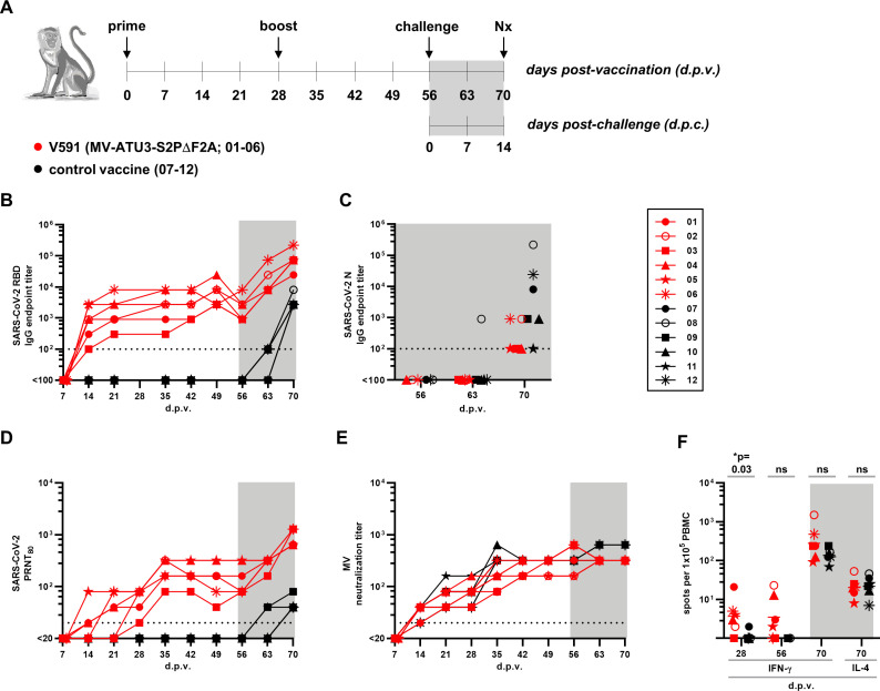
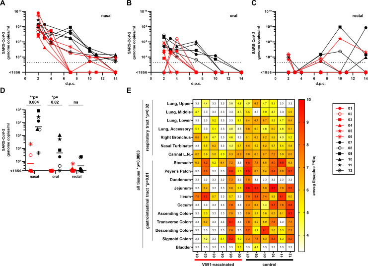
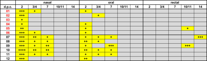
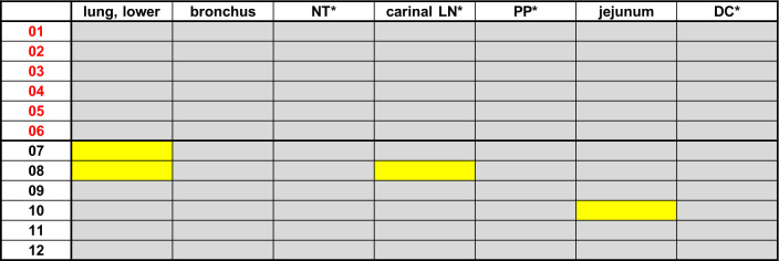
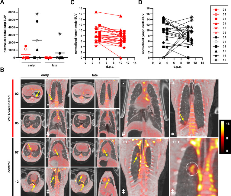
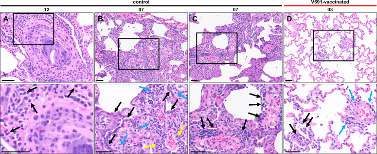
Severe acute respiratory syndrome coronavirus 2 (SARS-CoV-2) emerged at the end of 2019 and is responsible for the largest human pandemic in 100 years. Thirty-four vaccines are currently approved for use worldwide, and approximately 67% of the world population has received a complete primary series of one, yet countries are dealing with new waves of infections, variant viruses continue to emerge, and breakthrough infections are frequent secondary to waning immunity. Here, we evaluate a measles virus (MV)-vectored vaccine expressing a stabilized prefusion SARS-CoV-2 spike (S) protein (MV-ATU3-S2PΔF2A; V591) with demonstrated immunogenicity in mouse models (see companion article [J. Brunet, Z. Choucha, M. Gransagne, H. Tabbal, M.-W. Ku et al., J Virol 98:e01693-23, 2024, https://doi.org/10.1128/jvi.01693-23]) in an established African green monkey model of disease. Animals were vaccinated with V591 or the control vaccine (an equivalent MV-vectored vaccine with an irrelevant antigen) intramuscularly using a prime/boost schedule, followed by challenge with an early pandemic isolate of SARS-CoV-2 at 56 days post-vaccination. Pre-challenge, only V591-vaccinated animals developed S-specific antibodies that had virus-neutralizing activity as well as S-specific T cells. Following the challenge, V591-vaccinated animals had lower infectious virus and viral (v) RNA loads in mucosal secretions and stopped shedding virus in these secretions earlier. vRNA loads were lower in these animals in respiratory and gastrointestinal tract tissues at necropsy. This correlated with a lower disease burden in the lungs as quantified by PET/CT at early and late time points post-challenge and by pathological analysis at necropsy.IMPORTANCESevere acute respiratory syndrome coronavirus 2 (SARS-CoV-2) is responsible for the largest human pandemic in 100 years. Even though vaccines are currently available, countries are dealing with new waves of infections, variant viruses continue to emerge, breakthrough infections are frequent, and vaccine hesitancy persists. This study uses a safe and effective measles vaccine as a platform for vaccination against SARS-CoV-2. The candidate vaccine was used to vaccinate African green monkeys (AGMs). All vaccinated AGMs developed robust antigen-specific immune responses. After challenge, these AGMs produced less virus in mucosal secretions, for a shorter period, and had a reduced disease burden in the lungs compared to control animals. At necropsy, lower levels of viral RNA were detected in tissue samples from vaccinated animals, and the lungs of these animals lacked the histologic hallmarks of SARS-CoV-2 disease observed exclusively in the control AGMs.

Keywords: SARS-CoV-2; measles-vectored vaccine; non-human primates; vectored vaccines.


Conflict of interest statement

C.G., A.M., and N.E. are inventors on a patent application describing measles-vectored vaccine candidates against SARS-CoV-2. All other authors declare no financial or non-financial competing interests.

Figures

Fig 1
Fig 1
Immunogenicity of V591 in African green monkeys. (A) Schematic of the experimental set-up. Two groups of six AGMs were vaccinated (day 0) and boosted (28 days post-vaccination) with 105 TCID50 V591 (MV-ATU3-S2PΔF2A; red) or control vaccine (black). All animals were challenged with 8.35 × 10⁵ p.f.u. SARS-CoV-2 at 56 d.p.v (i.e., day 0 for challenge) and necropsies (Nx) were performed at 14 days post-challenge (i.e., 70 d.p.v.). The gray-shaded areas depict post-challenge data throughout this figure, and the individual animal symbols depicted in the key are used again in Fig. 2 and Fig. 6. (B and C) SARS-CoV-2-specific IgG detected by ELISA using RBD (B) or N (C) as antigen. (D and E) Plaque reduction neutralization test (PRNT80) titers or neutralization titers in serum against SARS-CoV-2 (D) and MV (E), respectively. The dotted lines (B–E) represent the limits of detection (100 for panels B and C and 20 for panels D and E) for the assays. (F) Numbers of spots representing peripheral blood mononuclear cells (PBMCs) secreting IFN-γ at 28, 56, and 70 d.p.v. or IL-4 at 70 d.p.v. after stimulation with SARS-CoV-2 S peptide pools 1–4 and measurement by ELISpot. Individual assays were performed with each peptide pool, and the number of spots (after subtraction of the number of spots for the negative control) was summed to generate the numbers graphed. Horizontal lines represent the geometric mean number of spots for each group. P-values were determined by Mann-Whitney tests.
Fig 2
Fig 2
Detection of SARS-CoV-2 viral (v) RNA in mucosal swabs and tissues. SARS-CoV-2 vRNA detected in nasal (A), oral (B), and rectal (C) swabs quantified by qRT-PCR; swab samples were collected between 2- and 14-days post-challenge (d.p.c.). The dotted lines represent the limit of detection (1,856) for the assay. The data for 7 d.p.c. from (A–C) are replotted in panel D for clarity. Horizontal lines represent the geometric mean SARS-CoV-2 genome copies/mL for each group. Symbols (A–D) as for Fig. 1. (E) SARS-CoV-2 vRNA detected in tissues collected at necropsy (14 d.p.c.) and quantified by qRT-PCR. The heat map shows the log-transformed vRNA copies/gram (g) of tissue. Any sample below the limit of detection of the qRT-PCR assay (white squares) was designated 103.3 (limit of detection for the assay). L.N., lymph node. P-values (D and E) were determined by Mann-Whitney tests. (A–E) V591-vaccinated, red; control-vaccinated, black.
Fig 3
Fig 3
Summary of live SARS-CoV-2 virus isolated from swabs in Vero E6 cells at indicated days post-challenge (d.p.c.). V591-vaccinated, red; control-vaccinated, black. Yellow shading, virus isolated; gray shading, no virus isolated; + to ++++ represent qualitative assessment of the number of plaques detected.
Fig 4
Fig 4
Summary of live SARS-CoV-2 virus isolated from tissues at necropsy in Vero E6 cells. V591-vaccinated, red; control-vaccinated, black. Yellow shading, virus isolated; gray shading, no virus isolated. NT, nasal turbinates; LN, lymph node; PP, Peyer’s patches; DC, descending colon.
Fig 5
Fig 5
CT disease scores at indicated days post-challenge (d.p.c.). V591-vaccinated, red; control-vaccinated, black. Yellow shading, CT score greater than 0; gray shading, CT score of 0. R/LUL, right/left upper lobe; R/LML, right/left middle lobe; R/LLL, right/left lower lobe; and ACC, accessory lobe. Scoring, 0, no significant findings; 1, 0%–5%; 2, 5%–25%; 3, 25%–50%; 4, 50%–75%; 5, >75%.
Fig 6
Fig 6
Positron emission tomography (PET)/computed tomography (CT) imaging of SARS-CoV-2 challenged AGMs. (A) and (B) Region-of-interest analysis on PET images of animals challenged with SARS-CoV-2. Total lung inflammation (A) measured by total 18F-fluoro-2-deoxy-D-glucose (18F-FDG) uptake in standard uptake values (SUV) at early and late days post-challenge (d.p.c.; 3 and 10 d.p.c. for animals 04–09 or 4 and 11 d.p.c. for animals 01–03 and 10–12). Horizontal lines represent the mean total SUV for each group. (B) Representative PET/CT scans collected at early and late d.p.c. (as above). Each time point for each animal depicts an axial (left) and coronal (right) section. Symbols *, #, and ‡ denote the same scans at higher magnification in the larger right panels. Yellow arrows/dotted lines indicate 18F-FDG uptake. Representative scans shown in the larger right panels depict no 18F-FDG uptake (−), minimal 18F-FDG uptake (+), and extensive 18F-FDG uptake (+++). A representative PET color scale depicting 0–15 SUV is shown on the far right. (C and D) Estimation of inflammation in individual lymph nodes for each animal over time by measuring 18F-FDG uptake of each lymph node in SUV. Data have been split into V591-vaccinated (C) and control (D) groups for clarity. Symbols (A, C, and D) as for Fig. 1. (A–D) V591-vaccinated, red; control-vaccinated, black.
Fig 7
Fig 7
V591 vaccination prevents histologic hallmarks of SARS-CoV-2 disease observed exclusively in control AGMs at necropsy. (A–D) Representative photomicrographs of hematoxylin and eosin-stained 5 µm lung tissue sections from control (A–C) and V591-vaccinated (D) animals after necropsy at 14 days post-challenge. Areas marked by black rectangles in the top panels are shown at higher magnification in the bottom panels. Scale bars represent 50 µm. V591-vaccinated, red; control-vaccinated, black. (A) Neutrophilic infiltrates (black arrows) in animal 12. (B) Multifocal alveolar type 2 cell hyperplasia/hypertrophy (black arrows) adjacent to areas of moderate mononuclear interstitial infiltrates (blue arrows) and perivascular cuffing (yellow arrows) in animal 07. (C) Moderate multifocal mononuclear perivascular infiltrates (black arrows) in animal 07. (D) Minimal interstitial/alveolar (black arrows) and perivascular mononuclear infiltrate (blue arrows) in animal 03.

Similar articles

Cited by

References

    1. Zhu N, Zhang D, Wang W, Li X, Yang B, Song J, Zhao X, Huang B, Shi W, Lu R, Niu P, Zhan F, Ma X, Wang D, Xu W, Wu G, Gao GF, Tan W, China Novel Coronavirus Investigating and Research Team . 2020. A novel coronavirus from patients with pneumonia in China, 2019. N Engl J Med 382:727–733. doi:10.1056/NEJMoa2001017 - DOI - PMC - PubMed
    1. World Health Organization . 2020. WHO director-general’s opening remarks at the media briefing on COVID-19. Available from: https://www.who.int/dg/speeches/detail/who-director-general-s-opening-re...
    1. World Health Organization . 2023. WHO coronavirus (COVID-19) dashboard. Available from: https://covid19.who.int/
    1. Shrotri M, Swinnen T, Kampmann B, Parker EPK. 2021. An interactive website tracking COVID-19 vaccine development. Lancet Glob Health 9:e590–e592. doi:10.1016/S2214-109X(21)00043-7 - DOI - PMC - PubMed
    1. World Health Organization . 2021. Classification of Omicron (B11529): SARS-CoV-2 variant of concern. Available from: https://www.who.int/news/item/26-11-2021-classification-of-omicron-(b.1....

MeSH terms

LinkOut - more resources