. 2024 May 14;98(5):e0169323.
doi: 10.1128/jvi.01693-23. Epub 2024 Apr 2.

A measles-vectored vaccine candidate expressing prefusion-stabilized SARS-CoV-2 spike protein brought to phase I/II clinical trials: candidate selection in a preclinical murine model

Affiliations

A measles-vectored vaccine candidate expressing prefusion-stabilized SARS-CoV-2 spike protein brought to phase I/II clinical trials: candidate selection in a preclinical murine model

Jérémy Brunet et al. J Virol. .

Abstract

In the early COVID-19 pandemic with urgent need for countermeasures, we aimed at developing a replicating viral vaccine using the highly efficacious measles vaccine as vector, a promising technology with prior clinical proof of concept. Building on our successful pre-clinical development of a measles virus (MV)-based vaccine candidate against the related SARS-CoV, we evaluated several recombinant MV expressing codon-optimized SARS-CoV-2 spike glycoprotein. Candidate V591 expressing a prefusion-stabilized spike through introduction of two proline residues in HR1 hinge loop, together with deleted S1/S2 furin cleavage site and additional inactivation of the endoplasmic reticulum retrieval signal, was the most potent in eliciting neutralizing antibodies in mice. After single immunization, V591 induced similar neutralization titers as observed in sera of convalescent patients. The cellular immune response was confirmed to be Th1 skewed. V591 conferred long-lasting protection against SARS-CoV-2 challenge in a murine model with marked decrease in viral RNA load, absence of detectable infectious virus loads, and reduced lesions in the lungs. V591 was furthermore efficacious in an established non-human primate model of disease (see companion article [S. Nambulli, N. Escriou, L. J. Rennick, M. J. Demers, N. L. Tilston-Lunel et al., J Virol 98:e01762-23, 2024, https://doi.org/10.1128/jvi.01762-23]). Thus, V591 was taken forward into phase I/II clinical trials in August 2020. Unexpected low immunogenicity in humans (O. Launay, C. Artaud, M. Lachâtre, M. Ait-Ahmed, J. Klein et al., eBioMedicine 75:103810, 2022, https://doi.org/10.1016/j.ebiom.2021.103810) revealed that the underlying mechanisms for resistance or sensitivity to pre-existing anti-measles immunity are not yet understood. Different hypotheses are discussed here, which will be important to investigate for further development of the measles-vectored vaccine platform.IMPORTANCESARS-CoV-2 emerged at the end of 2019 and rapidly spread worldwide causing the COVID-19 pandemic that urgently called for vaccines. We developed a vaccine candidate using the highly efficacious measles vaccine as vector, a technology which has proved highly promising in clinical trials for other pathogens. We report here and in the companion article by Nambulli et al. (J Virol 98:e01762-23, 2024, https://doi.org/10.1128/jvi.01762-23) the design, selection, and preclinical efficacy of the V591 vaccine candidate that was moved into clinical development in August 2020, 7 months after the identification of SARS-CoV-2 in Wuhan. These unique in-human trials of a measles vector-based COVID-19 vaccine revealed insufficient immunogenicity, which may be the consequence of previous exposure to the pediatric measles vaccine. The three studies together in mice, primates, and humans provide a unique insight into the measles-vectored vaccine platform, raising potential limitations of surrogate preclinical models and calling for further refinement of the platform.

Keywords: COVID-19 vaccine; measles vector; prefusion-stabilized spike; vectored vaccine.


Conflict of interest statement

J. Brunet, Z.C., M.G., H.S.-M., M.M., P.C., L. Majlessi, C.G., A.M., and N.E. are inventors of a patent application describing measles-vectored vaccine candidates against SARS-CoV-2. All other authors declare no financial or non-financial competing interests.

Figures

Fig 1
Fig 1
Production and characterization of MV-SARS-CoV-2 recombinant viruses expressing native SARS-CoV-2 spike protein from either ATU2 or ATU3 site. (A) Schematic representation of SARS-CoV-2 spike sequence (blue box) inserted as an additional transcription unit at either the ATU2 or the ATU3 position (represented by red triangles) of the Measles virus Schwarz vector (underneath). MV anti-genome is cloned downstream of T7 RNA polymerase promoter (T7) and framed at its 5' end by hammerhead ribozyme (hhR) and at its 3' end by hepatitis delta virus ribozyme (hδvR) followed by T7 RNA polymerase terminator (T7t). MV-encoded proteins are indicated: N (nucleoprotein), P (phosphoprotein), C and V accessory proteins, M (matrix protein), F (fusion protein), H (hemagglutinin), and L (polymerase). (B) Viral production obtained from cells infected with MV-ATU2-S and MV-ATU3-S recombinants. The plot shows infectious titers (TCID50/mL) of P0 viral stocks obtained for 10 viral clones randomly selected from 2 independent rescue experiments and expanded. Red dots point to outlier viruses with unexpectedly high titers compared to mean titers obtained for the other MV-ATU2-S recombinants. Bars show medians. (C) Cells were infected at an MOI of 0.05 TCID50/cell with two viral clones (# 1, 2) of recombinant MV-ATU3-S or the parental MVSchw or were not infected (NI). Protein extracts collected at 39 h post-infection were analyzed by Western blot and probed with anti-SARS-CoV spike polyclonal rabbit antibodies (upper panel) and anti-MV nucleoprotein polyclonal rabbit antibodies (lower panel). The left and right part of each panel are from the same membrane (unnecessary in-between lanes have been removed). The positions of SARS-CoV-2 native S, S1, and S2 cleaved products, and MV N protein are shown with respect to molecular weight markers (in kilodalton). (D) Images of cell monolayers infected at an MOI of 0.05 TCID50/cell with MV-ATU3-S (clones # 1, 2) or the parental MVSchw and observed at 39 h post-infection. Areas of fused cells are highlighted by white frames.
Fig 2
Fig 2
Fusion properties of various SARS-CoV-2 spike mutants encoded by MV-ATU3 recombinants. (A) Schematic representation of the primary structure of SARS-CoV-2 spike protein, which comprises a signal peptide (SP) and two subdomains, S1 and S2, separated by an FCS. In the S2 domain, the fusion peptide (FP), heptad repeats (HR) 1 and 2, connector domain (CD), transmembrane domain (TM), and C-terminal endoplasmic reticulum retrieval signal (ERRS) are shown. Positions and combinations of mutations introduced into S sequence (2P, 3F, ∆F, 2A) are indicated below the schemes in yellow, cyan, blue, and green, respectively, and described by the position and nature of the resulting amino acid changes. Mutated S sequences were all inserted in the pCI mammalian expression vector and in ATU3 of Measles Schwarz vector, as illustrated at the bottom of the panel. (B) Fusion activities of native S and mutated S proteins were measured in transfected HEK-293T cells using split-GFP system in the presence of hACE2 and TMPRSS2 protease. Cells transfected with an empty plasmid (pEmpty) provided a negative control. Images of the cell monolayers (examples shown on the right) were analyzed at 18 h post-transfection. Percentages of cells expressing the indicated native or mutated S, and percentages of cells exhibiting GFP, scored as GFP areas per cell area, are plotted as means ± SD from duplicates.
Fig 3
Fig 3
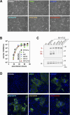
Comparative characterization of measles vectors expressing various SARS-CoV-2 S mutants. (A) Images of cell monolayers infected at an MOI of 0.05 TCID50/cell with MV-ATU3 expressing the indicated native or mutated S or with parental MVSchw and observed at 39 h post-infection. Areas of fused cells are highlighted by white frames. (B) Viral growth curves were established following infection of cells with recombinant MV-ATU3 expressing the indicated native or mutated S or with parental MVSchw at an MOI of 0.05. Cell-associated viral titers were determined in cell cultures collected at the indicated times post-infection. Geometric means of duplicate experiments are presented, and error bars indicate geometric SD. (C) Cells were infected at an MOI of 1 with the indicated recombinant MV, or the parental MVSchw, or were not infected (NI). Protein extracts collected at 24 h post-infection were analyzed by Western blot and probed with anti-SARS-CoV-2 spike polyclonal rabbit antibodies (upper panel) and anti-MV nucleoprotein antibodies (lower panel). The positions of SARS-CoV-2 full-length S, S1, and S2 domains, and MV N protein are shown with respect to molecular weight markers (in kilodalton). (D) Cell monolayers infected at an MOI of 0.05 with the same viruses as in (A) were fixed at 30 h post-infection and processed without permeabilization for indirect immunofluorescence with anti-SARS-CoV-2 spike polyclonal rabbit antibodies and AF488-conjugated anti-rabbit IgG antibodies. Cell nuclei were counterstained with DAPI. Representative Z-projection images recorded with a 40× objective are shown. Bar, 20 µm.
Fig 4
Fig 4
Antibody responses in IFNAR-KO mice after prime and boost immunization with recombinant MV expressing SARS-CoV-2 native or mutated spike. (A–F) Mice (n = 6 per group) were immunized with the parental MV Schwarz strain (Schw), one viral clone of MV-ATU3-S (S) and MV-ATU3-S2P (S2P), or either of two viral clones of MV-ATU3-S2P3F (S2P3F), MV-ATU3-S2P∆F (S2P∆F), and MV-ATU3-S2P∆F2A (S2P∆F2A). Antibody responses were measured by measles-specific ELISA (A, D), SARS-CoV-2 spike-specific ELISA (B, E), and SARS-CoV-2 microneutralization assay (C, F) in individual mouse sera collected following prime (left parts of the graphs) or boost (right parts of the graphs). Bars show medians. ELISA titers are expressed as reciprocals of endpoint serum dilutions. Neutralization titers are expressed as reciprocals of serum dilutions that resulted in the neutralization of 95% SARS-CoV-2 infectivity scored by cytopathic effect. Detection limits (dotted lines) in the anti-MV and anti-S ELISAs were 50 ELISA units and 20 in the microneutralization assay. Representative results of two or more independent experiments are shown. Statistical significance (B, C, E, F) was assessed using the non-parametric Kruskal-Wallis test with Dunn’s uncorrected post hoc analysis and is systematically indicated : *P < 0.05, **P < 0.005, ***P < 0.0005. Statistical power of the analysis has been increased by running the values obtained in the same experiment for two clones of the same construct (S2P3F, S2P∆F, S2P∆F2A) against those obtained for each of the parental constructs (S, S2P) . Non-significance (ns) is indicated whenever referred to in the text. (G) Comparative assessment of the SARS-CoV-2 microneutralization titers of sera from mice immunized with MV-ATU3-S2P∆F2A in three independent experiments (expt 1, 2, 3) and convalescent human sera 20/120, /122, /124, /126, /130 or negative control human serum 20/128 provided by the National Institute of Biological Standards and Controls (NIBSC, UK). The results for NIBSC research reagents are plotted as geometric means with geometric SD error bars of the titers obtained in all microneutralization assays in which they were tested together with mouse sera. The results for individual mice are represented per immunization experiment. Bars show medians. (H, I) Total IgG or isotype-specific (IgG1 and IgG2a) antibody responses against SARS-CoV-2 spike were measured by ELISA using recombinant SARS-CoV-2 trimeric S ectodomain produced in mammalian (H) or insect (I) cells as coating antigen. Bars show medians. Detection limit was 50 ELISA units. For comparison of the IgG2a/IgG1 ratio elicited by recombinant MV expressing S, wild-type 129/Sv mice were immunized with aluminum-adjuvanted trimerized spike ectodomain (tri-S + alum) produced in mammalian cells or aluminum adjuvant alone (alum).
Fig 5
Fig 5
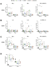
MV and S-specific T cell responses in mice immunized with recombinant MV expressing SARS-CoV-2 native or mutated spike. Mice (n = 6 per group) were immunized twice with MV-ATU3-S (S), MV-ATU3-S2P (S2P), MV-ATU3-S2P3F (S2P3F), MV-ATU3-S2P∆F (S2P∆F), MV-ATU3-S2P∆F2A (S2P∆F2A), or parental MV Schwarz (Schw). Splenocytes collected following boost were stimulated with peptide pools spanning the S1 or S2 domain of SARS-CoV-2 spike (summed responses to S1 and S2 pools are shown: SCoV-2, left parts of the graphs) or a pool of two H-2b class I-restricted measles hemagglutinin peptides (HMV, right parts of the graphs). Frequencies of cytokine-producing cells determined by intracellular cytokine staining followed by flow cytometry are shown for (A) splenic CD4+ T cells producing Th1 signature cytokines TNF-α, IFN-γ, or both IFN-γ and TNF-α, (B) splenic CD4+ T cells producing Th2 signature cytokines IL-5, IL-13, or both IL-5 and IL-13, and (C) splenic CD8+ T cells producing TNF-α, IFN-γ, or both IFN-γ and TNF-α. Frequencies of IFN-γ-producing T cells were enumerated by ELISpot analysis and shown as spot forming units (SPU) per 106 cells (D). Bars show medians. Statistical significance was assessed using the non-parametric Kruskal-Wallis test with Dunn’s uncorrected post hoc analysis: **P < 0.005.
Fig 6
Fig 6
Protection of mice against challenge with SARS-CoV-2 after prime/boost or single immunization. Schematic of the two experimental set-ups is shown. Mice (n = 5 or 6 per group) were immunized twice with MV-ATU3-S (S), MV-ATU3-S2P (S2P), MV-ATU3-S2P∆F (S2P∆F), MV-ATU3-S2P∆F2A (S2P∆F2A), or the parental MV Schwarz strain (Schw) (A) or once with MV-ATU3-S2P (S2P), MV-ATU3-S2P∆F2A (S2P∆F2A), or the parental Schw (B). SARS-CoV-2 microneutralization titers (NAb) in sera collected 3 weeks after boost (IS2, panel A) or 3 weeks after prime (IS1, panel B) were expressed as reciprocals of serum dilutions that resulted in inhibition of 95% of virus infectivity (left graphs). Following i.n. instillation with Ad5::hACE2 4 weeks after boost or prime and subsequent challenge with SARS-CoV-2, pulmonary viral loads were quantified 4 days after challenge as genomic RNA or infectious titer per lungs (middle graphs), and number of infection foci in IHC sections of the five lung lobes (right graph). Statistical significance was assessed using the non-parametric Kruskal-Wallis test with Dunn’s uncorrected post hoc analysis (A) or Mann-Whitney test (B). ns: not significant, *P < 0.05, **P < 0.005, ***P < 0.0005, ****P < 0.0001.
Fig 7
Fig 7
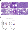
Reduction of lung histopathology in challenged mice after single immunization. (A) Routine H&E staining of lung sections from control, non-vaccinated-mice which were instilled with Ad5::hACE2 and subsequently mock infected (I) or challenged with SARS-CoV-2 (II–VI). Green stars: marked infiltrate around the bronchiole and/or accompanying vessel. Orange stars or arrows: eosinophilic exudate and cells debris filling the bronchiole lumen. Black arrows: predegenerative or degenerative lesions of the bronchiolar epithelium. Blue arrows: intranuclear patches of condensed chromatin, suggestive of apoptosis, or signs of kariolysis. Blue circles: fading of bronchiole-parenchyma delineation. (B) Inflammation (interstitial pneumonia) and histopathological (bronchiolar) lesions from vaccinated and SARS-CoV-2-challenged mice taken from the cohorts described in Fig. 6B. Interstitial pneumonia and bronchiolar lesions were evaluated and scored on H&E sections of the five lung lobes according to a semi-quantitative zero-to-five severity rating, as described in the Materials and Methods. Green and black bars show medians. The dotted lines indicate median baselines of inflammation and bronchiolar lesions, respectively, in a group of three control, non-vaccinated mice, following i.n. instillation with Ad5::hACE2 only, in the absence of subsequent challenge with SARS-CoV-2. Statistical significance was assessed using the Mann-Whitney test. *P < 0.05.
Fig 8
Fig 8
Longevity of antibody response and protection after prime/boost or prime-only immunization. CD46-IFNAR mice (n = 6 per group) were immunized twice with MV-ATU3-S (S), MV-ATU3-S2P (S2P), MV-ATU3-S2P3F (S2P3F), MV-ATU3-S2P∆F (S2P∆F), or the parental MV Schwarz strain (Schw) (A, B, C), and IFNAR-KO mice were immunized only once with parental Schw or MV-ATU3-S2P∆F2A (S2P∆F2A) (D). Antibody responses were measured in sera collected 3 weeks after prime, 3 weeks after boost, and 3 months after boost (A, B, C) or 6 months after prime (D) by measles-specific ELISA (anti-MV, panel A), SARS-CoV-2 spike-specific ELISA (anti-S, panel B), or microneutralization assay (NAb, panels C, D). The mice were instilled with Ad5::hACE2 and challenged with SARS-CoV-2 6 months after single immunization as described in the schematic (D). Pulmonary SARS-CoV-2 loads were quantified 4 days after challenge as genomic RNA (left graph) and infectious titer (right graph) per lungs. Statistical significance was assessed using the Mann-Whitney test. *P < 0.05, **P < 0.005.

Similar articles

Cited by

References

    1. Zhou P, Yang XL, Wang XG, Hu B, Zhang L, Zhang W, Si HR, Zhu Y, Li B, Huang CL, et al. . 2020. A pneumonia outbreak associated with a new coronavirus of probable bat origin. Nature 579:270–273. doi:10.1038/s41586-020-2012-7 - DOI - PMC - PubMed
    1. Zhu N, Zhang D, Wang W, Li X, Yang B, Song J, Zhao X, Huang B, Shi W, Lu R, Niu P, Zhan F, Ma X, Wang D, Xu W, Wu G, Gao GF, Tan W, China Novel Coronavirus Investigating and Research Team . 2020. A novel coronavirus from patients with pneumonia in China, 2019. N Engl J Med 382:727–733. doi:10.1056/NEJMoa2001017 - DOI - PMC - PubMed
    1. World Health Organization . 2023. WHO COVID-19 dashboard. Available from: https://covid19.who.int. Retrieved 27 Jul 2023.
    1. World Health Organization . 2023. WHO director-general's opening remarks at the media briefing. Available from: https://www.who.int/news-room/speeches/item/who-director-general-s-openi.... Retrieved 27 Jul 2023.
    1. Basta NE, Moodie EMM, on behalf of the VIPER (Vaccines IdP, and Epidemiology Research) Group COVID-19 Vaccine Development and Approvals Tracker Team . 2022. COVID-19 vaccine development and approvals tracker. Available from: https://covid19.trackvaccines.org/vaccines/approved/#vaccine-list. Retrieved 27 Jul 2023.

MeSH terms

LinkOut - more resources