. 2024 Jun;476(6):963-974.
doi: 10.1007/s00424-024-02954-9. Epub 2024 Apr 2.

The extracellular matrix protein fibronectin promotes metanephric kidney development

Affiliations

The extracellular matrix protein fibronectin promotes metanephric kidney development

Kathrin Skoczynski et al. Pflugers Arch. 2024 Jun.

Abstract

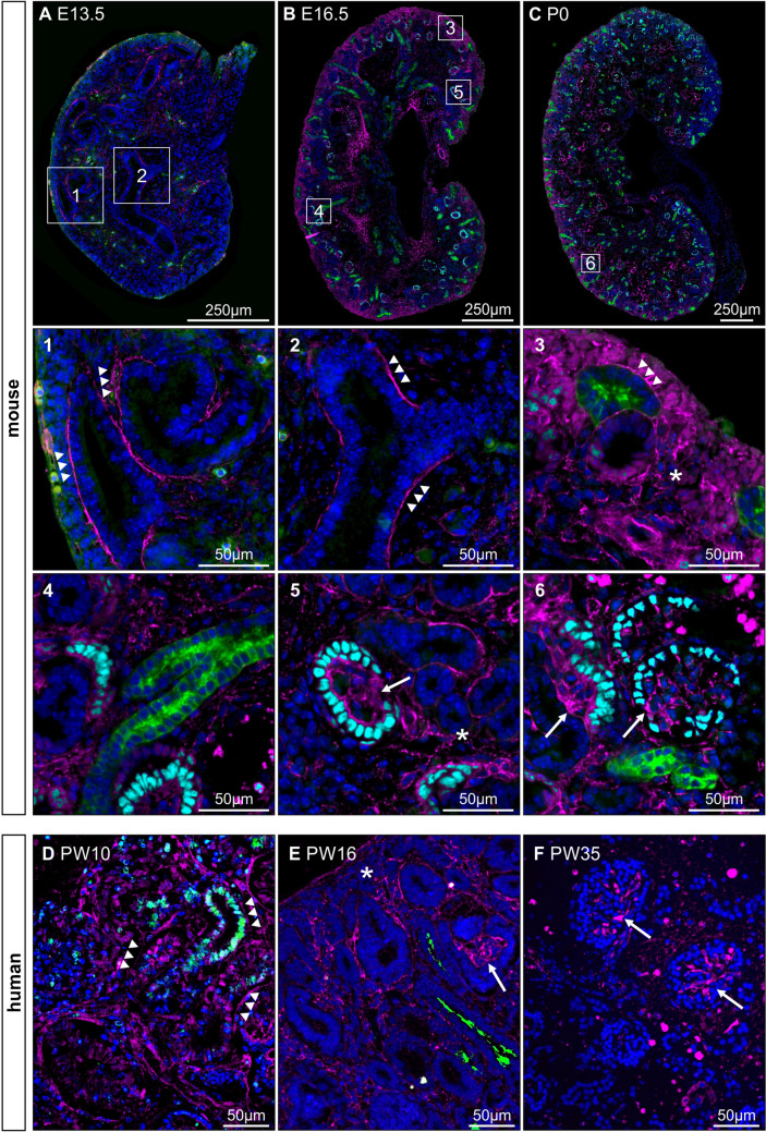
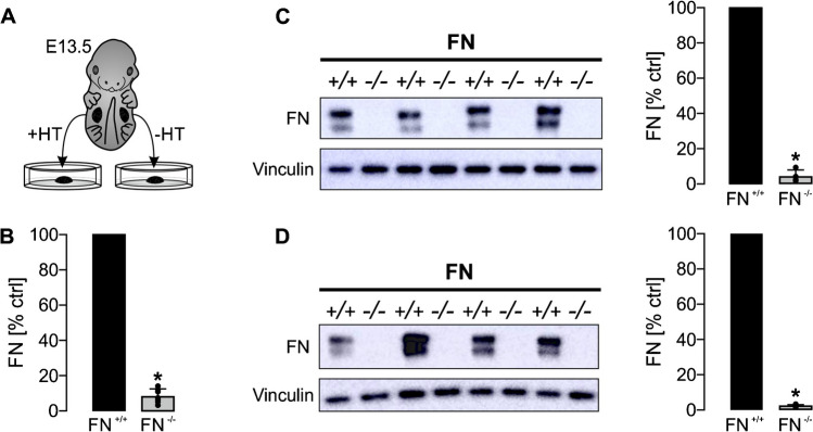
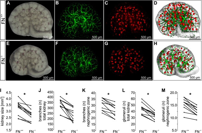
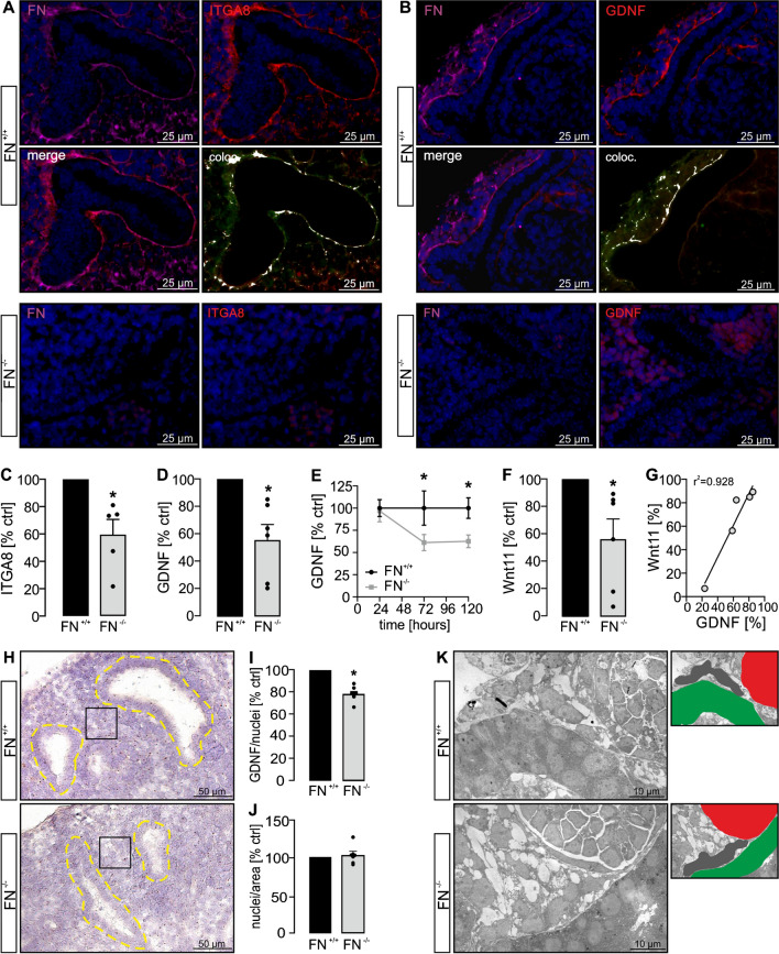
Complex interactions of the branching ureteric bud (UB) and surrounding mesenchymal cells during metanephric kidney development determine the final number of nephrons. Impaired nephron endowment predisposes to arterial hypertension and chronic kidney disease. In the kidney, extracellular matrix (ECM) proteins are usually regarded as acellular scaffolds or as the common histological end-point of chronic kidney diseases. Since only little is known about their physiological role in kidney development, we aimed for analyzing the expression and role of fibronectin. In mouse, fibronectin was expressed during all stages of kidney development with significant changes over time. At embryonic day (E) 12.5 and E13.5, fibronectin lined the UB epithelium, which became less pronounced at E16.5 and then switched to a glomerular expression in the postnatal and adult kidneys. Similar results were obtained in human kidneys. Deletion of fibronectin at E13.5 in cultured metanephric mouse kidneys resulted in reduced kidney sizes and impaired glomerulogenesis following reduced cell proliferation and branching of the UB epithelium. Fibronectin colocalized with alpha 8 integrin and fibronectin loss caused a reduction in alpha 8 integrin expression, release of glial-derived neurotrophic factor and expression of Wnt11, both of which are promoters of UB branching. In conclusion, the ECM protein fibronectin acts as a regulator of kidney development and is a determinant of the final nephron number.

Keywords: Alpha 8 integrin; Branching; Fibronectin; Kidney development; Nephron number.


Conflict of interest statement

All the authors declared no competing interests.

Figures

Fig. 1
Fig. 1
Expression and localization of fibronectin in kidney development. Fibronectin (magenta), dolichos biflorus agglutinin (DBA; green), as a marker for UB cells and collecting ducts, nuclei (DAPI; blue), and Wilms tumor protein (WT1; cyan) representing (pre-) glomerular structures (AC) were stained at different stages of kidney development. AC shows sections of mouse kidneys from embryonic day E13.5, E16.5 and postnatal day P0 which represent the change of fibronectin expression obtained from E12.5 until adulthood (extended in supplemental Fig. 1). Numbers (16) show magnifications indicated by squares in AC. (1) Fibronectin lining ureteric bud epithelial cells (tips and trunks) indicated by arrowheads within the nephrogenic zone at E13.5 (and E12.5). (2) At the time from E12.5 to E13.5, fibronectin also lines ureteric bud cells that are not situated exclusively in the nephrogenic zone. (3) At E16.5, fibronectin can only rarely be found lining ureteric buds limited to the nephrogenic zone but becomes present in the primary interstitium (asterisk). (4) At E16.5, fibronectin is no longer detectable lining ureteric bud cells outside the nephrogenic zone. (5) At E16.5, fibronectin cannot only be detected within the primary interstitium (asterisk) but also becomes present within pre-glomerular structures (arrow). (6) At P0 (and adulthood), fibronectin is predominantly expressed in glomeruli. DF show representative stainings of fetal human kidney sections at week 10 (D), 16 (E), and 35 (F) of pregnancy (PW). Arrowheads indicate fibronectin lining ureteric bud epithelial cells, asterisk marks interstitial fibronectin expression, and arrows indicate glomerular staining pattern
Fig. 2
Fig. 2
Fibronectin deletion in ex vivo cultured metanephric mouse kidneys. E13.5 metanephric kidney pairs (n = 11) were cultured ex vivo for 5 days. (Z)-4-hydroxytamoxifen (HT) was applied and the contralateral kidneys were maintained under control condition (illustrated in A). Application of HT resulted in a significant loss of fibronectin (FN−/−) 4 days later (day 5 of experiments) as indicated by real-time PCR (B) and western blot analysis (C). C Left shows representative western blot; right shows statistical analysis of fibronectin expression normalized for vinculin with FN+/+ set = 100%. + / + represents control kidneys (not induced with HT); − / − represents contralateral kidneys that were induced with HT to delete fibronectin. D Significant reduction of fibronectin was already noticed 48 h after application of HT by western blot analysis (n = 5 kidney pairs). *Significant compared to FN+/+
Fig. 3
Fig. 3
Loss of fibronectin resulted in reduced kidney size, impaired UB branching, and lower number of glomeruli. E13.5 metanephric kidney pairs (n = 11) were cultured ex vivo for 5 days. Deletion of fibronectin was induced at day 1 (FN−/−) and compared with contralateral kidneys cultured under control conditions (FN+/+) after 5 days of culture. Thereafter, kidney size, UB branches, and glomeruli were visualized by the use of extended depth of field images of whole mount kidneys in combination with bright-field microscopy (A, E), DBA staining (green) (B, F), and staining for Wilms tumor protein (WT1; red) (C, G), respectively. D and H show segmented images of the whole mount kidneys of AC and EG. IM show quantification of kidney sizes (I), number of branches within the total kidney (J), number of branches within the nephrogenic zone (K), number of glomeruli within the total kidney (L), and number of glomeruli within the nephrogenic zone (M). *Significant compared to FN+/+
Fig. 4
Fig. 4
Loss of fibronectin resulted in reduced epithelial cell proliferation. Metanephric mouse kidneys (n = 6 kidney pairs) were harvested at E13.5 and cultured ex vivo ± application of HT for 5 days. A Kidney sections were stained for the proliferation marker ki67 (green signals) and nuclei (DAPI; blue). Upper row shows whole kidney sections; squares indicate magnifications depicted below with the corresponding numbers. (14) The white dotted lines illustrate representative UB branches localized within the nephrogenic zone of FN+/+ (1, 2) and FN−/− (3, 4) kidneys. B Quantification of ki67-positive cells in relation to total kidney tissue area and normalized to FN+/+. C Quantification of ki67-positive cells within UB branches (tips and trunks) in the nephrogenic zone and normalized to the total number of UB branching cells. D Quantification of ki67-positive cells within UB tips in the nephrogenic zone and normalized to the total number of cells within the UB tips. *Significant compared to FN+/+
Fig. 5
Fig. 5
Fibronectin affects ITGA8-dependent signaling. Metanephric mouse kidneys (n = 5 kidney pairs) were harvested at E13.5 and cultured ex vivo ± HT for 5 days. A Fibronectin-competent kidneys (FN+/+) were stained for fibronectin (FN; magenta), ITGA8 (red), and nuclei (DAPI; blue). Both, fibronectin and ITGA8 staining lined the UB branches and showed significant colocalization (white signals). No fibronectin, but also no significant ITGA8 signal could be detected in the nephrogenic zone of FN-deficient kidneys (FN−/−). B Fibronectin-competent kidneys (FN+/+) were stained for fibronectin (FN; magenta), GDNF (red), and nuclei (DAPI; blue). Fibronectin and GDNF stainings lined the UB branches and were significantly colocalized (white signals). No fibronectin, but also no significant GDNF signal could be detected in FN-deficient kidneys (FN−/−). C ITGA8 mRNA level was significantly reduced in fibronectin-deleted kidneys (FN−/−) (FN+/+ set = 100%; n = 5 kidney pairs). D GDNF mRNA level was significantly reduced in fibronectin-deficient kidneys (FN−/−) (FN+/+ set = 100%; n = 6 kidney pairs). E ELISA assays performed after 24 h, 72 h, and 120 h of kidney culture revealed reduced concentration of GDNF in the medium of FN−/− kidneys (FN+/+ set = 100%; n = 6 kidney pairs). F Wnt11 mRNA expression was significantly reduced in FN−/− kidneys (FN+/+ set = 100%; n = 6 kidney pairs). G Correlation of GDNF and Wnt11 mRNA expression (n = 5 identical FN−/− kidneys in D and F). H In situ hybridization of GDNF (n = 7 kidney pairs) was performed. Regions of interest (ROIs) covering the interstitial space between UB branches within the nephrogenic zone of each kidney were analyzed for GDNF signals and the number of nuclei. The square (black line) illustrates one exemplary ROI. Yellow dotted lines indicate UB branches. I FN−/− kidneys showed a significantly lower expression of GDNF normalized to cell number. Each dot represents the mean value of all ROIs of one kidney. J The cell number (number of nuclei) was identical in FN+/+ and FN−/− kidneys. Each dot represents the mean value of all ROIs of one kidney. K Electron microscopy images revealed a similar appearance of mesenchymal cells in FN+/+ and FN−/− kidneys surrounding UB epithelial cells. Small photos on the right illustrate the structures displayed in the EM photo on the left. Gray: metanephric mesenchymal cells; green: UB epithelial branch; red: glomerular structure. *Significant compared to FN+/+

Similar articles

Cited by

References

    1. Affolter M, Bellusci S, Itoh N, Shilo B, Thiery J-P, Werb Z. Tube or not tube: remodeling epithelial tissues by branching morphogenesis. Dev Cell. 2003;4:11–18. doi: 10.1016/s1534-5807(02)00410-0. - DOI - PubMed
    1. Airik R, Kispert A. Down the tube of obstructive nephropathies: the importance of tissue interactions during ureter development. Kidney Int. 2007;72:1459–1467. doi: 10.1038/sj.ki.5002589. - DOI - PubMed
    1. Alcorn D, Maric C, McCausland J. Development of the renal interstitium. Pediatr Nephrol Berl Ger. 1999;13:347–354. doi: 10.1007/s004670050624. - DOI - PubMed
    1. Brandenberger R, Schmidt A, Linton J, Wang D, Backus C, Denda S, Müller U, Reichardt LF. Identification and characterization of a novel extracellular matrix protein nephronectin that is associated with integrin alpha8beta1 in the embryonic kidney. J Cell Biol. 2001;154:447–458. doi: 10.1083/jcb.200103069. - DOI - PMC - PubMed
    1. Brenner BM, Garcia DL, Anderson S. Glomeruli and blood pressure. Less of one, more the other? Am J Hypertens. 1988;1:335–347. doi: 10.1093/ajh/1.4.335. - DOI - PubMed

LinkOut - more resources