. 2024 Apr 2;52(1):52.
doi: 10.1007/s00240-024-01559-2.

Cymbopogon proximus and Petroselinum crispum seed ethanolic extract/Gum Arabic nanogel emulsion: Preventing ethylene glycol and ammonium chloride-induced urolithiasis in rats

Affiliations

Cymbopogon proximus and Petroselinum crispum seed ethanolic extract/Gum Arabic nanogel emulsion: Preventing ethylene glycol and ammonium chloride-induced urolithiasis in rats

Hend A Essa et al. Urolithiasis. .

Abstract

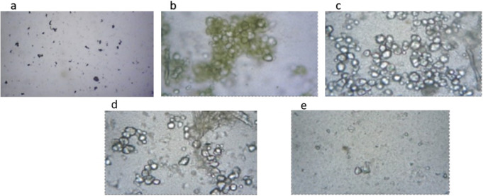
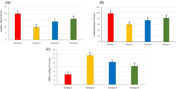
Urolithiasis is a prevalent urological disorder that contributes significantly to global morbidity. This study aimed to assess the anti-urolithic effects of Cymbopogon proximus (Halfa Bar) and Petroselinum crispum (parsley) seed ethanolic extract /Gum Arabic (GA) emulsion, and its nanogel form against ethylene glycol (EG) and ammonium chloride (AC)-induced experimental urolithiasis in rats. Rats were divided into four groups: group 1 served as the normal control, group 2 received EG with AC in drinking water for 14 days to induce urolithiasis, groups 3 and 4 were orally administered emulsion (600 mg/kg/day) and nanogel emulsion (600 mg/kg/day) for 7 days, followed by co-administration with EG and AC in drinking water for 14 days. Urolithiatic rats exhibited a significant decrease in urinary excreted magnesium, and non-enzymic antioxidant glutathione and catalase activity. Moreover, they showed an increase in oxalate crystal numbers and various urolithiasis promoters, including excreted calcium, oxalate, phosphate, and uric acid. Renal function parameters and lipid peroxidation were intensified. Treatment with either emulsion or nanogel emulsion significantly elevated urolithiasis inhibitors, excreted magnesium, glutathione levels, and catalase activities. Reduced oxalate crystal numbers, urolithiasis promoters' excretion, renal function parameters, and lipid peroxidation while improving histopathological changes. Moreover, it decreased renal crystal deposition score and the expression of Tumer necrosis factor-α (TNF-α) and cleaved caspase-3. Notably, nanogel emulsion showed superior effects compared to the emulsion. Cymbopogon proximus (C. proximus) and Petroselinum crispum (P. crispum) seed ethanolic extracts/GA nanogel emulsion demonstrated protective effects against ethylene glycol induced renal stones by mitigating kidney dysfunction, oxalate crystal formation, and histological alterations.

Keywords: Cymbopogon proximus; Ethylene glycol; Gum Arabic; Parsley seed; Renal dysfunction; Urolithiasis.


Conflict of interest statement

The authors declare no competing interests.

Figures

Fig. 1
Fig. 1
Macroscopic studies of (a) GA, (b) C. proximus and P. crispum seeds extracts, (c) C. proximus and P. crispum seeds extracts/GA emulsion, (d) C. proximus and P. crispum seeds/GA nanogel emulsion
Fig. 2
Fig. 2
Illustrates a schematic diagram depicting the experimental design for inducing urolithiasis in rats through EG and AC in drinking water for 14 days. Daily oral administration of either the emulsion (600 mg/kg) and nanogel emulsion (600 mg/kg) was conducted for 21 days. Following the last treatment, urine samples were collected from each rat. Twenty-four hours later (day 22), blood samples were obtained. Finally, the rats were sacrificed for kidney harvesting
Fig. 3
Fig. 3
DLS size distribution curve of C. proximus and P. crispum seed extract/GA nanogel emulsion
Fig. 4
Fig. 4
Zeta potential in mV of (a) GA (b) C. Proximus and P. crispum seeds extracts/GA and (c) C. proximus and P. crispum seeds extracts/ GA nanogel emulsion
Fig. 5
Fig. 5
Microscopic analysis of urine among various experimental groups; A droplet of urine is applied onto a glass slide and examined under an optical light microscope to quantify the number of calcium oxalate crystals. a) Group 1, Demonstrates absence of oxalate crystals, b & c) Group 2, Indicates a high presence of oxalate crystals (+++), d) Group 3, Exhibits a moderate count of oxalate crystals (“++”)., e) Group 4, Shows a lower count of oxalate crystals (“+”)
Fig. 6
Fig. 6
Illustrate the impact of oral administration of emulsion and nanogel emulsion on renal oxidative stress markers in renal tissue across the different studied groups: Group 1 (negative control), Group 2 (urolithatic group), Group 3 (emulsion), and Group 4 (nanogel emulsion). (A) Renal catalase, (B) Renal glutathione, (C) Renal malondialdehyde. Values are presented as mean ± SEM (n = 6). Different letters within the columns indicate a significant difference at p < 0.05
Fig. 7
Fig. 7
Illustrates representative photomicrographs displaying H & E stained renal tissue of rats, a) Negative Control (untreated rats), showing the normal tissue architecture of renal parenchyma, glomerulus and renal tubules, b & c) positive control showing diffuse renal crystal deposition (green arrows), congested blood vessels and diffuse interstitial nephritis with high magnification in upper right corner showing renal crystal deposition inside renal tubule, d) Group 3 (rats treated with emulsion) showing multifocal renal crystal deposition with moderate inflammatory cells infiltration, e) Group 4 (rats treated with nanogel emulsion) showing significant decrease in crestal deposition and mild inflammatory cells infiltration, f) Renal crystal score in different experimental groups represented as median. (scale bar 50 and 100 μm). CO: renal cortex, M; renal medulla and CMJ; cortico-medullary junction
Fig. 8
Fig. 8
Immunohistochemical staining of cleaved (active) caspase-3 and TNF-α in renal tissues across different groups. Representative photomicrographs (scale bar 100 μm) of renal tissues exhibit brown immunostaining expression of caspase-3 (a-d) and TNF-α (f-i) as follows: (a and f) Show absence of staining in the negative control rats, (b and g) Demonstrate strong immune-expression in the positive control group, (c and h) Depict moderate expression in group 3, and (d and i) Reveal mild immune reactive cells in group 4. Furthermore, (i and j) indicate the immunostaining area (%) of (i) cleaved caspase-3 and (j) TNF-α expression in the renal tissue of rats treated with emulsion and nanogel emulsion. Data analysis used One-way analysis of variance (ANOVA) followed by the Duncan post-hoc test. *Significant differences were considered at p ≤ 0.05

References

    1. Wagner CA (2021) Etiopathogenic factors of urolithiasis. Arch Esp Urol 74:16–23 PMID: 33459618 - PubMed
    1. Thongprayoon C, Krambeck AE, Rule AD (2020) Determining the true burden of kidney stone disease. Nat Rev Nephrol 16:736–746. 10.1038/s41581-020-0320-7 - PubMed
    1. Geraghty RM, Cook P, Walker V, Somani BK (2020) Evaluation of the economic burden of kidney stone disease in the UK: a retrospective cohort study with a mean follow-up of 19 years. BJU Int 125:586–594. 10.1111/bju.14991 - PubMed
    1. Tefekli A, Cezayirli F (2013) The history of urinary stones: in parallel with civilization. Sci. World J. 2013 - PMC - PubMed
    1. Stamatelou K, Goldfarb DS (2023) Epidemiology of kidney stones. Healthcare 11:424. 10.3390/healthcare11030424 - PMC - PubMed

MeSH terms