. 2024 Apr 2;19(1):60.
doi: 10.1186/s11671-024-04005-9.

Focused ultrasound on the substantia nigra enables safe neurotensin-polyplex nanoparticle-mediated gene delivery to dopaminergic neurons intranasally and by blood circulation

Affiliations

Focused ultrasound on the substantia nigra enables safe neurotensin-polyplex nanoparticle-mediated gene delivery to dopaminergic neurons intranasally and by blood circulation

Juan U Mascotte-Cruz et al. Discov Nano. .

Abstract

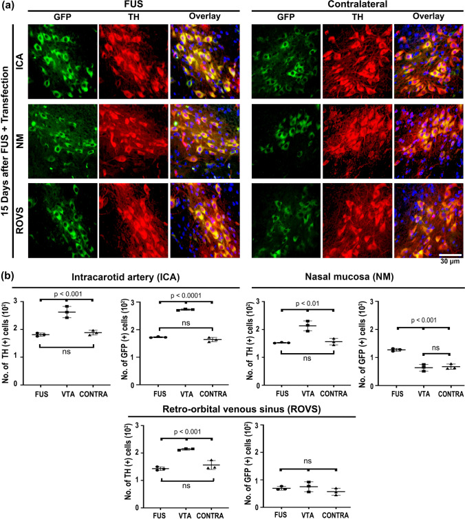
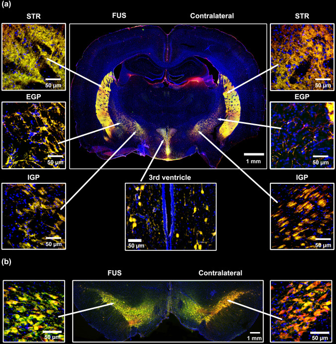
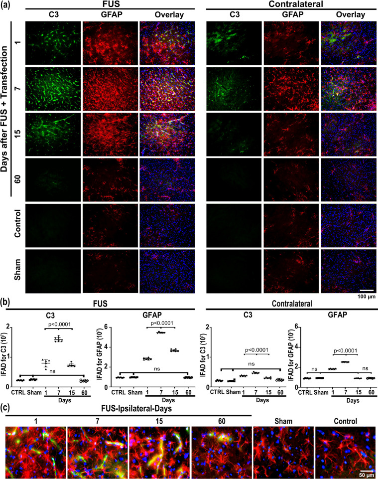
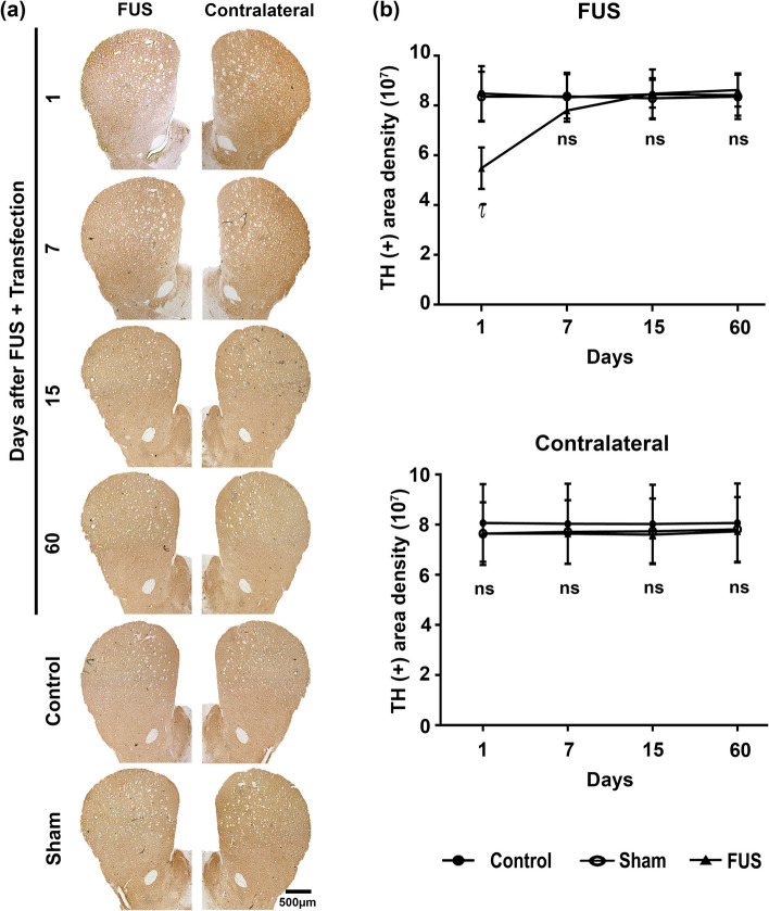
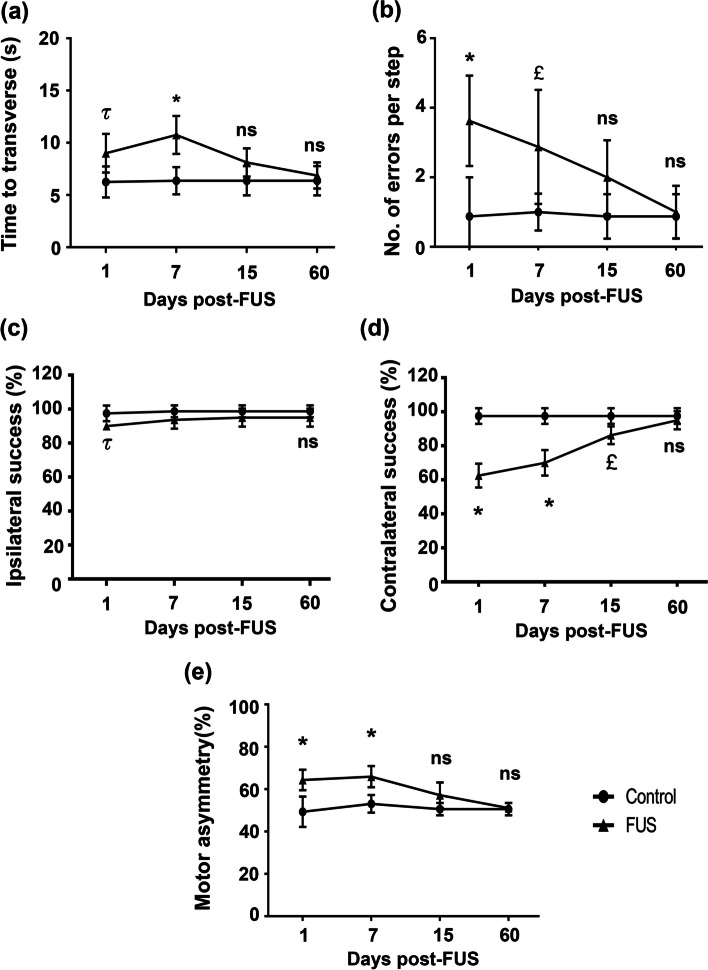
Neurotensin-polyplex nanoparticles provide efficient gene transfection of nigral dopaminergic neurons when intracerebrally injected in preclinical trials of Parkinson's disease because they do not cross the blood-brain barrier (BBB). Therefore, this study aimed to open BBB with focused ultrasound (FUS) on the substantia nigra to attain systemic and intranasal transfections and evaluate its detrimental effect in rats. Systemically injected Evans Blue showed that a two-pulse FUS opened the nigral BBB. Accordingly, 35 μL of neurotensin-polyplex nanoparticles encompassing the green fluorescent protein plasmid (79.6 nm mean size and + 1.3 mV Zeta-potential) caused its expression in tyrosine hydroxylase(+) cells (dopaminergic neurons) of both substantiae nigrae upon delivery via internal carotid artery, retro-orbital venous sinus, or nasal mucosa 30 min after FUS. The intracarotid delivery yielded the highest transgene expression, followed by intranasal and venous administration. However, FUS caused neuroinflammation displayed by infiltrated lymphocytes (positive to cluster of differentiation 45), activated microglia (positive to ionized calcium-binding adaptor molecule 1), neurotoxic A1 astrocytes (positive to glial fibrillary acidic protein and complement component 3), and neurotrophic A2 astrocytes (positive to glial fibrillary acidic protein and S100 calcium-binding protein A10), that ended 15 days after FUS. Dopaminergic neurons and axonal projections decreased but recuperated basal values on day 15 after transfection, correlating with a decrease and recovery of locomotor behavior. In conclusion, FUS caused transient neuroinflammation and reversible neuronal affection but allowed systemic and intranasal transfection of dopaminergic neurons in both substantiae nigrae. Therefore, FUS could advance neurotensin-polyplex nanotechnology to clinical trials for Parkinson's disease.

Keywords: Bionanotechnology; Gene transfection; Motor behavior; Nanomedicine; Parkinson’s disease; Reversible neurodegeneration; Transient neuroinflammation.


Conflict of interest statement

The authors declare no competing interests.

Figures

Fig. 1
Fig. 1
Size and Z-potential of NTS-polyplex nanoparticles harboring the plasmid pEGFP-N1. The nanoparticles were prepared with 6 nM pDNA, 144 nM NTS carrier, and 3 µM karyophilic peptides (KPRa or KPSV40) or without karyophilic peptides (-KPs). a Representative Cryo-electron microscopy (Cryo-SEM) micrographs of NTS-polyplex NPs. b Hydrodynamic radius of size distribution by Dynamic light scattering (DLS) and c Zeta potential by Electrophoretic light scattering (ELS)
Fig. 2
Fig. 2
Focus ultrasound (FUS) enables the targeted green fluorescent protein (GFP) gene delivery to nigral dopamine neurons via different administration routes. Thirty minutes after FUS, 35 μL of NPs harboring the pEGFP-N1 plasmid were injected into the internal carotid artery, the retro-orbital venous sinus, or deposited through a capillary tube on the nasal mucosa. a Representative micrographs of the substantia nigra 15 days after transfection. The merged micrographs also include the Hoechst nuclear counterstaining. The value of the calibration bar is valid for all micrographs. b Quantification of GFP(+) cells and TH(+) cells using ImageJ software in the conditions shown by the micrographs of panel a). The values are the mean ± SD from three anatomical levels (n = 3 independent rats per experimental condition). GFP (green) immunoreactivity in TH (red) cells with nuclear Hoechst counterstaining (blue) in the substantia nigra. One-way ANOVA and post hoc Tukey tests. ns Not significant
Fig. 3
Fig. 3
Bilateral distribution of green fluorescent protein (GFP) in dopaminergic cells and axonal ramifications in their target innervation nuclei after transfection via the internal carotid artery in FUS-treated rats. Representative micrographs 15 days after transfection of a coronal brain slice at − 2.16 mm from bregma and b mesencephalon slice immunostained against GFP and TH and counterstained with Hoechst. The amplifications show details of GFP expression in cells and axonal projections. STR Striatum, EGP External globus pallidus, IGP Internal globus pallidus. GFP (green) immunoreactivity in TH (red) cells with nuclear Hoechst counterstaining (blue). The scale value is equal for the respective set of micrographs
Fig. 4
Fig. 4
Focus ultrasound (FUS) causes transient microglia activation in the substantia nigra. a Representative micrographs of the most injured site (where FUS was applied) of the substantia nigra double immunostained with positive to ionized calcium-binding adaptor molecule 1 (Iba1) and tyrosine hydroxylase (TH) over time after FUS. The merged micrographs also include the Hoechst nuclear counterstaining. b Area density (IFAD) for Iba1 and TH immunofluorescence measured from panel a micrographs using ImageJ software. The values are the mean ± SD from three anatomical levels (n = 6 independent rats per experimental condition). One-way ANOVA and post hoc Tukey tests. ns Not significant. Iba1 (green) immunoreactivity in TH (red) cells with nuclear Hoechst counterstaining (blue) in the substantia nigra over time. c Amplified micrographs show Iba1(+) cells with phenotypic changes according to their activation state. The scale value is equal for the respective set of micrographs
Fig. 5
Fig. 5
Transient induction of neurotoxic A1 astrocyte after FUS and gene delivery. a Representative micrographs of the most injured site of the substantia nigra double immunostained with complement 3 (C3) and glial fibrillary acidic protein (GFAP) over time after FUS. The merged micrographs also include the Hoechst nuclear counterstaining. b C3 and GFAP immunofluorescence area density (IFAD) measured from micrographs of panel A using ImageJ software. The values are the mean ± SD from three anatomical levels (n = 6 independent rats per experimental condition). One-way ANOVA and post hoc Tukey tests. ns Not significant. c Amplified merged images show the reduction of C3 (green) immunoreactivity in GFAP(+) (red) cells with nuclear Hoechst counterstaining (blue) in the substantia nigra over time. The scale value is equal for the respective set of micrographs
Fig. 6
Fig. 6
Transient neurotrophic A2 activation after focus ultrasound (FUS) and gene delivery. a Representative micrographs of the most injured site of the substantia nigra double immunostained with calcium-binding protein A10 (S100A10) and GFAP over time after FUS. The merged micrographs also include the Hoechst nuclear counterstaining. b S100A10 and GFAP immunofluorescence area density (IFAD) measured from micrographs of panel A using ImageJ software. The values are the mean ± SD from three anatomical levels (n = 6 independent rats per experimental condition). One-way ANOVA and post hoc Tukey tests. ns Not significant. c Amplified merged images show the reduction of S100A10 (green) immunoreactivity in GFAP (red) cells with nuclear Hoechst counterstaining (blue) in the substantia nigra over time. The scale value is equal for the respective micrograph set
Fig. 7
Fig. 7
Transient leucocyte infiltration after focus ultrasound (FUS) and gene delivery. a Representative micrographs of the substantia nigra with double immunostaining against the cluster of differentiation CD45 and tyrosine hydroxylase (TH) over time after FUS. The merged micrographs also include the Hoechst nuclear counterstaining. b CD45 and TH immunofluorescence area density (IFAD) measured from micrographs of panel A using ImageJ software. The values are the mean ± SD from three anatomical levels (n = 6 independent rats per experimental condition). One-way ANOVA and post hoc Tukey tests. ns Not significant. CD45 (green) immunoreactivity in TH (red) cells with nuclear Hoechst counterstaining (blue) in the substantia nigra over time. c Amplified micrographs show morphological details of CD45(+) cells with nuclear counterstaining and their disappearance after 60 days post-FUS. The scale value is equal for the respective micrograph set
Fig. 8
Fig. 8
Reversible drop of dopaminergic neuron population of the substantia nigra after focus ultrasound (FUS) and transfection. a Representative micrographs of the substantia nigra with tyrosine hydroxylase (TH) immunohistochemistry over time after FUS. The scale value is equal for the respective set of micrographs. b TH(+) cell counting and c immunoreactivity area density measured from micrographs of panel a using ImageJ software. The values are the mean ± SD from average measurements in five anatomical levels (n = 3 independent rats per experimental condition. p < 0.0001 = *, p < 0.001 = τ and p < 0.01 = £ compared with the control and sham groups in each time-point. Two-way ANOVA and post hoc Tukey tests. ns Not significant
Fig. 9
Fig. 9
Reversible decrease in dopaminergic innervation of the striatum after focus ultrasound (FUS) and transfection. a Representative micrographs of the striatum with tyrosine hydroxylase (TH) immunohistochemistry over time after FUS. The scale value is equal for all micrographs. b TH(+) immunoreactivity density from micrographs of panel a using ImageJ software. The values are the mean ± SD from the average measurements in five anatomical levels per rat (n = 3 independent rats per experimental condition). p < 0.001 = τ, compared with the control and sham groups at each time point. Two-way ANOVA and post hoc Tukey tests. ns Not significant
Fig. 10
Fig. 10
Reversible sensorimotor deficits after FUS and transfection. Rats were subjected to focus ultrasound (FUS) and transfected with NTS-polyplex NPs with pEGFP-N1 at 30 min after FUS and evaluated over time with the behavioral tests. a and b Beam walking test, c and d Vibrissae-evoked forelimb placing test, e Cylinder test. Values are expressed as the mean ± SD of 6 independent rats for each experimental condition. p < 0.001 = £, p < 0.01 = τ and p < 0.05 = *. Control group compared with FUS group. Two-way ANOVA and Tukey post hoc test. ns Not significant

Similar articles

References

    1. Martinez-Fong D, Bannon MJ, Trudeau LE, Gonzalez-Barrios JA, Arango-Rodriguez ML, Hernandez-Chan NG, Reyes-Corona D, Armendáriz-Borunda J, Navarro-Quiroga I. NTS-Polyplex: a potential nanocarrier for neurotrophic therapy of Parkinson’s disease. Nanomed Nanotechnol Biol Med. 2012;8:1052–1069. doi: 10.1016/j.nano.2012.02.009. - DOI - PMC - PubMed
    1. Hernandez-Chan NG, Bannon MJ, Orozco-Barrios CE, Escobedo L, Zamudio S, De La Cruz F, Gongora-Alfaro JL, Armendáriz-Borunda J, Reyes-Corona D, Espadas-Alvarez AJ, Flores-Martínez YM, Ayala-Davila J, Hernandez-Gutierrez ME, Pavón L, García-Villegas R, Nadella R, Martinez-Fong D. Neurotensin-polyplex-mediated brain-derived neurotrophic factor gene delivery into nigral dopamine neurons prevents nigrostriatal degeneration in a rat model of early Parkinson’s disease. J Biomed Sci. 2015;22:1–4. doi: 10.1186/s12929-015-0166-7. - DOI - PMC - PubMed
    1. Arango-Rodriguez ML, Navarro-Quiroga I, Gonzalez-Barrios JA, Martinez-Arguelles DB, Bannon MJ, Kouri J, Forgez P, Rostene W, Garcia-Villegas R, Jimenez I, Martinez-Fong D. Biophysical characteristics of neurotensin polyplex for in vitro and in vivo gene transfection. Biochim Biophys Acta Gen Subj. 2006;1760:1009–1020. doi: 10.1016/j.bbagen.2006.02.021. - DOI - PubMed
    1. Fernandez-Parrilla MA, Reyes-Corona D, Flores-Martinez YM, Nadella R, Bannon MJ, Escobedo L, Maldonado-Berny M, Santoyo-Salazar J, Soto-Rojas LO, Luna-Herrera C, Ayala-Davila J, Gonzalez-Barrios JA, Flores G, Gutierrez-Castillo ME, Espadas-Alvarez AJ, Martinez-Davila IA, Nava P, Martinez-Fong D. Cerebral dopamine neurotrophic factor transfection in dopamine neurons using neurotensin-polyplex nanoparticles reverses 6-hydroxydopamine-induced nigrostriatal neurodegeneration. Neural Regen Res. 2022;17:854–866. doi: 10.4103/1673-5374.321001. - DOI - PMC - PubMed
    1. Espadas-Alvarez AJ, Bannon MJ, Orozco-Barrios CE, Escobedo-Sanchez L, Ayala-Davila J, Reyes-Corona D, Soto-Rodriguez G, Escamilla-Rivera V, De Vizcaya-Ruiz A, Eugenia Gutierrez-Castillo M, Padilla-Viveros A, Martinez-Fong D. Regulation of human GDNF gene expression in nigral dopaminergic neurons using a new doxycycline-regulated NTS-polyplex nanoparticle system. Nanomed Nanotechnol Biol Med. 2017;13:1363–1375. doi: 10.1016/j.nano.2017.02.006. - DOI - PubMed

Grants and funding

LinkOut - more resources