

Microglia in retinal angiogenesis and diabetic retinopathy
- PMID: 38564108
- PMCID: PMC11303477
- DOI: 10.1007/s10456-024-09911-1
Microglia in retinal angiogenesis and diabetic retinopathy
Abstract
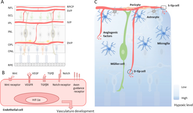
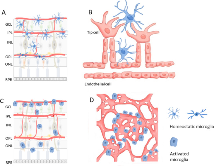
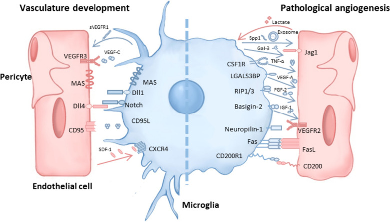
Diabetic retinopathy has a high probability of causing visual impairment or blindness throughout the disease progression and is characterized by the growth of new blood vessels in the retina at an advanced, proliferative stage. Microglia are a resident immune population in the central nervous system, known to play a crucial role in regulating retinal angiogenesis in both physiological and pathological conditions, including diabetic retinopathy. Physiologically, they are located close to blood vessels and are essential for forming new blood vessels (neovascularization). In diabetic retinopathy, microglia become widely activated, showing a distinct polarization phenotype that leads to their accumulation around neovascular tufts. These activated microglia induce pathogenic angiogenesis through the secretion of various angiogenic factors and by regulating the status of endothelial cells. Interestingly, some subtypes of microglia simultaneously promote the regression of neovascularization tufts and normal angiogenesis in neovascularization lesions. Modulating the state of microglial activation to ameliorate neovascularization thus appears as a promising potential therapeutic approach for managing diabetic retinopathy.
Keywords: Angiogenesis; Diabetic retinopathy; Microglia modulation; Microglia state; Retinal neovascularization.
© 2024. The Author(s).
Conflict of interest statement
The authors declare no competing interests.
Figures
Similar articles
-
Heterotypic macrophages/microglia differentially contribute to retinal ischaemia and neovascularisation.Diabetologia. 2024 Oct;67(10):2329-2345. doi: 10.1007/s00125-024-06215-3. Epub 2024 Jul 8. Diabetologia. 2024. PMID: 38977459
-
Single and Compound Knock-outs of MicroRNA (miRNA)-155 and Its Angiogenic Gene Target CCN1 in Mice Alter Vascular and Neovascular Growth in the Retina via Resident Microglia.J Biol Chem. 2015 Sep 18;290(38):23264-81. doi: 10.1074/jbc.M115.646950. Epub 2015 Aug 4. J Biol Chem. 2015. PMID: 26242736 Free PMC article.
-
DDR2 alleviates retinal vaso-obliteration and pathological neovascularization by modulating microglia M1/M2 phenotypic polarization in a mouse model of proliferative retinopathy.Biochim Biophys Acta Mol Basis Dis. 2025 Jun;1871(5):167787. doi: 10.1016/j.bbadis.2025.167787. Epub 2025 Mar 9. Biochim Biophys Acta Mol Basis Dis. 2025. PMID: 40064060
-
The Role of Microglia in Diabetic Retinopathy: Inflammation, Microvasculature Defects and Neurodegeneration.Int J Mol Sci. 2018 Jan 1;19(1):110. doi: 10.3390/ijms19010110. Int J Mol Sci. 2018. PMID: 29301251 Free PMC article. Review.
-
Placental growth factor and its potential role in diabetic retinopathy and other ocular neovascular diseases.Acta Ophthalmol. 2018 Feb;96(1):e1-e9. doi: 10.1111/aos.13325. Epub 2016 Nov 22. Acta Ophthalmol. 2018. PMID: 27874278 Free PMC article. Review.
Cited by
-
Ferroptosis in ocular diseases: mechanisms, crosstalk with other cell death pathways, and therapeutic prospects.Front Med (Lausanne). 2025 Jul 8;12:1608975. doi: 10.3389/fmed.2025.1608975. eCollection 2025. Front Med (Lausanne). 2025. PMID: 40697914 Free PMC article. Review.
-
BARD1-mediated stabilization of METTL14 promotes retinal neovascularization by m6A-modifying MXD1 mRNA on a YTHDF2-dependent manner.Theranostics. 2025 Apr 13;15(11):5481-5498. doi: 10.7150/thno.110122. eCollection 2025. Theranostics. 2025. PMID: 40303324 Free PMC article.
-
Single-cell analysis identifies MKI67+ microglia as drivers of neovascularization in proliferative diabetic retinopathy.J Transl Med. 2025 Mar 11;23(1):310. doi: 10.1186/s12967-025-06320-w. J Transl Med. 2025. PMID: 40069725 Free PMC article.
-
Proliferative Diabetic Retinopathy Microenvironment Drives Microglial Polarization and Promotes Angiogenesis and Fibrosis via Cyclooxygenase-2/Prostaglandin E2 Signaling.Int J Mol Sci. 2024 Oct 21;25(20):11307. doi: 10.3390/ijms252011307. Int J Mol Sci. 2024. PMID: 39457089 Free PMC article.
-
Enhancing Retinal Resilience: The Neuroprotective Promise of BDNF in Diabetic Retinopathy.Life (Basel). 2025 Feb 9;15(2):263. doi: 10.3390/life15020263. Life (Basel). 2025. PMID: 40003672 Free PMC article. Review.
References
-
- Yang QH, Zhang Y, Zhang XM, Li XR (2019) Prevalence of diabetic retinopathy, proliferative diabetic retinopathy and non-proliferative diabetic retinopathy in Asian t2dm patients: a systematic review and metaanalysis. Int J Ophthalmol 12:302–311. 10.18240/ijo.2019.02.19 10.18240/ijo.2019.02.19 - DOI - PMC - PubMed
-
- Sardarinia M, Asgari S, Hizomi Arani R et al (2022) Incidence and risk factors of severe non-proliferative/proliferative diabetic retinopathy: More than a decade follow up in the Tehran Lipids and Glucose Study. J Diabetes Investig 13:317–327. 10.1111/jdi.13647 10.1111/jdi.13647 - DOI - PMC - PubMed
Publication types
MeSH terms
LinkOut - more resources
Full Text Sources
Medical
