. 2024 Apr 1;65(4):6.
doi: 10.1167/iovs.65.4.6.

Expression of Collagen XIII in Tissues of the Thyroid and Orbit With Relevance to Thyroid-Associated Ophthalmopathy

Affiliations

Expression of Collagen XIII in Tissues of the Thyroid and Orbit With Relevance to Thyroid-Associated Ophthalmopathy

Oula Norman et al. Invest Ophthalmol Vis Sci. .

Abstract

Purpose: Antibodies against collagen XIII have previously been identified in patients with active thyroid-associated ophthalmopathy (TAO). Although collagen XIII expression has been described in extraocular muscles and orbital fat, its detailed localization in extraocular and thyroid tissues and the connection to autoimmunity for collagen XIII remain unclear. Our objective was to map the potential targets for these antibodies in the tissues of the orbit and thyroid.

Methods: We evaluated the expression of collagen XIII in human patient and mouse orbital and thyroid tissues with immunostainings and RT-qPCR using Col13a1-/- mice as negative controls. COL13A1 expression in Graves' disease and goiter thyroid samples was compared with TGF-β1 and TNF, and these were also studied in human thyroid epithelial cells and fibroblasts.

Results: Collagen XIII expression was found in the neuromuscular and myotendinous junctions of extraocular muscles, blood vessels of orbital connective tissue and fat and the thyroid, and in the thyroid epithelium. Thyroid expression was also seen in germinal centers in Graves' disease and in neoplastic epithelium. The expression of COL13A1 in goiter samples correlated with levels of TGF-B1. Upregulation of COL13A1 was reproduced in thyroid epithelial cells treated with TGF-β1.

Conclusions: We mapped the expression of collagen XIII to various locations in the orbit, demonstrated its expression in the pathologies of the Graves' disease thyroid and confirmed the relationship between collagen XIII and TGF-β1. Altogether, these data add to our understanding of the targets of anti-collagen XIII autoantibodies in TAO.


Conflict of interest statement

Disclosure: O. Norman, None; T. Vornanen, None; H. Franssila, None; J. Liinamaa, None; E. Karvonen, None; T. Kotkavaara, Santen (R), Allergan (R), Chiesi Pharma (R); V.-M. Pohjanen, None; R. Ylikärppä, None; T. Pihlajaniemi, None; M. Hurskainen, Santen (R); A. Heikkinen, None

Figures

Figure 1.
Figure 1.
Collagen XIII expression patterns in human and mouse extraocular muscles. (A, B) Whole mount immunofluorescent stainings of Col13a1+/+ (wild-type) and Col13a/ (knockout) mouse rectus muscle identifying myotendinous (A), and neuromuscular junctions (B) labeled with antibodies against collagen XIII (yellow) and laminin β1 (Col13a1+/+; A; magenta) or laminin γ1 (Col13a/; A; magenta), or with α-BTX (B; magenta). (CE) Whole mount immunofluorescent stainings of human samples labeled with antibodies against collagen XIII (yellow) and laminin γ1 (C; magenta) or with α-BTX (D, E; magenta), showing collagen XIII expression in the human extraocular muscle MTJ (C), in NMJs of human orbicularis oculi muscle (D) and in MTJs and en grappe NMJs at the distal end of a human rectus muscle (E). MTJs marked with arrows, NMJs marked with asterisks. Scalebars are 10 µm in A-B and 20 µm in C-E. The images are maximum intensity projections of confocal stacks.
Figure 2.
Figure 2.
Collagen XIII expression in orbital blood vessels. (A) immunofluorescent labeling of a human cryo section with antibodies against collagen XIII (yellow), CD31 (magenta), and Lyve-1 (cyan), with hematoxylin-eosin (H&E) staining of the same section, showing collagen XIII expression in cross-sections of the blood vessels of human extraocular muscles. (BD) Whole mount immunofluorescent labelings of human samples with antibodies against collagen XIII (yellow), α-SMA (B, D; magenta) and laminin γ1 (C; magenta), showing collagen XIII expression in the blood vessels of human extraocular muscles (B, C) and in extraocular fat (D). Blood vessels marked with arrows, adipocytes marked with asterisks and a lymphatic vessel marked with an arrowhead. Scalebars are 200 µm in A, 10 µm in B, and 50 µm in C and D. The images B–D are maximum intensity projections of confocal stacks. In the H&E image, white balance and brightness have been adjusted for easier viewing.
Figure 3.
Figure 3.
Collagen XIII expression in thyroid follicles and blood vessels. (A, B) Immunofluorescent labeling for collagen XIII expression (yellow) and laminin γ1 (magenta) with DAPI (cyan) to stain nuclei in Col13a1+/+ and Col13a1−/− mouse thyroid whole mounts, showing collagen XIII expression in the basal surfaces/basement membranes of follicular cells in the wild-type (A). (C–F) Immunofluorescent labeling for collagen XIII expression (yellow) and CD31 (D, magenta; F, cyan) or perlecan (C, magenta) or α-SMA (E, magenta) together with DAPI (C, D, cyan) or laminin γ1 (F, magenta) in human thyroid samples (C, D, thin paraffin sections, E, F, whole mounts), showing collagen XIII expression in blood vessels (D–F) and selected follicles (C). GD, Graves’ disease. (G, H) CLEM of human thyroid samples, where collagen XIII is colored yellow and DAPI cyan, showing collagen XIII location in a blood vessel wall. (I–K) Immunohistochemical staining of collagen XIII in a goiter sample with (I) and without vessel signal (J), primary antibody omitted in negative control (K). Blood vessels marked with arrows, thyroid follicles marked with asterisks, and nuclei marked with arrowheads. Scale bars: 20 µm in A–D, F; 50 µm in E, I–K; 10 µm in G; and 2 µm in H. The images E–F are maximum intensity projections of confocal stacks.
Figure 4.
Figure 4.
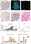
Collagen XIII in goiter and Graves’ disease thyroids. (A) Immunohistochemistry of a germinal center reaction in a Graves’ disease thyroid examined for collagen XIII. Germinal center marked with dashed line. (B, C) Immunofluorescence of a lymphocytic infiltrate in a Graves’ disease thyroid section examined for collagen XIII (yellow). Co-labeling with DAPI (cyan) and perlecan (magenta). Lymphocytic infiltrate marked with dashed line in both images. (D) Collagen XIII immunohistochemistry of connective tissue in a goiter thyroid, immunopositive fibroblasts indicated by arrows. (E) Collagen XIII immunohistochemistry of an incidental papillary microcarcinoma marked with dashed line in a goiter thyroid. (F) Hematoxylin-eosin staining of the corresponding area on an adjacent section. Scalebars: 100 µm. Thyroid follicles marked with asterisks in all images. (G) Expression of COL13A1 in thyroid cohort samples detected by RT-qPCR. GD, Graves’ disease. (H) Correlation between TGFB1 and COL13A1 expression in goiter thyroids, linear regression. (I, J) Expression of COL13A1 detected by RT-qPCR in human thyroid epithelial cells after treatment with TGF-β1 (positive control TGFBI) or TNF (positive control ICAM-1) either in growth (I) or depleted (J) medium (median with range, N = 3).

Similar articles

References

    1. Bartalena L, Kahaly GJ, Baldeschi L, et al. .. The 2021 European Group on Graves’ orbitopathy (EUGOGO) clinical practice guidelines for the medical management of Graves’ orbitopathy. Eur J Endocrinol. 2021; 185(4): G43–G67. - PubMed
    1. Lazarus JH. Epidemiology of Graves’ orbitopathy (GO) and relationship with thyroid disease. Best Pract Res Clin Endocrinol Metab. 2012; 26: 273–279. - PubMed
    1. Parrilla C, Mele DA, Gelli S, et al. .. Multidisciplinary approach to orbital decompression. A review. Acta Otorhinolaryngol Ital. 2021; 41(2 Suppl 1): S90–S101. - PMC - PubMed
    1. Smith TJ. TSH-receptor-expressing fibrocytes and thyroid-associated ophthalmopathy. Nat Rev Endocrinol. 2015; 11: 171–181. - PMC - PubMed
    1. Bahn RS. Current insights into the pathogenesis of Graves’ ophthalmopathy. Horm Metab Res. 2015; 47: 773–778. - PubMed