. 2024 Apr 16;18(15):10509-10526.
doi: 10.1021/acsnano.3c12064. Epub 2024 Apr 2.

Iron Oxide Nanoparticles Inhibit Tumor Progression and Suppress Lung Metastases in Mouse Models of Breast Cancer

Affiliations

Iron Oxide Nanoparticles Inhibit Tumor Progression and Suppress Lung Metastases in Mouse Models of Breast Cancer

Preethi Korangath et al. ACS Nano. .

Abstract

Systemic exposure to starch-coated iron oxide nanoparticles (IONPs) can stimulate antitumor T cell responses, even when little IONP is retained within the tumor. Here, we demonstrate in mouse models of metastatic breast cancer that IONPs can alter the host immune landscape, leading to systemic immune-mediated disease suppression. We report that a single intravenous injection of IONPs can inhibit primary tumor growth, suppress metastases, and extend survival. Gene expression analysis revealed the activation of Toll-like receptor (TLR) pathways involving signaling via Toll/Interleukin-1 receptor domain-containing adaptor-inducing IFN-β (TRIF), a TLR pathway adaptor protein. Requisite participation of TRIF in suppressing tumor progression was demonstrated with histopathologic evidence of upregulated IFN-regulatory factor 3 (IRF3), a downstream protein, and confirmed in a TRIF knockout syngeneic mouse model of metastatic breast cancer. Neither starch-coated polystyrene nanoparticles lacking iron, nor iron-containing dextran-coated parenteral iron replacement agent, induced significant antitumor effects, suggesting a dependence on the type of IONP formulation. Analysis of multiple independent clinical databases supports a hypothesis that upregulation of TLR3 and IRF3 correlates with increased overall survival among breast cancer patients. Taken together, these data support a compelling rationale to re-examine IONP formulations as harboring anticancer immune (nano)adjuvant properties to generate a therapeutic benefit without requiring uptake by cancer cells.

Keywords: Breast cancer metastasis; Iron oxide nanoparticles; T cell signaling; Toll-like receptors; Toll/interleukin-1 receptor-domain-containing adaptor-inducing interferon-β (TRIF).


Conflict of interest statement

The authors declare the following competing financial interest(s): R.I. is an inventor on several issued and pending patents. All patents are assigned to Johns Hopkins University or Aduro Biotech, Inc. All other authors declare no competing interests.

Figures

Figure 1
Figure 1
A single intravenous injection of BP nanoparticles significantly inhibits tumor growth and metastasis in HER2+ transgenic mice. (A) MMTV-HuHER2 (human HER2)-overexpressing transgenic FVB/NJ mice were assigned randomly to one of two groups. When a tumor was detected, designated primary, mice received a single injection of either PBS (control) or BP (5 mg of Fe/mouse) into tail vein (n = 16–17/group). A feature of this model is that approximately 40% of mice develop pulmonary metastasis and some develop second primary tumors in the same or another mammary gland, with age. Once detected, volumes of all tumors were measured every 3 or 4 days with calipers. A cohort of mice (n = 5–6) from each group was sacrificed on day 31 and tumors and lungs were harvested for further analysis. All other mice were followed until the total measured tumor (single primary, or combined primary and other primary tumors) volumes reached 2000 mm3. (B) Significant (primary) tumor growth inhibition occurred in BP treated cohorts compared to PBS controls. (C) At 4 weeks after the first tumor was detected, 40% (2 out of 5) of mice in the control group presented with lung metastasis, whereas no metastases (0 out of 6) were detected in BP treated mice. (D) Tumors in BP treated mice grew slowly and took significantly longer time to reach the end point. (E) Despite living longer than the control cohorts, none of the BP-treated mice presented with lung metastatic nodules (0 out of 10 in BP; compared against 5 out of 12 in PBS) at the end point, defined by total tumor burden. (F) Nodules in PBS treated lungs surrounded pulmonary blood vessels, reflecting dissemination from primary tumors through blood vessels to the lungs; these cells stained positive for HER2 protein. *p < 0.05; **p < 0.01.
Figure 2
Figure 2
Systemic exposure to BP nanoparticles induced no discernible toxicity in healthy mice and distribution of nanoparticle accumulation in organs showed none in lungs. (A) Schematic of in vitro endotoxin testing and in vivo toxicity and biodistribution tests. (B) Endotoxin was either not detected or present only in negligible concentrations in different nanoparticle batches. (C) Body weight measurement in normal FVB/NJ mice showed a gradual increase over time, indicating overall good health. (D) Prussian blue analysis of organs showed nanoparticles were present in areas of organs and tumors populated by immune cells. No BP was detected in the lungs. (E) ICP-MS analysis, corroborated with Prussian blue data, showed that the liver and spleen had a significant accumulation of iron. Tumors, lymph nodes, and kidneys also had nanoparticles present, but in much lesser concentrations. Noticeably, neither lungs nor serum showed significant nanoparticle content. *p < 0.05; **p < 0.01.
Figure 3
Figure 3
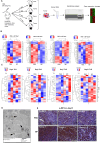
BP alters chemokine and cytokine signaling pathways exclusively in the tumor, lymph nodes, and nanoparticle-associated cells isolated from organs with high BP accumulation. (A) To elucidate molecular mechanisms of BP-mediated tumor inhibition, HuHER2 transgenic mice bearing primary mammary tumors were injected with either PBS or BP (n = 3/group). Seven days after injection, we collected tumor, lung, and the nanoparticle accumulating organs liver, spleen, lymph nodes, and bone marrow. RNA was isolated from these organs (PBS and BP). Portions of tissue samples were separately digested and dissociated to isolate single cells, which were magnetically sorted to isolate nanoparticle-associated cells (BP-NA), from which RNA was also isolated. RNA sequencing was performed on all RNA samples isolated from whole tumor, organs, and nanoparticle associated cells. (B) Despite substantial accumulation in other normal organs such as liver, spleen, and bone marrow, little difference between PBS- and BP-treated samples was detected. Similarly, gene expression measured in the lungs also showed no difference between control and BP-treated mice. (C) Heat map of differentially regulated genes measured in tumors and pathway analysis (D) showed inflammation-mediated chemokine and cytokine signaling among the top hits. Differentially expressed genes of FDR < 0.1 is considered significant. (E) In contrast to whole tissue analysis, significant changes in gene expression were observed in nanoparticle-associated cells (BP-NA). Lung tissues yielded no nanoparticle-associated cells.
Figure 4
Figure 4
Time-dependent alteration of immune gene expression in the tumor microenvironment by BP is mediated through endosomal accumulation and activation of Toll-like receptor (TLR) pathway. (A) HuHER2 transgenic tumor-bearing mice were treated with PBS or BP (5 mg of Fe) on the day of tumor detection (n = 12/group). Three mice were sacrificed from each group on days 1, 3, 7, and 31 to collect primary tumor for RNA isolation and gene expression analysis by Nanostring using the pan-immune cell marker panel. (B) Heat map of differentially regulated genes at each time point. (C) Heat map of differentially regulated TLR pathway at different time points showed that expression of most of the downstream of TLR pathway effectors was upregulated in the BP-treated tumors by day 31. TLR pathway is involved in innate immune regulation leading to antitumor immune stimulation. (D) Representative TEM image showing BP nanoparticle accumulation in endosomes and autophagosomes inside tumor immune cells. (E) IHC analysis of tumors collected on day 31 showed higher p-IRF3 in BP-treated mouse tumors than in PBS controls, confirming increased TLR3-mediated TRIF signaling culminating in phosphorylation and translocation of IRF3 to activate downstream Type 1 interferon signaling.
Figure 5
Figure 5
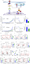
BP nanoparticle mediated primary tumor inhibition mediated through innate immune cell activation of T cells through TLR pathway. (A) Schema of the proposed mechanism of BP action. BP nanoparticles taken up by the innate immune cells are internalized and placed in the endosomes. There they activate TLR3/4 pathways via TRIF/MyD88, which in turn activates Type1 IFN signaling, leading to antiviral immune response and T cell stimulation, thereby affecting tumor growth. (B–D) No inhibition of tumor growth, metastasis, or survival was observed in BP treated TRIF–/– mice growing Py230 tumors. (E–G) Significant tumor growth inhibition was exhibited by BP treatment when the same tumors were grown in wild-type C57BL/6 mice with extended survival and a modest effect on metastasis. (H, I) Flow cytometry analysis of Py230 tumors in wild-type C57BL/6 mice treated as in (E) and analyzed on the 40th day showed an increase in activated CD8+/Gzmb+ T cells and NK T cells in tumor microenvironments with BP treatment. (J) CD11c+ dendritic cells are higher than in controls in the spleens of BP treated mice. (K) A significant reduction in granulocyte populations was observed in lungs of BP-treated mice compared to PBS controls. (L) When treated with BP, the effector cell population identified as CD44high/CD62low was significantly upregulated in CD8+ cells with a concomitant decrease in naïve (CD44low/CD62low) and central memory (CD44high/CD62high) cells in the liver. (M–O) Flow cytometry analysis of the liver showed significant increases in dendritic cell subsets in BP treated mice. *p < 0.05; **p < 0.01.
Figure 6
Figure 6
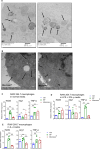
Pro-inflammatory gene induction in macrophages by nanoparticles. (A, B) Transmission electron microscopy (TEM) images showing internalized BP and Micromer nanoparticles in RAW264.7 cells after 24 h of treatment. (C) Real-time qRT-PCR analysis showed significant upregulation of pro-inflammatory genes iNOS and TNF-α, and downregulation of suppressive Arg1 24 h after RAW264.7 cells in normal media were exposed to BP nanoparticles. Micromer nanoparticles also induced iNOS and TNF-α, but to a lesser degree. (D) Relative gene expression changes 24 h after nanoparticle exposure of RAW264.7 cells grown in LPS + IFN-γ media showed further induction of iNOS and TNF-α by BP nanoparticles. (E) When the RAW264.7 cells were grown in IL-4 media and treated with nanoparticles for 24 h, they expressed iNOS and TNF-α, indicating a switch from the anti-inflammatory phenotype to a pro-inflammatory one. *p < 0.05; **p < 0.01.
Figure 7
Figure 7
Nanoparticle formulation determines antitumor effects. (A–D) HuHER2 transgenic mice were injected intravenously with hydroxyethyl starch-coated polystyrene nanoparticles (Micromer – 100 nm) or FDA approved iron oxide colloid (INFeD – 10 nm), as described in Figure 1, and followed for tumor growth, time to reach end point, presence of lung metastasis, and development of other mammary tumors. No significant difference was seen with INFeD treatment compared to control mice in tumor growth, survival, or metastasis. A slower tumor growth and reduction in number of mice with lung metastasis was observed in the Micromer-treated cohort, which was not statistically significant. *p < 0.05.
Figure 8
Figure 8
Survival advantage with higher expression of TLR3 and IRF3 in breast cancer patients. (A) Kaplan–Meier (KM) survival plot of breast cancer patients based on the gene expression of TLR3 in their tumors. High TLR3 expression was associated with a significant increase in recurrence free survival in all breast cancer patients and in both the luminal (ER+) and basal (triple negative) subgroups. (B) KM survival plot of all breast cancer patients based on the high or low expression of IRF3 in their tumors. KM plots of breast cancer patients separated by molecular subgroups show that IRF3 expression correlates with a longer recurrence free survival.

Similar articles

Cited by

References

    1. Siegel R. L.; Miller K. D.; Fuchs H. E.; Jemal A. Cancer statistics, 2022. CA Cancer J. Clin 2022, 72 (1), 7–33. 10.3322/caac.21708. - DOI - PubMed
    1. Allemani; Matsuda C.; Di Carlo T.; Harewood V.; Matz R.; Nikšić M.; Bonaventure M.; Valkov A.; Johnson M.; Estève C. J.; Ogunbiyi J.; Azevedo O. J.; Silva E.; Chen W. Q.; Eser S.; Engholm G.; Stiller C. A.; Monnereau A.; Woods R. R.; Visser O.; Lim G. H.; et al. Global surveillance of trends in cancer survival 2000–14 (CONCORD-3): analysis of individual records for 37513025 patients diagnosed with one of 18 cancers from 322 population-based registries in 71 countries. Lancet 2018, 391 (10125), 1023–1075. 10.1016/S0140-6736(17)33326-3. - DOI - PMC - PubMed
    1. Jagannathan G.; White M. J.; Xian R. R.; Emens L. A.; Cimino-Mathews A. A New Landscape of Testing and Therapeutics in Metastatic Breast Cancer. Clin Lab Med. 2023, 43 (2), 299–321. 10.1016/j.cll.2023.03.004. - DOI - PubMed
    1. Hanahan D.; Weinberg R. A. Hallmarks of cancer: the next generation. Cell 2011, 144 (5), 646–674. 10.1016/j.cell.2011.02.013. - DOI - PubMed
    1. Zitvogel L.; Apetoh L.; Ghiringhelli F.; André F.; Tesniere A.; Kroemer G. The anticancer immune response: indispensable for therapeutic success?. J. Clin Invest 2008, 118 (6), 1991–2001. 10.1172/JCI35180. - DOI - PMC - PubMed

Publication types