. 2024 May 8;15(5):e0033024.
doi: 10.1128/mbio.00330-24. Epub 2024 Apr 2.

Characterization of the transcriptionally active form of dephosphorylated DctD complexed with dephospho-IIAGlc

Affiliations

Characterization of the transcriptionally active form of dephosphorylated DctD complexed with dephospho-IIAGlc

Sebin Kang et al. mBio. .

Abstract

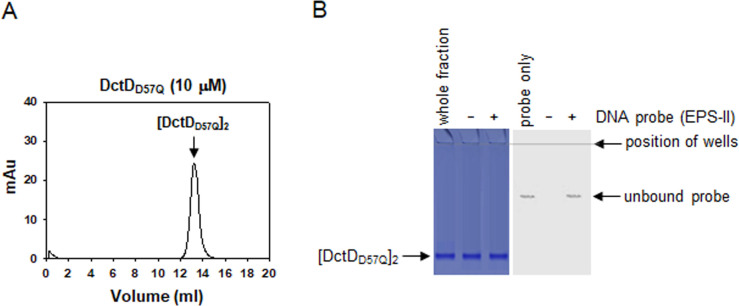
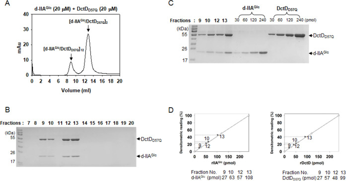
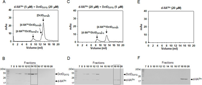
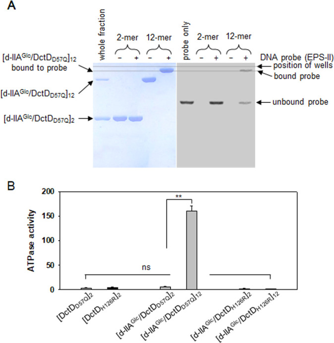
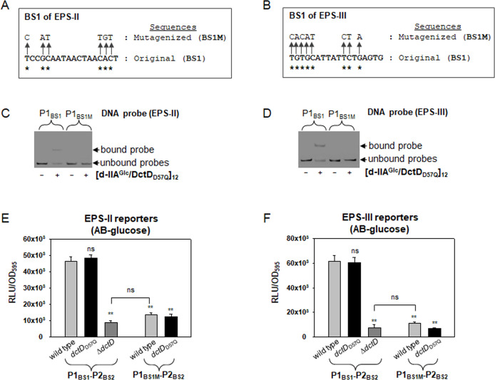
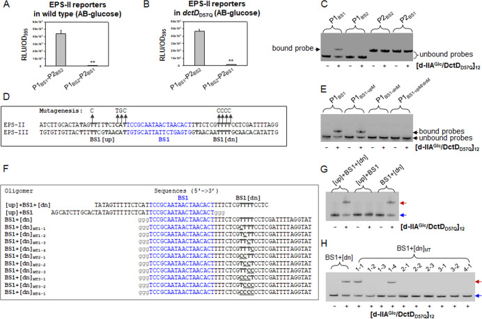
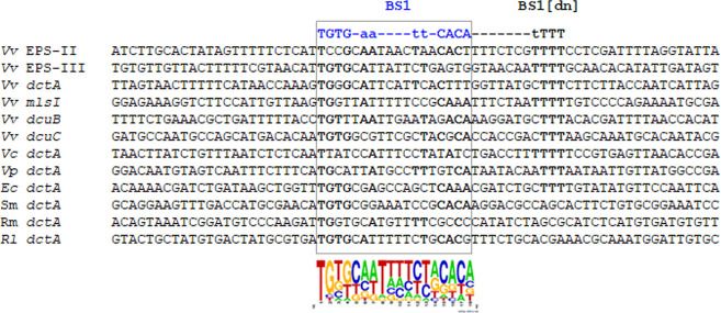
Bacterial enhancer-binding proteins (bEBPs) acquire a transcriptionally active state via phosphorylation. However, transcriptional activation by the dephosphorylated form of bEBP has been observed in DctD, which belongs to Group I bEBP. The formation of a complex between dephosphorylated DctD (d-DctD) and dephosphorylated IIAGlc (d-IIAGlc) is a prerequisite for the transcriptional activity of d-DctD. In the present study, characteristics of the transcriptionally active complex composed of d-IIAGlc and phosphorylation-deficient DctD (DctDD57Q) of Vibrio vulnificus were investigated in its multimeric conformation and DNA-binding ability. DctDD57Q formed a homodimer that could not bind to the DNA. In contrast, when DctDD57Q formed a complex with d-IIAGlc in a 1:1 molar ratio, it produced two conformations: dimer and dodecamer of the complex. Only the dodecameric complex exhibited ATP-hydrolyzing activity and DNA-binding affinity. For successful DNA-binding and transcriptional activation by the dodecameric d-IIAGlc/DctDD57Q complex, extended upstream activator sequences were required, which encompass the nucleotide sequences homologous to the known DctD-binding site and additional nucleotides downstream. This is the first report to demonstrate the molecular characteristics of a dephosphorylated bEBP complexed with another protein to form a transcriptionally active dodecameric complex, which has an affinity for a specific DNA-binding sequence.IMPORTANCEResponse regulators belonging to the bacterial two-component regulatory system activate the transcription initiation of their regulons when they are phosphorylated by cognate sensor kinases and oligomerized to the appropriate multimeric states. Recently, it has been shown that a dephosphorylated response regulator, DctD, could activate transcription in a phosphorylation-independent manner in Vibrio vulnificus. The dephosphorylated DctD activated transcription as efficiently as phosphorylated DctD when it formed a complex with dephosphorylated form of IIAGlc, a component of the glucose-phosphotransferase system. Functional mimicry of this complex with the typical form of transcriptionally active phosphorylated DctD led us to study the molecular characteristics of this heterodimeric complex. Through systematic analyses, it was surprisingly determined that a multimer constituted with 12 complexes gained the ability to hydrolyze ATP and recognize specific upstream activator sequences containing a typical inverted-repeat sequence flanked by distinct nucleotides.

Keywords: bacterial enhancer-binding protein; dephospho-DctD/dephospho-IIAGlccomplex; dodecameric conformation; upstream activator sequences.


Conflict of interest statement

The authors declare no conflict of interest.

Figures

Fig 1
Fig 1
Characterization of the multimeric conformation of DctDD57Q (A) Chromatographic profile of DctDD57Q. Recombinant protein of DctDD57Q (7), whose calculated molecular weight is 53.53 kDa, was subjected to GPC as described in the Materials and Methods section. The resultant profile showing a single peak at 13.4 mL was presented with the absorbance at 280 nm (mAu). Its size was determined using a regression equation of the standard proteins, as shown in Fig. S1. The fractions (13 and 14 mL) containing this peak were collected and used for electrophoretic mobility shift assay (EMSA) in Fig. 1B. (B) DNA-binding characteristic of the dimeric DctDD57Q. Approximately 130 nM of the labeled probe from −418 to +62 relative to the transcription initiation site 1 of the EPS-II cluster (6) was incubated with 250 nM dimeric DctDD57Q. Reaction mixtures were run on two identical 8% native polyacrylamide gels; one was stained with Coomassie Blue to localize the proteins (left panel), and the other was observed under a phosphoimager to localize the DNA probes (right panel). The first lanes in each gel were run with the whole fraction of DctDD57Q preparation (left panel) and the labeled probe only (right panel). The second and third lanes in each gel were run with the mixtures containing [DctDD57Q]2 in the absence (−) and presence (+) of DNA probe, respectively.
Fig 2
Fig 2
Characterization of the multimeric conformation of the complex composed of dephospho-IIAGlc and DctDD57Q (d-IIAGlc/DctDD57Q complex). (A and B) Chromatographic and electrophoretic analyses of d-IIAGlc/DctDD57Q complex. A mixture of the same concentrations of two recombinant proteins (20 µM each), d-IIAGlc and DctDD57Q (7), was subjected to GPC (A), as described in Fig. 1A. Each fraction for the peaks at 8.9 and 12.8 mL was run in an SDS-polyacrylamide gel (B). (C and D) Compositional analysis of the d-IIAGlc/DctDD57Q complex in the two peaks. The fractions for the peaks at 8.9 mL (fractions 9 and 10) and 12.8 mL (fractions 12 and 13) were separated in an SDS-polyacrylamide gel (C). For quantitative analysis, the known amounts (30–240 pmol) of each recombinant protein were included in the same gel. Densitometric readings of each band were plotted against the protein amounts to produce the standard curves (D, open circles). Using the standard curves for d-IIAGlc (left graph) and DctDD57Q (right graph), the amounts of each protein in the four fractions were extrapolated and the resultant values were provided below the graphs.
Fig 3
Fig 3
Verification of the 1:1 binding ratio of IIAGlc and DctD in the d-IIAGlc/DctDD57Q complexes. Mixtures containing 5 µM d-IIAGlc and 20 µM DctDD57Q (A and B) or 20 µM d-IIAGlc and 5 µM DctDD57Q (C and D) were analyzed as described in Fig. 2A and B. Both chromatographic and electrophoretic profiles showed the presence of both proteins in the fractions 9, 10, 12, and 13 mL, as shown in Fig. 2. An extra peak corresponding to dimeric DctDD57Q (A) was formed in a mixture of 5 µM d-IIAGlc and 20 µM DctDD57Q, which was evidenced in an SDS-polyacrylamide gel (B, the fractions 14 and 15 mL were marked with a dashed box). SDS-PAGE analysis of a mixture of 20 µM d-IIAGlc and 5 µM DctDD57Q showed that the fractions 18 and 19 mL contained a protein with the MW of IIAGlc (D, marked with a dashed box). Its identity was verified by profiling d-IIAGlc (20 µM) using chromatographic and electrophoretic analyses (E and F).
Fig 4
Fig 4
Identification of the transcriptionally active form of d-IIAGlc/DctDD57Q complex. (A) DNA-binding ability of the multimeric forms of the d-IIAGlc/DctDD57Q complex. Approximately 100 nM of a labeled DNA probe used in Fig. 1B was incubated with 2-mer ([d-IIAGlc/DctDD57Q]2) and 12-mer ([d-IIAGlc/DctDD57Q]12) of the complex (500 nM [d-IIAGlc/DctDD57Q]1-equivalents). Reaction mixtures were run on two identical 8% native polyacrylamide gels: one was for localizing the proteins (left panel) and the other was for localizing the DNA probes (right panel). For comparison of the bands corresponding to the d-IIAGlc/DctDD57Q complex bound to DNA (left panel) with the probe bound by d-IIAGlc/DctDD57Q complex (right panel), a dashed line was drawn parallel to the line positioning the loading wells. The first lanes in each gel were run with the whole fraction of the d-IIAGlc/DctDD57Q complex (left panel) and the labeled probe only (right panel). (B) ATPase activity of the multimeric forms of the d-IIAGlc/DctDD57Q complex. Both [d-IIAGlc/DctDD57Q]2 and [d-IIAGlc/DctDD57Q]12 were fractionated and aliquots containing 2 µM complexes ([d-IIAGlc/DctDD57Q]1-equivalents) were subjected to the ATPase activity assay (11). One micromolar dimeric DctDD57Q ([DctDD57Q]2) was included in the assay. As a negative control, an ATPase-deficient mutant DctD, DctDH216R, was purified as a dephosphorylated state, and then its multimeric forms ([DctDH216R]2, [d-IIAGlc/DctDH216R]2, and [d-IIAGlc/DctDH216R]12) were added to the ATPase reaction mixture (black bars). The activity was presented as μM phosphate produced by μg of proteins per minute. P-values were indicated (**P < 0.001; ns, not significant).
Fig 5
Fig 5
Localization of [d-IIAGlc/DctDD57Q]12-binding sites in the upstream regions of EPS-II and EPS-III clusters. (A and B) Two putative DctD-binding sites in the EPS-II and EPS-III clusters. Two sites (designated as BS1 for the remote site and BS2 for the close site from TIS-1) showing moderate identity to the DctD-binding sequences previously found in R. meliloti (5′-TGTGCGgaaatCCGCACA-3′; 15) were localized in the upstream regions of two gene clusters: the nucleotides positioned at −254 to −237 (BS1) and −142 to −125 (BS2) relative to RpoN-dependent TIS-1 of EPS-II (A) and the nucleotides positioned at −451 to −434 (BS1) and −160 to −143 (BS2) relative to the TIS-1 of EPS-III (B). Homologous nucleotides in each site were marked by boldfaces. In case of the EPS-II cluster (or brp operon), the regulatory sites interacting with the other transcription factors, such as BrpS and BrpT, were marked (16, 17). (C–F) Binding of [d-IIAGlc/DctDD57Q]12 to the probes containing either BS1 or BS2. DNA probes containing BS1 of EPS-II (C) or EPS-III (D), which were designated as P1BS1 (a 210 bp DNA fragment from −418 to −209 of EPS-II and a 272 bp DNA fragment from −602 to −331 of EPS-III), were prepared for EMSA. Similarly, the probes containing BS2 of EPS-II (E) or EPS-III (F) were prepared, which were designated as P2BS2 (a 270 bp DNA fragment from −208 to +62 of EPS-II and a 366 bp DNA fragment from −330 to +36 of EPS-III). Each probe was labeled and 100 nM was incubated with various concentrations of [d-IIAGlc/DctDD57Q]12 up to 100 nM. To verify the specific binding to P1BS1 (C and D), the identical but unlabeled DNA fragments (cold probes) and the noncompetitive gap DNA were included. The resultant reaction mixtures were subjected to 8% native polyacrylamide gel electrophoresis, and DNA bands corresponding to the unbound or bound probes were indicated by arrows. Lanes 1, probe only; lanes 2, probe with 10 nM of [d-IIAGlc/DctDD57Q]12; lanes 3, probe with 20 nM of [d-IIAGlc/DctDD57Q]12; lanes 4, probe with 40 nM of [d-IIAGlc/DctDD57Q]12; lanes 5, probe with 60 nM of [d-IIAGlc/DctDD57Q]12; lanes 6, probe with 80 nM of [d-IIAGlc/DctDD57Q]12; lanes 7, probe with 100 nM of [d-IIAGlc/DctDD57Q]12; lanes 8, probe with 100 nM of [d-IIAGlc/DctDD57Q]12 and 0.5 µM of cold probe; lanes 9, probe with 100 nM of [d-IIAGlc/DctDD57Q]12 and 1.0 µM of cold probe; and lanes 10, probe with 100 nM of [d-IIAGlc/DctDD57Q]12 and 0.5 µM of gap DNA.
Fig 6
Fig 6
Effect of the mutations in the BS1s on binding by [d-IIAGlc/DctDD57Q]12 and transcription of the EPS clusters. (A and B) Mutagenesis of the BS1s of EPS-II and EPS-III clusters. The nucleotides in BS1s of EPS-II (A) and EPS-III (B), which are conserved in the R. meliloti DctD-binding consensus sequences (5′-TGTGCGgnnntCCGCACA-3′; 15), were marked with asterisks. These conserved nucleotides were substituted with other nucleotides, as indicated by arrows, to produce BS1M. (C and D) DNA-binding affinity of [d-IIAGlc/DctDD57Q]12 to the probes containing BS1M. DNA fragments of P1 containing BS1 (P1BS1) or BS1M (P1BS1M) were prepared from the EPS-II (C) and EPS-III (D) clusters. Then, EMSA was performed as described in Fig. 5. Labeled DNA bands corresponding to the unbound or bound probes, which were resolved in 6% native polyacrylamide gel, were indicated by arrows. (E and F) Expression of the BS1M-containing transcription reporters of EPS-II and EPS-III clusters. The luxAB-based transcription reporters fused with the original upstream regulatory regions (P1BS1-P2BS2; 18) or the mutagenized upstream regions (P1BS1M-P2BS2) of EPS-II (E) and EPS-III (F) were transferred to the wild type and dctDD57Q (7). Then, V. vulnificus cells were grown in AB-glucose supplemented with tetracycline (3 µg/mL). At an OD595nm of 0.4, aliquots of bacterial cells were harvested, and their luciferase activities were measured, as described in the Materials and Methods section. The expression of each reporter was presented as normalized values: the relative light unit (RLU) divided by the cell mass (OD595nm) of each sample. As a negative control, ΔdctD strain carrying the original reporter was included in each assay. P-values for comparison with the P1BS1-P2BS2 reporter in the wild type or ΔdctD were indicated (**P < 0.001; ns, not significant).
Fig 7
Fig 7
Characterization of the flanking region of BS1 for binding by [d-IIAGlc/DctDD57Q]12 and successful expression of EPS-II cluster. (A and B) Expression of the BS-switched transcription reporter of EPS-II. A mutagenized reporter was constructed by switching the positions of the original BS1 and BS2 of the EPS-II cluster, to produce P1BS2-P2BS1. Its expression in wild type (A) and dctDD57Q (B) was compared to that of the original reporter (P1BS1-P2BS2). At an OD595nm of 0.4 in AB-glucose supplemented with tetracycline (3 µg/mL), the expression of each reporter was estimated and presented with normalized values, RLU per OD595nm, as described in Fig. 6. P-values for comparison with the original reporter were indicated (**P < 0.001). (C) DNA-binding affinity of [d-IIAGlc DctDD57Q]12 to BS-switched probes. Various DNA fragments of the EPS-II cluster, including the original P1 (P1BS1), switched P1 (P1BS2), the original P2 (P2BS2), and switched P2 (P2BS1), were prepared for EMSA using [d-IIAGlc/DctDD57Q]12. Each probe (100 nM) was mixed with 40 nM [d-IIAGlc/DctDD57Q]12. DNA bands corresponding to the unbound probes and the probe bound by [d-IIAGlc/DctDD57Q]12 were indicated by brackets and arrow, respectively. (D) Mutagenesis of BS1-flanking regions. The nucleotide sequences flanking the BS1s are conserved in the EPS-II and EPS-III clusters (marked with boldfaces). Nucleotides upstream of the EPS-II BS1 (BS1[up]) and downstream of the EPS-II BS1 (BS1[dn]) were mutagenized as indicated by arrows and substituted nucleotides, to produce BS1-dnM and BS1-upM, respectively. (E) DNA-binding affinity of [d-IIAGlc/DctDD57Q]12 to the probes containing BS1-dnM and BS1-upM. DNA fragments of the EPS-II cluster, including the original P1 (P1BS1), upstream-mutagenized P1 (P1BS1-upM), downstream-mutagenized P1 (P1BS1-dnM), and P1 mutagenized both upstream and downstream (P1BS1-upM/dnM), were prepared for EMSA using [d-IIAGlc/DctDD57Q]12. Each probe (100 nM) was mixed with 40 nM [d-IIAGlc/DctDD57Q]12, and resultant reaction mixtures were run on a native gel, as described above. (F) Preparation of various oligomers containing BS1. Nucleotide oligomers containing BS1 with both flanking regions ([up] + BS1 + [dn]), BS1 with upstream sequences only ([up] + BS1), and BS1 with downstream sequences only (BS1 + [dn]) were prepared. Series of BS1 + [dn] derivatives, BS1 + [dn]MT, whose Ts in BS1[dn] were mutagenized in a combinatorial way, were prepared. Eighteen nucleotides corresponding to the BS1 of the EPS-II cluster were blue-colored, and the extra three Gs linked to the 3’-end of [up] + BS1 and the 5’-end of BS1 + [dn] oligomers were gray-colored. Four nucleotides in BS1 + [dn] and its derivatives were bold-faced. (G, H) DNA-binding affinity of [d-IIAGlc/DctDD57Q]12 to various oligomers. Each oligomer (100 nM) was labeled and used for EMSA. Oligomers of [up] + BS1 + [dn], [up] + BS1, and BS1 + [dn] were mixed to reaction buffers without (−) and with (+) 40 nM [d-IIAGlc/DctDD57Q]12 (G). Ten mutant oligomers covering from BS1+[dn]MT1-1 to BS1+[dn]MT4-1 were mixed with [d-IIAGlc/DctDD57Q]12, and resultant reaction mixtures were subjected to 8% native polyacrylamide gel electrophoresis (lanes 3–12, H). As controls, BS1 + [dn] only (lane 1) and BS1 + [dn] with [d-IIAGlc/DctDD57Q]12 (lane 2) were included in the same gel. Oligomer bands corresponding to the unbound and bound probes were indicated by blue and red arrows, respectively.
Fig 8
Fig 8
Proposed [d-IIAGlc/DctDD57Q]12-binding sequences in the DctD-regulons. The BS1 and BS1[dn] sequences of the EPS-II and EPS-III clusters were aligned with the putative BS1 and BS1[dn] in the upstream regions of dctA genes of V. vulnificus (Vv; 6), Vibrio cholerae (Vc; 21), Vibrio parahaemolyticus (Vp; VPY51_04360), E. coli (Ec; 20), S. meliloti (Sm; 14), R. meliloti (Rm; 15), and R. leguminosarum (Rl; 14). In addition, the upstream regions of the putative DctD-regulons, whose expression was assumed to be related to the metabolisms of dicarboxylic acids in V. vulnificus, were included in this analysis: malate synthase (mlsI, VVMO6_03414) and two C4-dicarboxylate transporters (dcuB, VVMO6_03835; dcuC, VVMO6_03887). Conserved regions among 12 BSs were marked by a dashed box and a nucleotide-frequency plot, and the highly conserved nucleotides among 12 BSs and 9 BS[dn]s were indicated with bold-faced letters.

References

    1. Webster N, Jin JR, Green S, Hollis M, Chambon P. 1988. The yeast UASG is a transcriptional enhancer in human HeLa cells in the presence of the GAL4 Trans-activator. Cell 52:169–178. doi:10.1016/0092-8674(88)90505-3 - DOI - PubMed
    1. Wigneshweraraj SR, Burrows PC, Bordes P, Schumacher J, Rappas M, Finn RD, Cannon WV, Zhang X, Buck M. 2005. The second paradigm for activation of transcription. Prog Nucleic Acid Res Mol Biol 79:339–369. doi:10.1016/S0079-6603(04)79007-8 - DOI - PubMed
    1. Bush M, Dixon R. 2012. The role of bacterial enhancer binding proteins as specialized activators of Σ54-dependent transcription. Microbiol Mol Biol Rev 76:497–529. doi:10.1128/MMBR.00006-12 - DOI - PMC - PubMed
    1. Engelke T, Jording D, Kapp D, Pühler A. 1989. Identification and sequence analysis of the Rhizobium meliloti dctA gene encoding the C4-dicarboxylate carrier. J Bacteriol 171:5551–5560. doi:10.1128/jb.171.10.5551-5560.1989 - DOI - PMC - PubMed
    1. Golby P, Davies S, Kelly DJ, Guest JR, Andrews SC. 1999. Identification and characterization of a two-component sensor-kinase and response-regulator system (DcuS-DcuR) controlling gene expression in response to C4-dicarboxylates in Escherichia coli. J Bacteriol 181:1238–1248. doi:10.1128/JB.181.4.1238-1248.1999 - DOI - PMC - PubMed

Publication types

MeSH terms

LinkOut - more resources