

Ocular blood flow in preterm neonates
- PMID: 38565630
- PMCID: PMC10987658
- DOI: 10.1038/s41598-024-58523-8
Ocular blood flow in preterm neonates
Abstract
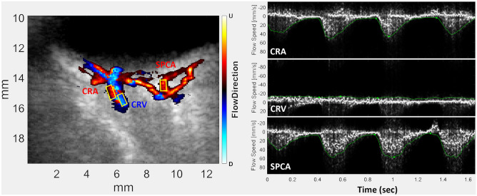
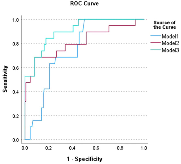
Retinopathy of prematurity (ROP) is a disorder affecting low birthweight, preterm neonates. In the preterm eye, the retina is not fully developed and neovascularization may occur at the margin between the developed vascular retina and undeveloped avascular retina. Without timely treatment by laser or intravitreal anti-vascular endothelial growth factor (VEGF) therapy, this can lead to tractional retinal detachment and blindness. Visualization of the retina in regular examinations by indirect ophthalmoscopy is hence the current standard of care, but the exams are stressful and interpretation of images is subjective. The upregulation of VEGF in ROP would suggest an increase in ocular blood flow. In this report, we evaluate the potential of ultrafast plane-wave Doppler ultrasound (PWU) to detect increased flow velocities in the orbital vessels supplying the eye in a gentle exam with objective findings. We imaged both eyes of 50 low-birthweight preterm neonates using 18 MHz PWU. Flow velocity in the central retinal artery (CRA) and vein (CRV), and the short posterior ciliary arteries were determined and values at each ROP Stage compared. We found significantly increased velocities in the CRA and CRV in Stage 3 ROP eyes, where intervention would be considered. We compared multivariate models for identifying Stage 3 eyes comprised solely of clinical factors, solely of Doppler parameters, and clinical plus Doppler parameters. The respective models provided areas under their respective ROC curves of 0.760, 0.812, and 0.904. PWU Doppler represents a gentle, objective means for identifying neonates at risk for ROP that could complement ophthalmoscopy.
© 2024. The Author(s).
Conflict of interest statement
The authors declare no competing interests.
Figures
References
MeSH terms
Substances
Grants and funding
LinkOut - more resources
Full Text Sources
