. 2024 May;16(5):1143-1161.
doi: 10.1038/s44321-024-00059-5. Epub 2024 Apr 2.

ImmunoPET imaging of Trop2 in patients with solid tumours

Affiliations

ImmunoPET imaging of Trop2 in patients with solid tumours

Wei Huang et al. EMBO Mol Med. 2024 May.

Abstract

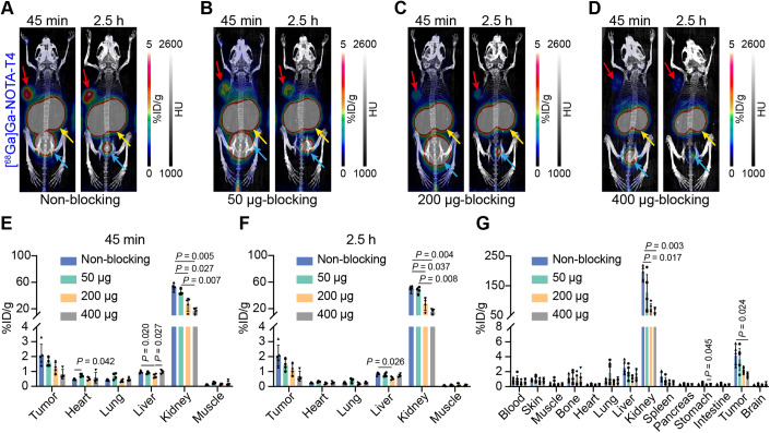
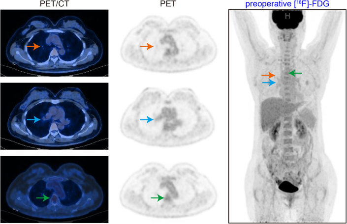
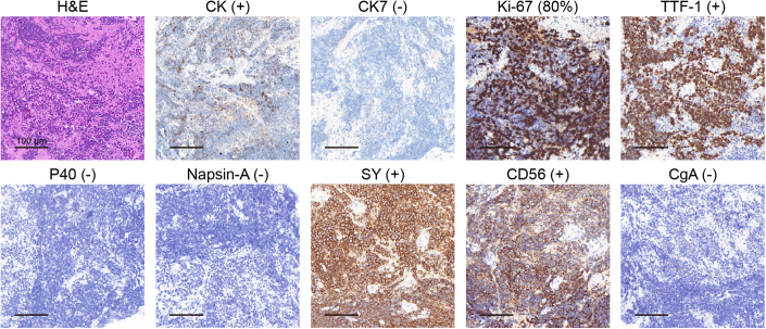
Accurately predicting and selecting patients who can benefit from targeted or immunotherapy is crucial for precision therapy. Trophoblast cell surface antigen 2 (Trop2) has been extensively investigated as a pan-cancer biomarker expressed in various tumours and plays a crucial role in tumorigenesis through multiple signalling pathways. Our laboratory successfully developed two 68Ga-labelled nanobody tracers that can rapidly and specifically target Trop2. Of the two tracers, [68Ga]Ga-NOTA-T4, demonstrated excellent pharmacokinetics in preclinical mouse models and a beagle dog. Moreover, [68Ga]Ga-NOTA-T4 immuno-positron emission tomography (immunoPET) allowed noninvasive visualisation of Trop2 heterogeneous and differential expression in preclinical solid tumour models and ten patients with solid tumours. [68Ga]Ga-NOTA-T4 immunoPET could facilitate clinical decision-making through patient stratification and response monitoring during Trop2-targeted therapies.

Keywords: Companion Diagnostics; ImmunoPET; Nanobody; Theranostics; Trop2.


Conflict of interest statement

W Wei, W Huang and J Liu are co-inventors on a provisional patent application encompassing the technology reported in the manuscript. W Wei is a consultant of Alpha Nuclide (Ningbo) Medical Technology Co., Ltd. The remaining authors declare no competing interests.

Figures

Figure 1
Figure 1. Characteristics of two nanobodies (T4 and T5) targeting human Trop2.
(A) SDS-PAGE tested the purity of T4. R: reducing condition; N-R: non-reducing condition. (B) Characterisation of purity of T4 by HPLC. (C) SPR study demonstrated T4 affinity and kinetic towards recombinant human Trop2 protein. The purity of another nanobody, T5, was also determined by SDS-PAGE (D) and HPLC (E). R reducing condition, N-R non-reducing condition. (F) The binding affinity of T5 with human Trop2 protein. Source data are available online for this figure.
Figure 2
Figure 2. [68Ga]Ga-NOTA-T4 and [68Ga]Ga-NOTA-T5 immunoPET imaging in T3M-4 models.
(A) Representative immunoPET/CT images of [68Ga]Ga-NOTA-T4 (n = 4) in T3M-4 tumour xenografts models at 45 min p.i. Red arrows: tumour; green arrows: liver; blue arrows: bladder. (B) ROI analysis of [68Ga]Ga-NOTA-T4 (mean ratio ± SD, n = 4). (C) Ex vivo Bio-D data presented the detailed distribution patterns of [68Ga]Ga-NOTA-T4 (mean ratio ± SD, n = 3) in the tumours and other major organs/tissues. (D) Coronal immunoPET/CT images of [68Ga]Ga-NOTA-T5 in T3M-4 models at 45 min p.i. Red arrows: tumour; yellow arrows: kidneys; blue arrows: bladder. (E) ROI analysis and (F) Bio-D results of [68Ga]Ga-NOTA-T5 after imaging (mean ratio ± SD, n = 4). (G, H) Trop2 IHC staining of T3M-4 tumours after the termination of immunoPET imaging. Source data are available online for this figure.
Figure 3
Figure 3. Effect of varying blocking doses on radioactivity uptake of [68Ga]Ga-NOTA-T4 in tumours and non-target tissues/organs.
(AD) Representative MIP images of the non-blocking and three blocking (50, 200, and 400 μg) groups at two time points. Red arrows: tumour; yellow arrows: kidneys; blue arrows: bladder. (E, F) ROI analysis results of the non-blocking and various blocking groups at 45 min (E) and 2.5 h (F). Groups divided into non-blocking group (n = 5), 50 μg-blocking group (n = 4), 200 μg-blocking group (n = 4), and 400 μg-blocking group (n = 4). (G) Bio-D data of the non-blocking and various blocking groups after the termination of [68Ga]Ga-NOTA-T4 immunoPET imaging. Groups divided into non-blocking (n = 5), 50 μg-blocking (n = 6), 200 μg-blocking (n = 6), and 400 μg-blocking groups (n = 6). Two‐way ANOVA, mean ratio ± SD. Source data are available online for this figure.
Figure 4
Figure 4. ABDT4 blocked tumour uptake of [68Ga]Ga-NOTA-T4 in tumour-bearing mice models.
(A) Representative images showed the results of blocking Trop2 on the surface of tumour cells with ABDT4. Red arrows: tumour; yellow arrows: kidneys; blue arrows: bladder. (B) ROI quantitative analysis of [68Ga]Ga-NOTA-T4 immunoPET imaging results in the ABDT4-blocking group (mean ratio ± SD, n = 4). (C) Ex vivo Bio-D results after termination of [68Ga]Ga-NOTA-T4 immunoPET imaging (mean ratio ± SD, n = 4). (D) Comparison of radioactivity uptake in tumour and major tissues/organs between the non-blocking (n = 5) and ABDT4-blocking groups (n = 4). t test, mean ratio ± SD. (E) Comparison of tumour-to-organ ratios between the non-blocking (n = 5) and ABDT4-blocking groups (n = 4). t test, mean ratio ± SD. TMR tumour-to-muscle, THR tumour-to-heart, TLR tumour-to-liver, TKR tumour-to-kidney. Source data are available online for this figure.
Figure 5
Figure 5. Dynamic [68Ga]Ga-NOTA-T4 immunoPET imaging in an adult male beagle dog.
(A) Whole-body maximum intensity projection (MIP) images of [68Ga]Ga-NOTA-T4 immunoPET imaging at different time points (15 s, 25 s, 45 s, 2 min, 5 min, and 60 min) in a beagle dog. Orange arrow: heart; green arrow: liver; yellow arrow: kidneys; blue arrows: bladder. Scale bar: 0–6. (B) TAC curves of [68Ga]Ga-NOTA-T4 in the major tissues/organs, including the brain, heart, lung, liver, pancreas, spleen, large intestine, kidneys, bladder, bone, and muscle. (C) MIP images of [68Ga]Ga-NOTA-T4 imaging at 15–20 s, 23 s, and 25 s time points present the process of kidney accumulation. Yellow arrows: kidneys. Scale bar: 0–6. (D) VOI curve of [68Ga]Ga-NOTA-T4 in the kidney during 30 s. Source data are available online for this figure.
Figure 6
Figure 6. [18F]-FDG and [68Ga]Ga-NOTA-T4 PET/CT imaging in a patient with Trop2-positive metastatic nasopharyngeal carcinomas.
A patient diagnosed with nasopharyngeal carcinoma and multiple metastases in the liver, bones, and lymph nodes underwent sequential [18F]-FDG (A−C) and [68Ga]Ga-NOTA-T4 PET/CT (D−F) scans. While the primary tumour and several metastases with intense [18F]-FDG uptake, [68Ga]Ga-NOTA-T4 immunoPET/CT identified bone metastases and multiple liver metastases, indicating positive expression of Trop2 in these metastatic lesions. Notably, a newly formed bone metastatic lesion (yellow arrows) with [68Ga]Ga-NOTA-T4 uptake was identified. H&E (G) and Trop2 IHC (H) staining of the biopsied liver metastasis. Scale bar: 2500 μm and 100 μm. Source data are available online for this figure.
Figure 7
Figure 7. [18F]-FDG and [68Ga]Ga-NOTA-T4 PET/CT imaging in a patient with suspicious lung cancer.
A patient with postoperative ovarian cancer with enlarged right hilar and mediastinal lymph nodes underwent [18F]-FDG (AC) and [68Ga]Ga-NOTA-T4 (DF) PET/CT examinations. Suspicious lesions/lymph nodes in the hilum of the right lung and mediastinum, as well as a suspicious bone uptake, were found on [18F]-FDG PET/CT images. In contrast, no abnormal [68Ga]Ga-NOTA-T4 uptake was seen in these suspicious lesions. (G, H) IHC staining of the biopsied right hilar lymph node tissue showed low Trop2 expression with a positive rate of 30%. The left panel (G) showed a low expression of Trop2, and the right panel (H) presented a negative expression. Scale bar: 2500 μm and 200 μm. Source data are available online for this figure.
Figure 8
Figure 8. [18F]-FDG and [68Ga]Ga-NOTA-T4 PET/CT imaging in a patient with small-cell lung cancer.
A patient with small-cell lung cancer with multiple systemic metastases underwent sequential [18F]-FDG (AC) and [68Ga]Ga-NOTA-T4 (D−F) PET/CT within a week. The four lesions, including primary and metastatic lesions labelled, were noted with high [18F]-FDG uptake but did not show significant [68Ga]Ga-NOTA-T4 uptake. H&E (G) and Trop2 IHC (H) staining of the biopsied right lung mass. IHC staining result shows negative Trop2 expression. Scale bar: 1000 μm and 200 μm. Source data are available online for this figure.
Figure EV1
Figure EV1. [68Ga]Ga-NOTA-T4 uptake in the non-blocking and three blocking groups.
Comparison of radioactivity uptake in tumour and major organs at two time points (45 min and 2.5 h) in the non-blocking group (A, n = 5), 50 μg-blocking (B, n = 6), 200 μg-blocking (C, n = 6), and 400 μg-blocking groups (D, n = 6). t test, mean ratio ± SD.
Figure EV2
Figure EV2. The initial baseline [18F]-FDG PET/CT examination of patient 1 with nasopharyngeal carcinoma.
The primary tumour (A, orange arrows) and multiple metastases, including the right parapharyngeal space lymph node (B, yellow arrows), right neck lymph node (C, blue arrows), bone and liver metastases (Fig. 6A–C in the main manuscript), and a suspicious right axillary lymph node metastasis (D, green arrows) were presented. Note: the [18F]-FDG MIP image here is the same MIP image in Fig. 6.
Figure EV3
Figure EV3. Histopathological examination of the biopsied liver nodule.
The tested biomarkers confirmed the liver metastasis from poorly differentiated nasopharyngeal carcinoma. “+“ means positive expression, and “–“ means negative expression. Scale bar: 100 μm.
Figure EV4
Figure EV4. The preoperative [18F]-FDG PET/CT examination of patient 2.
The lesions and metastasis with enlarged uptake detected on the second [18F]-FDG PET/CT were absent on the first preoperative [18F]-FDG PET/CT. Source data are available online for this figure.
Figure EV5
Figure EV5. Histopathological examination of the biopsied right upper lung mass.
The tested biomarkers indicated characteristics of small-cell lung cancer. “+“ means positive expression, and “–“ means negative expression. Scale bar: 100 μm.

References

    1. Ametamey SM, Honer M, Schubiger PA. Molecular Imaging with PET. Chem Rev. 2008;108:1501–1516. doi: 10.1021/cr0782426. - DOI - PubMed
    1. An S, Zhang D, Zhang Y, Wang C, Shi L, Wei W, Huang G, Liu J. GPC3-targeted immunoPET imaging of hepatocellular carcinomas. Eur J Nucl Med Mol Imaging. 2022;49:2682–2692. doi: 10.1007/s00259-022-05723-x. - DOI - PubMed
    1. Balar AV, Galsky MD, Rosenberg JE, Powles T, Petrylak DP, Bellmunt J, Loriot Y, Necchi A, Hoffman-Censits J, Perez-Gracia JL, et al. Atezolizumab as first-line treatment in cisplatin-ineligible patients with locally advanced and metastatic urothelial carcinoma: a single-arm, multicentre, phase 2 trial. The Lancet. 2017;389:67–76. doi: 10.1016/S0140-6736(16)32455-2. - DOI - PMC - PubMed
    1. Bardia A, Messersmith WA, Kio EA, Berlin JD, Vahdat L, Masters GA, Moroose R, Santin AD, Kalinsky K, Picozzi V, et al. Sacituzumab govitecan, a Trop-2-directed antibody-drug conjugate, for patients with epithelial cancer: final safety and efficacy results from the phase I/II IMMU-132-01 basket trial. Ann Oncol. 2021;32:746–756. doi: 10.1016/j.annonc.2021.03.005. - DOI - PubMed
    1. Bardia A, Tolaney SM, Punie K, Loirat D, Oliveira M, Kalinsky K, Zelnak A, Aftimos P, Dalenc F, Sardesai S, et al. Biomarker analyses in the phase III ASCENT study of sacituzumab govitecan versus chemotherapy in patients with metastatic triple-negative breast cancer. Ann Oncol. 2021;32:1148–1156. doi: 10.1016/j.annonc.2021.06.002. - DOI - PubMed