

Clinical efficacy of intraoral ultrasonography versus transgingival probing for measurement of gingival thickness in different gingival biotypes: a clinical trial
- PMID: 38566169
- PMCID: PMC10985878
- DOI: 10.1186/s13005-024-00422-4
Clinical efficacy of intraoral ultrasonography versus transgingival probing for measurement of gingival thickness in different gingival biotypes: a clinical trial
Abstract
Background: Transgingival probing is conventionally used for gingival thickness (GT) measurement. However, invasiveness is a major drawback of transgingival probing. Thus, researchers have been in search of alternative methods for measurement of GT. This study compared the clinical efficacy of intraoral ultrasonography and transgingival probing for measurement of GT in different biotypes.
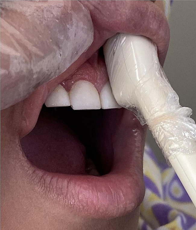
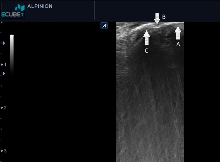
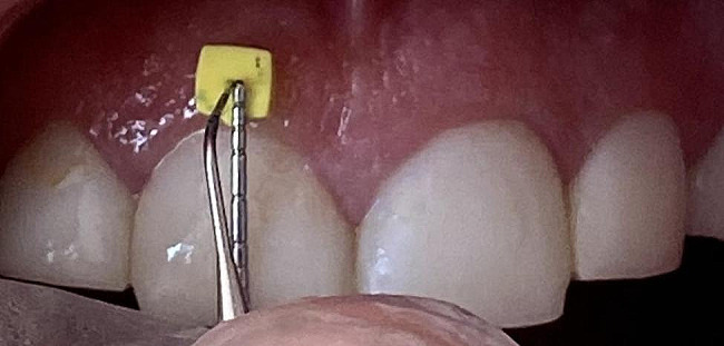
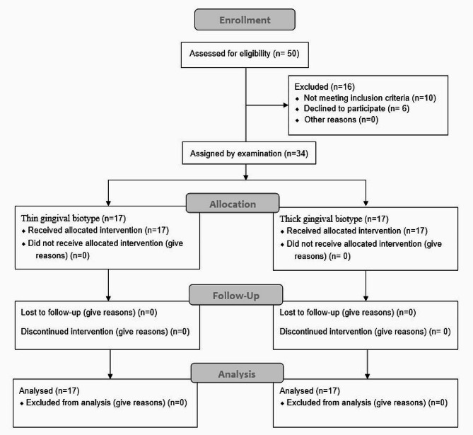
Materials and methods: This clinical trial was conducted on 34 patients requiring crown lengthening surgery. GT was measured at 40 points with 2- and 4-mm distances from the free gingival margin (FGM) of anterior and premolar teeth of both jaws in each patient by an intraoral ultrasound probe. For measurement of GT by the transgingival probing method, infiltration anesthesia was induced, and a #25 finger spreader (25 mm) was vertically inserted into the soft tissue until contacting bone. The inserted length was measured by a digital caliper with 0.01 mm accuracy. All measurements were made by an operator with high reliability under the supervision of a radiologist. Data were analyzed by t-test, Power and Effect Size formula, and intraclass correlation coefficient (ICC).
Results: The two methods were significantly different in measurement of GT in both thick and thin biotypes at 2- and 4-mm distances (P < 0.001). The two methods had a significant difference in both the mandible (P < 0.001) and maxilla (P < 0.001) and in both the anterior (P < 0.003) and premolar (P < 0.003) regions. Although the difference was statistically significant in t-tests, the power and effect formula proved it to be clinically insignificant. Also, the ICC of the two methods revealed excellent agreement.
Conclusion: The results showed optimal agreement of ultrasound and transgingival probing for measurement of GT.
Trial registration: The study was approved by the ethics committee of Shahid Beheshti University of Medical Sciences on 2021-12-28 (IR.SBMU.DRC.REC.1400.138) and registered in the Iranian Registry of Clinical Trials on 2022-03-14 (IRCT20211229053566N1).
Keywords: Clinical trial; Gingiva; Intraoral; Thickness; Ultrasonography.
© 2024. The Author(s).
Conflict of interest statement
The authors declare no competing interests.
Figures

References
-
- Esfahrood ZR, Kadkhodazadeh M, Ardakani MR. Gingival biotype: a review. Gen Dent. 2013;61(4):14–7. - PubMed
Publication types
MeSH terms
Supplementary concepts
LinkOut - more resources
Full Text Sources
Miscellaneous
