. 2024 Apr 2;22(1):211.
doi: 10.1186/s12964-024-01596-9.

EP300-ZNF384 transactivates IL3RA to promote the progression of B-cell acute lymphoblastic leukemia

Affiliations

EP300-ZNF384 transactivates IL3RA to promote the progression of B-cell acute lymphoblastic leukemia

Zhijie Hou et al. Cell Commun Signal. .

Abstract

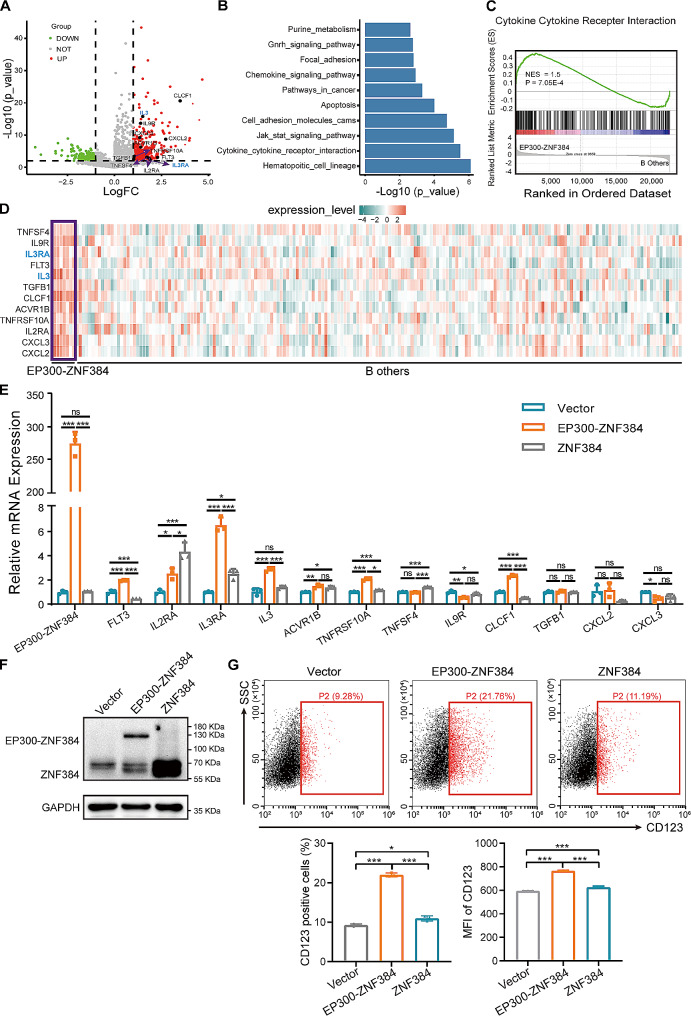
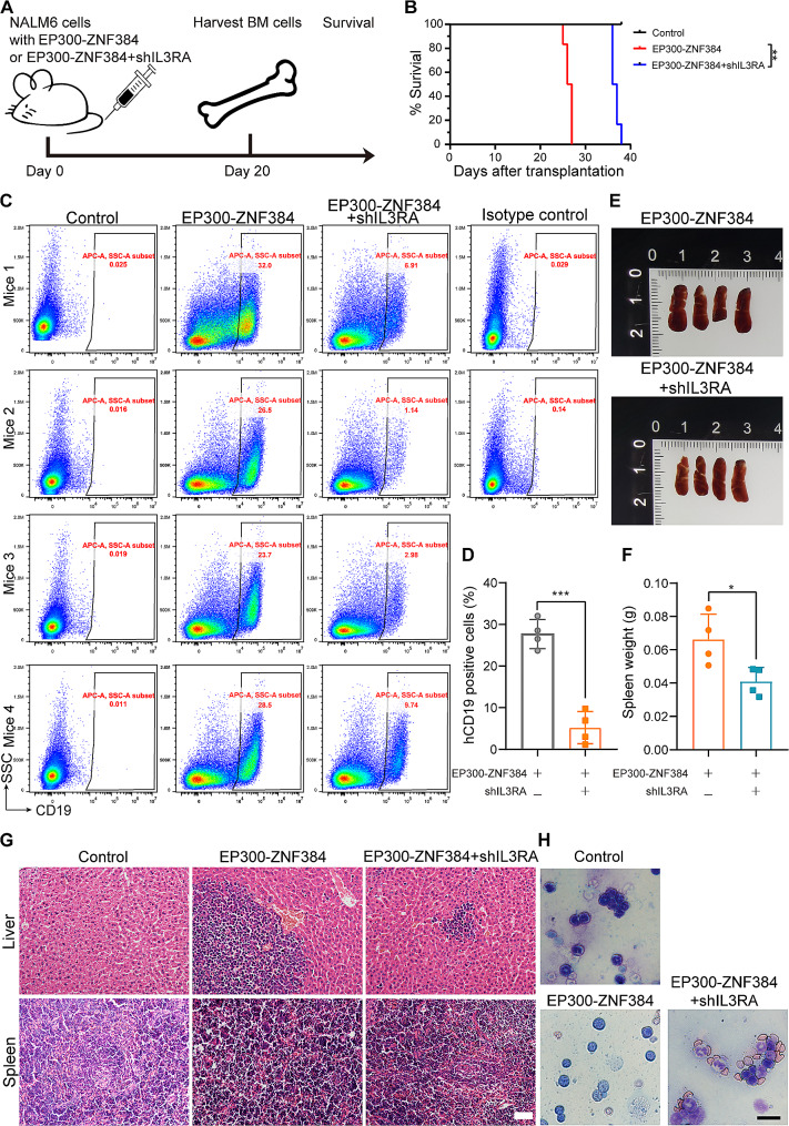
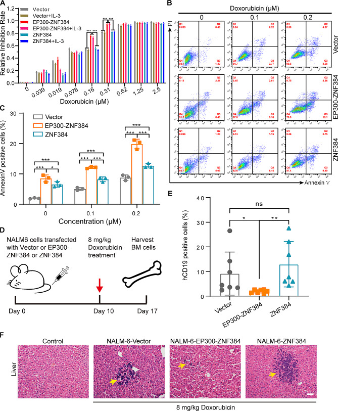
The EP300-ZNF384 fusion gene is an oncogenic driver in B-cell acute lymphoblastic leukemia (B-ALL). In the present study, we demonstrated that EP300-ZNF384 substantially induces the transcription of IL3RA and the expression of IL3Rα (CD123) on B-ALL cell membranes. Interleukin 3 (IL-3) supplementation promotes the proliferation of EP300-ZNF348-positive B-ALL cells by activating STAT5. Conditional knockdown of IL3RA in EP300-ZF384-positive cells inhibited the proliferation in vitro, and induced a significant increase in overall survival of mice, which is attributed to impaired propagation ability of leukemia cells. Mechanistically, the EP300-ZNF384 fusion protein transactivates the promoter activity of IL3RA by binding to an A-rich sequence localized at -222/-234 of IL3RA. Furthermore, forced EP300-ZNF384 expression induces the expression of IL3Rα on cell membranes and the secretion of IL-3 in CD19-positive B precursor cells derived from healthy individuals. Doxorubicin displayed a selective killing of EP300-ZNF384-positive B-ALL cells in vitro and in vivo. Collectively, we identify IL3RA as a direct downstream target of EP300-ZNF384, suggesting CD123 is a potent biomarker for EP300-ZNF384-driven B-ALL. Targeting CD123 may be a novel therapeutic approach to EP300-ZNF384-positive patients, alternative or, more likely, complementary to standard chemotherapy regimen in clinical setting.

Keywords: B-cell acute lymphoblastic leukemia; EP300-ZNF384; IL-3; IL3RA; Leukemogenesis.


Conflict of interest statement

The authors declare no conflict of interest.

Figures

Fig. 1
Fig. 1
EP300-ZNF384 promotes the expression of IL3RA in B-cell acute lymphoblastic leukemia (B-ALL). (A) Volcano plot of comparative analysis between EP300-ZNF384 positive (n = 7) and negative (n = 190) patients in the TARGET-ALL-P2 cohort. Upregulated (n = 346) and downregulated (n = 216) genes are colored by red and green (fold change > 2 and P < 0.01). The labeled texts showed the upregulated genes in the cytokine-cytokine receptor interaction pathway. (B) Bar plot of enriched Kyoto Encyclopedia of Genes and Genomes gene sets using the differentially expressed genes (fold change > 2 and P < 0.01) from the comparison of the EP300-ZNF384 positive and negative patients in the TARGET-ALL-P2 cohort. (C) Representative GSEA plot of EP300-ZNF384-positive patients compared with EP300-ZNF384-negative patients. The normalized enrichment score (NES) and nominal P-values are shown in the graph. (D) Heatmap of 12 upregulated genes in the cytokine-cytokine receptor interaction pathway in EP300-ZNF384-positive patients. IL3 and IL3RA are labeled with blue. (E) The empty vector, EP300-ZNF384, and ZNF384 were overexpressed in NALM-6 cells through lentivirus-mediated gene transfer. Puromycin (2 µg/mL)-selected cells were collected for RT-qPCR. The expression of EP300-ZNF384 together with those of 12 upregulated genes in the cytokine and cytokine receptor interaction pathway were determined relative to ACTB expression. Data represent the mean ± SD. *P < 0.05, **P < 0.01, ***P < 0.001. (F) Immunoblotting analysis of the EP300-ZNF384 fusion and wild-type ZNF384 proteins in the empty vector, EP300-ZNF384, and ZNF384-overexpressing NALM-6 cells. (G) Surface expression of CD123 was evaluated by flow cytometry on NALM-6 cells with the empty vector, EP300-ZNF384 fusion gene, and ZNF384. The proportion of CD123-positive cells and the median fluorescence intensity of CD123 were quantified. *P < 0.05, ***P < 0.001
Fig. 2
Fig. 2
Increased expression of IL3RA by EP300-ZNF384 promotes B-cell acute lymphoblastic leukemia (B-ALL) cell proliferation with or without IL-3. (A) NALM-6 cells overexpressing EP300-ZNF384, ZNF384, and the empty vector were subjected to a colony forming cell (CFC) assay in the presence or absence of IL-3 (10 ng/mL). Scale bar: 50 μm. Data derived from A was used for quantitative analysis of colony size (B) and colony number (C). *P < 0.05, **P < 0.01, ***P < 0.001, ns, no significance. (D) The NALM-6 cells were co-transfected with the shRNA vector (shIL3RA or empty vector) and overexpression vector (EP300-ZNF384 or empty vector) via lentivirus-mediated gene transfer. Hygromycin B (400 µg/mL) and puromycin (2 µg/mL) double selected cells were subjected to CFC assay. Scale bar: 100 μm. (E) Data derived from D were used for quantitative analysis of colony size (diameter) and number. *P < 0.05, **P < 0.01, ***P < 0.001, ns, no significance
Fig. 3
Fig. 3
Conditional knockdown of IL3RA impairs the engraftment of EP300-ZNF384-positive B-cell acute lymphoblastic leukemia (B-ALL) cells in mice. (A) The schematic diagram of the in vivo experiment. NALM-6 cells expressing EP300-ZNF384 or EP300-ZNF384 and shIL3RA were transplanted into immune-deficient NOD⁃PrkdcscidIl2rgnull mice. Mice were then treated with doxycycline (Dox, 2 mg/mL) in drinking water. After 20 d, four mice were sacrificed and femur bone marrow cells were collected for leukemia cell analysis. The remaining six mice were euthanized upon signs of disease. (B) Kaplan–Meier survival curve of mice injected with EP300-ZNF384 or EP300-ZNF384 and shIL3RA-expressing NALM-6 cells. **P < 0.01 (C) hCD19 fractions within the femur bone marrow of each mouse. (D) Column plots summarizing data from all animals analyzed as in C. ***P < 0.001. (E) Changes in the spleen in EP300-ZNF384-expressing NALM-6-transplantable mice in response to IL3RA knockdown. (F) Spleen weights are presented by column plots. ***P < 0.001. (G) The leukemic invasions in spleen and liver were analyzed by hematoxylin and eosin staining. Scale bar: 50 μm. (H) Wright–Giemsa staining of BM cells from mice with NALM-6 cells expressing EP300-ZNF384 or EP300-ZNF384 and shIL3RA. Scale bar: 20 μm
Fig. 4
Fig. 4
EP300-ZNF384 fusion protein transactivates the promoter activity of IL3RA (A) EP300-ZNF384- or ZNF384-expressing vectors were co-transfected with the PGL3-IL3RA-promoter and pRL-TK into HEK-293T cells. After 24 h, dual luciferase assay was performed. Data represents the mean ± SD. **P < 0.01, ***P < 0.001. (B) Increasing doses of EP300-ZNF384-expressing vectors and the luciferase reporter vectors were co-transfected into HEK-293T cells. Luciferase analysis for the IL3RA promoter was performed. ***P < 0.001. (C) Diagram showing the putative binding sites of ZNF384 in IL3RA promoter (orange). (D) Truncated mutations of the IL3RA promoter are shown. Numbering is indicated with respect to the transcriptional start site. (E) Luciferase analysis for the truncated IL3RA promoters was performed. *P < 0.05, **P < 0.01, ***P < 0.001, ns, no significance. (F) Schematic presentation of the IL3RA promoter mutations used in this study. The mutated IL3RA promoter reporter along with pRL-TK were co-transfected with the empty vector, EP300-ZNG384, or ZNF384-expressing vectors into HEK-293T cells for 24 h. Dual-luciferase assay was performed. ***P < 0.001, ns, no significance. (G) HEK-293T cells were transfected with flag-tagged EP300-ZNF384- and ZNF384- expressing vectors for 48 h. Chromatin fragments of these cells were immunoprecipitated with a flag or nonspecific antibody (ns-Ab). PCR was conducted with primers corresponding to the genomic IL3RA and MMP7 promoter sequences. MMP7 served as a positive control
Fig. 5
Fig. 5
EP300-ZNF384 elevates the expression of CD123 in B precursor cells. (A) CD19-positive cells were enriched with magnetic beads from peripheral blood mononuclear cells of healthy volunteers. Empty vector, EP300-ZNF384-, and ZNF384-expressing vectors were transfected into CD19-positive cells by lentivirus-mediated gene transfer. After 48 h, cells were collected for reverse-transcription quantitative PCR assay to assess the transcription of IL3RA. Data are represented as mean ± SD. *P < 0.05, **P < 0.01, ***P < 0.001, ns, no significance. (B) Representative flow cytometry dot plots illustrating CD123 fractions in CD19 positive B precursor cells in response to EP300-ZNF384 or ZNF384 expression
Fig. 6
Fig. 6
EP300-ZNF384 positive B-cell acute lymphoblastic leukemia (B-ALL) cells are more sensitive to doxorubicin in vitro and in vivo. (A) Empty vector-, EP300-ZNF384-, and ZNF384-expressing NALM-6 cells were treated with increasing doses of doxorubicin for 48 h in the presence or absence of IL-3. Cell viability was determined by CCK-8 assay. Data are represented as mean ± SD. ***P < 0.001. (B) Empty vector-, EP300-ZNF384-, and ZNF384-expressing NALM-6 cells were treated with indicated doses of doxorubicin for 48 h. Cell apoptosis was detected by dual Annexin V-FITC/PI staining. Representative flow cytometry dot plots are presented (C) Column plots represents the proportion of Annexin V-positive cells. Data were represented as mean ± SD (n = 3). Unpaired t-test. *P < 0.05, ***P < 0.001. (D) Schematic describing generation of CDX mouse model by engrafting NOD⁃PrkdcscidIl2rgnull (NYG) mice with NALM-6 cells harboring the empty vector, EP300-ZNF384, or ZNF384, followed directly by in vivo treatment with doxorubicin (8 mg/kg, intraperitoneally). (E) Percentage of human CD19+ subpopulations in the bone marrow of NYG mice engrafted with empty vector or EP300-ZNF384 or ZNF384 expressing NALM-6 cells, followed by in vivo doxorubicin treatment. (F) Leukemia involvement in the liver was determined by H&E staining. Scale bar indicates 50 μm

Similar articles

Cited by

References

    1. Gocho Y, Kiyokawa N, Ichikawa H, Nakabayashi K, Osumi T, Ishibashi T, et al. A novel recurrent EP300-ZNF384 gene fusion in B-cell precursor acute lymphoblastic leukemia. Leukemia. 2015;29(12):2445–8. doi: 10.1038/leu.2015.111. - DOI - PubMed
    1. Alexander TB, Gu Z, Iacobucci I, Dickerson K, Choi JK, Xu B, et al. The genetic basis and cell of origin of mixed phenotype acute leukaemia. Nature. 2018;562(7727):373–9. doi: 10.1038/s41586-018-0436-0. - DOI - PMC - PubMed
    1. Zhao X, Wang P, Diedrich JD, Smart B, Reyes N, Yoshimura S, et al. Epigenetic activation of the FLT3 gene by ZNF384 fusion confers a therapeutic susceptibility in acute lymphoblastic leukemia. Nat Commun. 2022;13(1):5401. doi: 10.1038/s41467-022-33143-w. - DOI - PMC - PubMed
    1. Chen X, Wang F, Zhang Y, Ma X, Cao P, Yuan L, et al. Fusion gene map of acute leukemia revealed by transcriptome sequencing of a consecutive cohort of 1000 cases in a single center. Blood Cancer J. 2021;11(6):112. doi: 10.1038/s41408-021-00504-5. - DOI - PMC - PubMed
    1. Hirabayashi S, Ohki K, Nakabayashi K, Ichikawa H, Momozawa Y, Okamura K, et al. ZNF384-related fusion genes define a subgroup of childhood B-cell precursor acute lymphoblastic leukemia with a characteristic immunotype. Haematologica. 2017;102(1):118–29. doi: 10.3324/haematol.2016.151035. - DOI - PMC - PubMed

Publication types

LinkOut - more resources