Review
. 2024 Apr 2;17(1):16.
doi: 10.1186/s13045-024-01535-8.

Nanoparticles in tumor microenvironment remodeling and cancer immunotherapy

Affiliations
Review

Nanoparticles in tumor microenvironment remodeling and cancer immunotherapy

Qiang Lu et al. J Hematol Oncol. .

Abstract

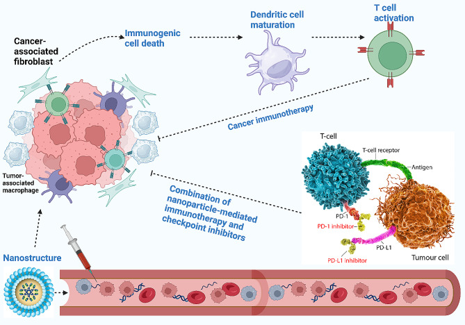
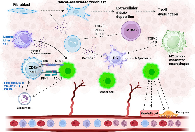
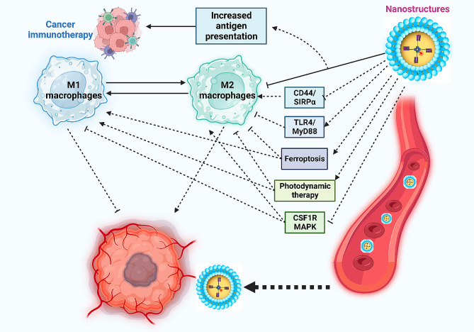
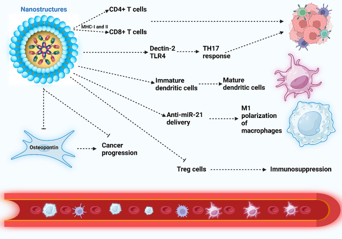
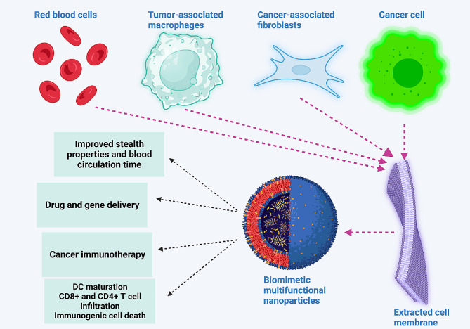
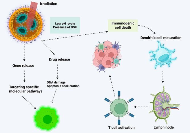
Cancer immunotherapy and vaccine development have significantly improved the fight against cancers. Despite these advancements, challenges remain, particularly in the clinical delivery of immunomodulatory compounds. The tumor microenvironment (TME), comprising macrophages, fibroblasts, and immune cells, plays a crucial role in immune response modulation. Nanoparticles, engineered to reshape the TME, have shown promising results in enhancing immunotherapy by facilitating targeted delivery and immune modulation. These nanoparticles can suppress fibroblast activation, promote M1 macrophage polarization, aid dendritic cell maturation, and encourage T cell infiltration. Biomimetic nanoparticles further enhance immunotherapy by increasing the internalization of immunomodulatory agents in immune cells such as dendritic cells. Moreover, exosomes, whether naturally secreted by cells in the body or bioengineered, have been explored to regulate the TME and immune-related cells to affect cancer immunotherapy. Stimuli-responsive nanocarriers, activated by pH, redox, and light conditions, exhibit the potential to accelerate immunotherapy. The co-application of nanoparticles with immune checkpoint inhibitors is an emerging strategy to boost anti-tumor immunity. With their ability to induce long-term immunity, nanoarchitectures are promising structures in vaccine development. This review underscores the critical role of nanoparticles in overcoming current challenges and driving the advancement of cancer immunotherapy and TME modification.

Keywords: Bioengineered nanostructures; cancer immunotherapy; Immune evasion nanoparticles; Tumor microenvironment.


Conflict of interest statement

The authors declare no competing interests.

Figures

Fig. 1
Fig. 1
An overview of using nanoparticles in cancer immunotherapy. The nanoparticles circulate in blood, and upon reaching the tumor site, they re-educate several tumor microenvironment components, including cancer-associated fibroblasts and tumor-associated macrophages, to finally activate the immune system. Moreover, nanoparticles can stimulate immunogenic cell death to enhance the maturation of dendritic cells for the activation of immune cells, such as T cells, to enhance cancer immunotherapy. The co-application of nanoparticles with immune checkpoint inhibitors, such as PD-L1 blockers, can augment the potential of cancer immunotherapy
Fig. 2
Fig. 2
Cellular components that influence the tumor microenvironment (TME). Interactions within the TME play a crucial role in accelerating cancer progression. Cancer cells activate the PD-L1/PD-1 axis, leading to T cell exhaustion and impairment of T cell function. In addition, cancer cell-secreted exosomes that carry PD-1 contribute to T cell dysfunction, reducing proliferation and hindering proper function. Natural killer cells counteract tumorigenesis by secreting perforin and granular enzymes. Increased infiltration of Treg cells in the TME secretes TGF-β, inducing fibroblast transformation into cancer-associated fibroblasts (CAFs), promoting extracellular matrix deposition, and causing T cell dysfunction. Myeloid-derived suppressor cells (MDSCs) induce Treg cell formation in the TME through the secretion of PGE-2, IL-10, and TGF-β. Regulatory T cells (Treg), in turn, suppress the function of dendritic cells (DCs) by secreting perforin, leading to DC cell apoptosis. M2-polarized macrophages secrete TGF-β and IL-10, disrupting DC cell function. The interaction between endothelial cells and cancer cells results in angiogenesis, further enhancing cancer progression (created by Biorender.com)
Fig. 3
Fig. 3
The impact of nanoparticles on macrophages, showcasing their potential to re-educate and impede cancer progression. These nanoparticles effectively target key mechanisms associated with the M2 polarization of tumor-associated macrophages. They inhibit CD44/SIRPα, CS1R, and MAPK, prompting the M1 polarization of macrophages. Additionally, nanocarriers activate the TLR4/MyD88 axis, contributing to increased M1 polarization of the tumor-associated macrophages. The nanoparticles further induce ferroptosis and photodynamic therapy, disrupting the polarization of these macrophages into the M2 phenotype (Created by Biorender.com)
Fig. 4
Fig. 4
Nanoparticles orchestrating immune cells and cancer-associated fibroblasts (CAFs). Nanoparticles elevate antigen presentation via MHC-I and MHC-II, stimulating CD4+ and CD8+ T cells, thereby facilitating cancer immunotherapy. The nanostructures amplify Dectin-2 and TLR-4 levels, fostering TH17 responses for effective cancer immunotherapy. Additionally, they boost dendritic cell maturation and, through the delivery of anti-miR-21, induce polarization of macrophages into the M1 phenotype. The nanoparticles’ downregulation of osteopontin in CAFs disrupts cancer progression. Moreover, these nanoparticles suppress Treg cells, preventing immunosuppression (Created by Biorender.com)
Fig. 5
Fig. 5
Biomimetic nanoparticles may be developed by the extraction of membranes from red blood cells, cancer cells, TAMs and CAFs. These modifications improve the potential of nanoparticles in cancer immunotherapy. Biomimetic nanoparticles may be utilized for drug and gene delivery by improving stealth properties. They demonstrate prolonged blood circulation and can induce maturation of dendritic cells, increase infiltration of CD4+ and CD8+ T cells, and cause immunogenic cell death. (Created by Biorender.com)
Fig. 6
Fig. 6
Stimuli-responsive nanocarriers in cancer immunotherapy. Nanoparticles that respond to pH, redox, or light can release cargo to induce apoptosis, DNA damage, and regulation of molecular pathways. Moreover, stimuli-responsive nanocarriers stimulate immunogenic cell death to increase dendritic cell maturation. They migrate into lymph nodes and increase activation of T cells

References

    1. Liu Y, et al. Emerging phagocytosis checkpoints in cancer immunotherapy. Signal Transduct Target Ther. 2023;8(1):104. doi: 10.1038/s41392-023-01365-z. - DOI - PMC - PubMed
    1. Kennedy LB, Salama AKS. A review of cancer immunotherapy toxicity. CA Cancer J Clin. 2020;70(2):86–104. doi: 10.3322/caac.21596. - DOI - PubMed
    1. Mahoney KM, Rennert PD, Freeman GJ. Combination cancer immunotherapy and new immunomodulatory targets. Nat Rev Drug Discov. 2015;14(8):561–84. doi: 10.1038/nrd4591. - DOI - PubMed
    1. Webster RM. The immune checkpoint inhibitors: where are we now? Nat Rev Drug Discov. 2014;13(12):883–4. doi: 10.1038/nrd4476. - DOI - PubMed
    1. Zhang Q, et al. Immune and Clinical features of CD96 expression in Glioma by in silico analysis. Front Bioeng Biotechnol. 2020;8:592. doi: 10.3389/fbioe.2020.00592. - DOI - PMC - PubMed

Publication types