. 2024 Apr 1;17(4):dmm050671.
doi: 10.1242/dmm.050671. Epub 2024 Apr 25.

Neurodevelopmental defects in a mouse model of O-GlcNAc transferase intellectual disability

Affiliations

Neurodevelopmental defects in a mouse model of O-GlcNAc transferase intellectual disability

Florence Authier et al. Dis Model Mech. .

Abstract

The addition of O-linked β-N-acetylglucosamine (O-GlcNAc) to proteins (referred to as O-GlcNAcylation) is a modification that is crucial for vertebrate development. O-GlcNAcylation is catalyzed by O-GlcNAc transferase (OGT) and reversed by O-GlcNAcase (OGA). Missense variants of OGT have recently been shown to segregate with an X-linked syndromic form of intellectual disability, OGT-linked congenital disorder of glycosylation (OGT-CDG). Although the existence of OGT-CDG suggests that O-GlcNAcylation is crucial for neurodevelopment and/or cognitive function, the underlying pathophysiologic mechanisms remain unknown. Here we report a mouse line that carries a catalytically impaired OGT-CDG variant. These mice show altered O-GlcNAc homeostasis with decreased global O-GlcNAcylation and reduced levels of OGT and OGA in the brain. Phenotypic characterization of the mice revealed lower body weight associated with reduced body fat mass, short stature and microcephaly. This mouse model will serve as an important tool to study genotype-phenotype correlations in OGT-CDG in vivo and for the development of possible treatment avenues for this disorder.

Keywords: O-GlcNAcylation; Intellectual disability; Vertebrate development.


Conflict of interest statement

Competing interests The authors declare no competing or financial interests.

Figures
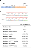
Fig. 1.
Fig. 1.
Genome editing to introduce the OGTC921Y mutation leads to viable mice. (A) Schematic of the OGT protein with position of the OGTC921Y variant marked. TPR (blue), TPR-like (TLR; gray) and catalytic (orange) domains are represented. (B) Sequencing of genomic DNA of male OGTWT and male OGTC921Y mice confirms the presence of the C921Y point mutation in the transgenic animals. DNA sequence chromatograms for a representative OGTWT and OGTC921Y animal are shown alongside the corresponding DNA and amino acid sequences. The G-to-A point mutation for the C921Y variant is marked in red, as are four silent mutations intended to eliminate gRNA recognition sequences. (C) Table showing numbers and percentages of female (OGT+/+), male (OGT+/y), female OgtC921Y/+ (OGTC921Y/+) and male OgtC921Y/y (OGTC921Y/y) animals generated from breeding pairs of female OgtC921Y/+ and male Ogt+/y mice. Litter size is shown as mean±s.d.
Fig. 2.
Fig. 2.
OGTC921Y mutation causes changes in brain O-GlcNAc homeostasis. (A) Western blot analysis of O-GlcNAc, OGA and OGT levels in adult brain of OGTWT and OGTC921Y male mice. Anti-lamin B antibodies raised in rabbit (rLamin B) or mouse (mLamin B) were used as loading controls. Each lane represents independent biological replicates. Molecular masses of markers are shown in kDa. (B) Quantification of total O-GlcNAcylated proteins from the western blot shown in A. (C) Quantification of OGA protein levels from the western blot shown in A. (D) Quantification of OGT protein levels from the western blot shown in A. (E) Quantification of Oga mRNA levels in whole adult brain of OGTWT and OGTC921Y male mice by RT-PCR. (F) Quantification of Ogt mRNA levels in whole adult brain of OGTWT and OGTC921Y male mice by RT-PCR. Protein and mRNA levels are normalized to the mean of the corresponding OGTWT replicate set (a.u., arbitrary units). Data in B-F are represented as mean±s.d., n=3 for all genotypes. **P<0.01; ***P<0.001 (two-tailed unpaired t-test used).
Fig. 3.
Fig. 3.
OGTC921Y mutation leads to changes in mass and size. (A) Measurement of body weight of 77- to 91-day-old OGTWT (n=9) and OGTC921Y (n=7) male mice. (B) Measurement of body length (nose-to-tail length) of 77- to 91-day-old OGTWT (n=10) and OGTC921Y (n=10) male mice. (C) Fat mass:body weight ratio (expressed as a percentage) of 6- to 7-month-old OGTWT (n=5) and OGTC921Y (n=6) male mice. (D) Lean mass:body weight ratio (expressed as a percentage) of 6- to 7-month-old OGTWT (n=5) and OGTC921Y (n=6) male mice. (E) Basal glycemia levels of 6- to 7-month-old OGTWT (n=5) and OGTC921Y (n=6) male mice. Data are represented as mean±s.d. *P<0.05; **P<0.01 (two-tailed unpaired t-test used).
Fig. 4.
Fig. 4.
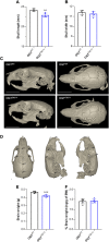
Microcephaly in OGT-CDG mice. (A) Measurement of skull length of 80- to 91-day-old OGTWT (n=4) and OGTC921Y (n=5) male mice. (B) Measurement of skull width of 80- to 91-day-old OGTWT (n=4) and OGTC921Y (n=5) male mice. (C) Lateral and superior views of representative microCT three-dimensional reconstructions of the skull of 80- to 91-day-old OGTWT and OGTC921Y male animals. Scale bars: 5 mm. (D) Lateral and superior views of a representative microCT three-dimensional reconstruction of a mouse skull presenting 14 landmarks (LM1-LM14) used for Euclidean distance matrix analysis (EDMA). (E) Absolute brain weight of 80- to 91-day-old OGTWT (n=5) and OGTC921Y (n=7) male mice, as determined using a precision weighing scale. (F) Brain weight:body weight (BW) ratio (expressed as a percentage) of 80- to 91-day-old OGTWT (n=5) and OGTC921Y (n=7) male mice. Data in A,B,E and F are represented as mean±s.d. **P<0.01; ***P<0.001 (two-tailed unpaired t-test used).

References

    1. Akimoto, Y., Comer, F. I., Cole, R. N., Kudo, A., Kawakami, H., Hirano, H. and Hart, G. W. (2003). Localization of the O-GlcNAc transferase and O-GlcNAc-modified proteins in rat cerebellar cortex. Brain Res. 966, 194-205. 10.1016/S0006-8993(02)04158-6 - DOI - PubMed
    1. Balana, A. T. and Pratt, M. R. (2021). Mechanistic roles for altered O-GlcNAcylation in neurodegenerative disorders. Biochem. J. 478, 2733. 10.1042/BCJ20200609 - DOI - PMC - PubMed
    1. Bond, M. R. and Hanover, J. A. (2013). O-GlcNAc cycling: a link between metabolism and chronic disease. Annu. Rev. Nutr. 33, 205-229. 10.1146/annurev-nutr-071812-161240 - DOI - PMC - PubMed
    1. Capotosti, F., Guernier, S., Lammers, F., Waridel, P., Cai, Y., Jin, J., Conaway, J. W., Conaway, R. C. and Herr, W. (2011). O-GlcNAc transferase catalyzes site-specific proteolysis of HCF-1. Cell 144, 376-388. 10.1016/j.cell.2010.12.030 - DOI - PubMed
    1. Castro, V. L. and Quintana, A. M. (2020). The role of HCFC1 in syndromic and non-syndromic intellectual disability. Med. Res. Arch. 8, mra.v8i6.212. 10.18103/mra.v8i6.2122 - DOI - PMC - PubMed

Publication types

MeSH terms

Substances