Review
. 2022 May 4;1(1):kyac001.
doi: 10.1093/discim/kyac001. eCollection 2022.

Unconventional modes of peptide-HLA-I presentation change the rules of TCR engagement

Affiliations
Review

Unconventional modes of peptide-HLA-I presentation change the rules of TCR engagement

Jade R Hopkins et al. Discov Immunol. .

Abstract

The intracellular proteome of virtually every nucleated cell in the body is continuously presented at the cell surface via the human leukocyte antigen class I (HLA-I) antigen processing pathway. This pathway classically involves proteasomal degradation of intracellular proteins into short peptides that can be presented by HLA-I molecules for interrogation by T-cell receptors (TCRs) expressed on the surface of CD8+ T cells. During the initiation of a T-cell immune response, the TCR acts as the T cell's primary sensor, using flexible loops to mould around the surface of the pHLA-I molecule to identify foreign or dysregulated antigens. Recent findings demonstrate that pHLA-I molecules can also be highly flexible and dynamic, altering their shape according to minor polymorphisms between different HLA-I alleles, or interactions with different peptides. These flexible presentation modes have important biological consequences that can, for example, explain why some HLA-I alleles offer greater protection against HIV, or why some cancer vaccine approaches have been ineffective. This review explores how these recent findings redefine the rules for peptide presentation by HLA-I molecules and extend our understanding of the molecular mechanisms that govern TCR-mediated antigen discrimination.

Keywords: T cells; antigen recognition; computational simulations; human leukocyte antigen (HLA); peptide presentation; protein flexibility.


Figures

Figure 1:
Figure 1:
Overview of the pHLA-I complex and the peptide-binding groove. (A) Side view of the HLA-I α1, α2 and α3 domains (blue cartoon) with β2m (pink cartoon). The HLA-I α1, α2 domains form the peptide-binding groove (peptide shown as yellow sticks). (B) Top view of the α1 and α2 domains (blue cartoon) showing the seven-stranded β-pleated sheets which form the floor of the binding groove. (C) Top view of the α1 and α2 domains as surface representation (cyan). The binding groove is labelled according to the different binding pockets (A-F) that interact with the peptide. (D) Top view of the α1 and α2 domains as surface representation (cyan) showing the peptide (yellow sticks) with the buried anchor residues in black circles.
Figure 2:
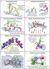
Figure 2:
Different modes of pHLA-I ‘rule breaking’. (A) The HLA-B*35:08-restricted 13-mer (LPEPLPQGQLTAY) peptide (yellow sticks) bulging out of the HLA-I groove (blue cartoon) limiting contacts between the SB27 TCR (green cartoon) and the HLA-I helices. (B) LEFT: C-terminal residue of the MLLSVPLLLG peptide (yellow sticks) extending out of the groove. C-terminal peptide residue usually points down into the groove as a primary anchor (pink sticks). RIGHT: 10-mer LYLVCGERGF peptide (yellow sticks) extends out of the side of the binding groove, mediated by residue Tyr84 (blue sticks) flipping into an open conformation and inducing a widening of the binding groove (black arrows). (C) The MEL5 TCR (green cartoon) ‘pulls’ the EAAGIGILTV peptide (yellow sticks) away from the HLA-I binding groove (shift shown by black dotted line and arrow) making optimised contacts with TCR residue Gln31 (green sticks) compared to the heteroclitic anchor residue optimised version of the peptide (ELAGIGILTV shown as pink sticks). (D) The TL9 (TPQDLNTML) peptide adopts unique conformations when presented by HLA-B*81:01 (yellow sticks) and HLA-B*41:01 (pink sticks). In HLA-B*81:01, residues Leu5 and The7 point down and Asn6 points up (indicated by coloured arrows above and below peptide sequence). In HLA-B*41:01, these residues are inverted, with residues Leu5 and The7 pointing up and Asn6 pointing down. These changes are attributed to micro-polymorphisms between HLA-B*81:01 and HLA-B*41:01. (E) The Tel1p (MLWGYLQYV) peptide (yellow sticks) induces a conformational change in the HLAα2 helix (blue cartoon to pink cartoon and labelled with a black arrow) upon A6 TCR binding (TCR not shown). (F) GVY01 TCR (green cartoon) residue Pro29 (green sticks) can detect alterations in the MAGE-A4 (GVYDGREHTV) peptide at the first residue (yellow and pink sticks) due a conformational change (black arrow) in HLA-I residue Trp167 (blue and pink sticks). (G) Abacavir (yellow sticks and circled in black) can bind into the F-pocket HLA-B*57:02 (blue surface), altering the chemical nature of the binding pocket. This blocks the binding of natural HLA-B*57:02-restricted peptides (pink sticks) due to steric repulsion, altering the nature of the presented peptide repertoire (peptide + abacavir shown in yellow sticks). (H) Representation of the correlated network of motions, found to be dependent on peptide sequence, analysed by molecular dynamics simulation. Networked communities shown as coloured spheres, with larger spheres indicating a greater number of residues within the community. Lines represent communication pathways between the nodes, with thicker lines indicating a greater degree of correlation between the two communities. It was found that different peptides, bound to the same HLA-I molecule, could modulate these networks (‘the tail wags the dog’) with implications for antigen processing, co-receptor binding, and TCR recognition.

Similar articles

Cited by

References

    1. Gorer PA. The detection of a hereditary antigenic difference in the blood of mice by means of human group a serum. J Genet 1936, 32, 17–31.
    1. Horton R, Wilming L, Rand V, Lovering RC, Bruford EA, Khodiyar VK, et al. . Gene map of the extended human MHC. Nat Rev Genet 2004, 5, 889–99. - PubMed
    1. Rothbard JB, Gefter ML.. Interactions between immunogenic peptides and MHC proteins. Annu Rev Immunol 1991, 9, 527–65. - PubMed
    1. Guillet J-G, Lai M-Z, Briner TJ, Smith JA, Gefter ML.. Interaction of peptide antigens and class II major histocompatibility complex antigens. Nature 1986, 324, 260–2. - PubMed
    1. Davis MM, Bjorkman PJ.. The T cell receptor genes and T cell recognition. Nature 1988, 334, 395–402. - PubMed

LinkOut - more resources