. 2024 Mar 19:15:1364036.
doi: 10.3389/fimmu.2024.1364036. eCollection 2024.

Uterine macrophages and NK cells exhibit population and gene-level changes after implantation but maintain pro-invasive properties

Affiliations

Uterine macrophages and NK cells exhibit population and gene-level changes after implantation but maintain pro-invasive properties

Sneha Mani et al. Front Immunol. .

Abstract

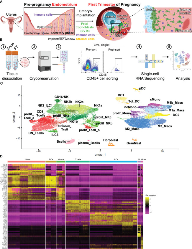
Introduction: Prior to pregnancy, hormonal changes lead to cellular adaptations in the endometrium allowing for embryo implantation. Critical for successful pregnancy establishment, innate immune cells constitute a significant proportion of uterine cells prior to arrival of the embryo and throughout the first trimester in humans and animal models. Abnormal uterine immune cell function during implantation is believed to play a role in multiple adverse pregnancy outcomes. Current work in humans has focused on uterine immune cells present after pregnancy establishment, and limited in vitro models exist to explore unique functions of these cells.

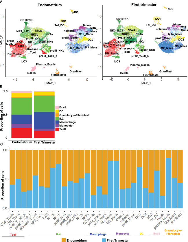
Methods: With single-cell RNA-sequencing (scRNAseq), we comprehensively compared the human uterine immune landscape of the endometrium during the window of implantation and the decidua during the first trimester of pregnancy.

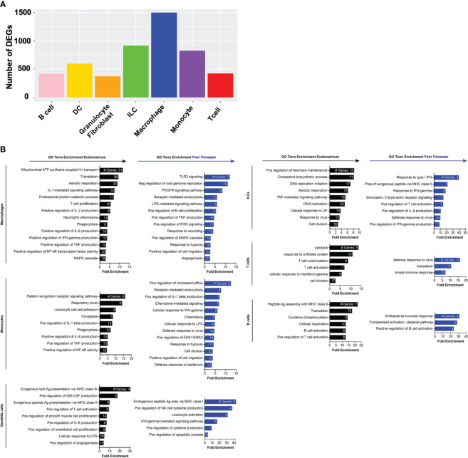
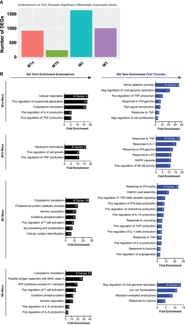
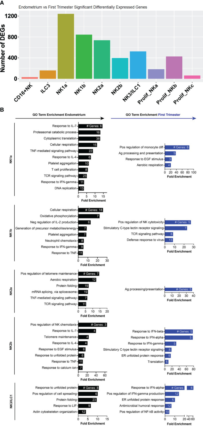
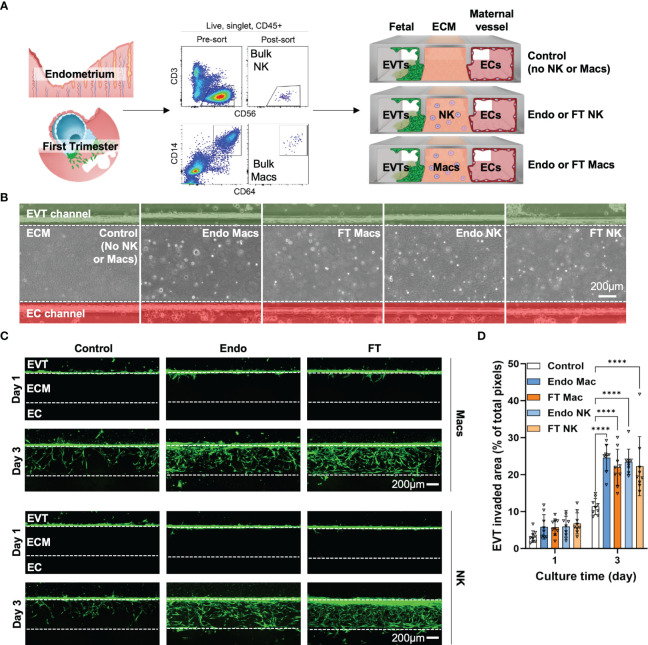
Results: We uncovered global and cell-type-specific gene signatures for each timepoint. Immune cells in the endometrium prior to implantation expressed genes associated with immune metabolism, division, and activation. In contrast, we observed widespread interferon signaling during the first trimester of pregnancy. We also provide evidence of specific inflammatory pathways enriched in pre- and post-implantation macrophages and natural killer (NK) cells in the uterine lining. Using our novel implantation-on-a-chip (IOC) to model human implantation ex vivo, we demonstrate for the first time that uterine macrophages strongly promote invasion of extravillous trophoblasts (EVTs), a process essential for pregnancy establishment. Pre- and post-implantation uterine macrophages promoted EVT invasion to a similar degree as pre- and post-implantation NK cells on the IOC.

Conclusions: This work provides a foundation for further investigation of the individual roles of uterine immune cell subtypes present prior to embryo implantation and during early pregnancy, which will be critical for our understanding of pregnancy complications associated with abnormal trophoblast invasion and placentation.

Keywords: placentation; decidua; endometrium; implantation; organ-on-a-chip; uterine NK cells; uterine macrophages.


Conflict of interest statement

The authors declare that the research was conducted in the absence of any commercial or financial relationships that could be construed as a potential conflict of interest.

Figures

Figure 1
Figure 1
Combined immune landscape of the window of implantation endometrium and first-trimester decidua at high resolution. (A) Graphical representation of cellular changes that occur during the menstrual cycle and early pregnancy, including recruitment of immune cells into the uterine lining. (B) Experimental workflow. Four endometrial biopsies and four first trimester deciduae were enzymatically digested into single cell suspensions and cryopreserved. Frozen cells were then thawed, stained, and FACS-purified as CD45+ cells prior to scRNAseq and downstream analyses. Shown are raw flow cytometry data of pre- and post-sort purity of CD45+ cells. Events shown have undergone forward and side scatter gating, singlet discrimination, and live/dead discrimination. (C) 30 total clusters of CD45+ cells were identified. Subclusters are shaded according to supercluster: red, T cells; green, ILCs; pink, B cells; yellow, DCs; purple, monocytes; blue, macropahges. “Prolif” denotes proliferating; “DN”, double-negative (CD4-CD8-); “ILC”, innate lymphoid cell; “NK”, natural killer cell; “DC”, dendritic cell; “Tol_DC”, tolerogenic DC; “pDC”, plasmacytoid DC; “cMono”, classical monocyte”; “ncMono”, nonclassical monocyte; “Macs”, macrophages. (D) Heatmap representation of supercluster-defining is shown.
Figure 2
Figure 2
Hallmark genes of immune subclusters in the combined window of implantation endometrium and first-trimester decidua. Heatmap visualizations of cluster-defining genes across subclusters of (A) lymphocytes and (C) myeloid cells. Additional dot plot visualizations (B, D–F) of selected genes and subclusters are shown. Size of the dots indicates percent of total cells in each subcluster that express the indicated genes. Color of the dots correlates to the average level of expression of the indicated genes. B cells, mast cells, and fibroblasts are not shown due to these being rare cells in our dataset.
Figure 3
Figure 3
Dynamic changes in subpopulations of immune superclusters and subclusters in response to embryo implantation. (A) UMAP plots showing subclustering of CD45+ cells meeting quality metrics, recovered from mid-secretory endometrium (left) and first trimester decidua (right). ILC, innate lymphoid cells. (B) Stacked bar charts showing proportion of total CD45+ cells represented by indicated superclusters in pre-implantation endometrium and first trimester decidua. (C) Stacked bar charts showing proportion of total cells in indicated subcluster (x-axis) recovered from endometrium (orange bars) or first trimester decidua (blue bars).
Figure 4
Figure 4
Gene expression changes across superclusters in response to embryo implantation. (A) Bar graph showing absolute number of significant differentially expressed genes (DEGs) between endometrial and first trimester decidual CD45+ cells in indicated superclusters. Significant genes are defined as having an adjusted p value of <0.05 and an absolute fold change of 25% or greater (either ≥25% enriched in endometrium or ≥25% in decidua). (B) Gene ontology (GO) analysis of DEGs enriched in indicated endometrial (black) or first trimester decidual (blue) superclusters. GO terms significantly enriched are shown (false discovery rate [FDR] value ≤0.05). The x-axis shows fold enrichment of the indicated GO term on the y-axis. Embedded in the bars are the number of DEGs contributing to each GO term. If applicable, GO terms were shortened due to space restrictions and/or combined into one term that contained the most enriched genes. For instance, “negative regulation of viral genome replication”, “response to virus”, “viral entry into host cell”, and “defense response to virus” are GO terms that contain highly overlapping genes. Thus, only one of the terms was included. Additionally, such broad GO terms as “immune system process” and “inflammatory response” were not included in a dataset consisting only of immune cells.
Figure 5
Figure 5
Inflammatory and metabolic pathways are enriched in pre-implantation endometrial macrophages, while interferon-stimulated, hypoxia, and inflammatory response genes are enriched in first trimester decidual macrophages. (A) Bar graph showing absolute number of significant differentially expressed genes (DEGs) between indicated endometrial and first trimester decidual macrophage subclusters. Significant genes are defined as having an adjusted p value of <0.05 and an absolute fold change of 25% or greater (either ≥25% enriched in fresh or ≥25% in frozen). (B) Gene ontology (GO) analysis of DEGs enriched in indicated endometrial (black) or first trimester decidual (blue) macrophage superclusters. Detailed information describing the GO term enrichment bar graphs is provided in the caption to Figure 4B .
Figure 6
Figure 6
Metabolic, chemotactic, and inflammatory response pathways are enriched in pre-implantation endometrial NK cells, while interferon-stimulated genes are enriched in first trimester decidual ILC subsets. (A) Bar graph showing absolute number of significant differentially expressed genes (DEGs) between indicated endometrial and first trimester decidual ILC subclusters. Significant genes are defined as having an adjusted p value of <0.05 and an absolute fold change of 25% or greater (either ≥25% enriched in fresh or ≥25% in frozen). (B) Gene ontology (GO) analysis of DEGs enriched in indicated endometrial (black) or first trimester decidual (blue) ILC superclusters. Detailed information describing the GO term enrichment bar graphs is provided in the caption to Figure 4B .
Figure 7
Figure 7
Pre- and post-implantation bulk CD14+CD64+ macrophages/monocytes promote invasion of primary EVTs as strongly as pre- and post-implantation bulk NK cells ex vivo on an implantation-on-a-chip device. (A) Graphical representation of the experimental approach taken to prepare and load purified endometrial and decidual bulk NK cells (CD56+CD3- cells, top flow cytometry plots) and bulk macrophages (CD14+CD64+, bottom flow cytometry plots) onto the implantation-on-a-chip (IOC). Shown are raw flow cytometry data of pre- and post-sort purity of NK cells and macrophages. Events shown have undergone forward and side scatter gating, singlet discrimination, live/dead discrimination, and CD45+ gating. “Fetal” denotes the channel of the IOC containing fluorescently labeled fetal EVTs, “ECM” denotes the channel of the IOC containing mock extracellular matrix, and “Maternal vessel” denotes the channel of the IOC containing endothelial cells, as described in the Methods section. Note the horizontal orientation of the actual devices. For purposes of data acquisition and interpretation, the IOC is imaged and shown in (B, C) in the vertical orientation, with the fetal channel at the top, ECM in the middle, and the maternal vessel channel at the bottom. (B) Brightfield imaging showing successful embedding of purified endometrial macrophages (Endo Mac), first trimester decidual macrophages (FT Mac), endometrial NK cells (Endo NK) and first trimester decidual NK cells (FT NK) onto the IOC. (C) Representative fluorescent imaging of labeled EVTs invading into the ECM and into the maternal vessel channel on the indicated post-seeding day. Days 1 and 3 post-seeding are shown. Channels on each device are separated by white dotted lines. After 1 day, note that most of the EVTs in any condition have not yet entered the ECM. After 3 days of culture, most of the EVTs are seen in the ECM channel in the macrophage and NK cell conditions. Scale bars represent 200μM. (D) Quantification of invasion area of EVTs beyond the fetal channel at day 3 of culture. One-way ANOVA with Tukey’s multiple comparison test was performed (n = 3 independent biological samples and 3 independent devices per biological sample). Data are presented as mean ± SD. Area of invasion at day 1 was not significantly different among groups. Invasion area of EVTs in the macrophage and NK cell conditions were all significantly different than the control (no immune cell) condition at day 3 of culture, ****, p<0.0001.

References

    1. Organization WH . Global Health Observatory Data (2015). Available online at: http://www.who.int/gho/maternal_health/en/.
    1. Tantbirojn P, Crum CP, Parast MM. Pathophysiology of placenta creta: the role of decidua and extravillous trophoblast. Placenta. (2008) 29:639–45. doi: 10.1016/j.placenta.2008.04.008 - DOI - PubMed
    1. Kaufmann P, Black S, Huppertz B. Endovascular trophoblast invasion: implications for the pathogenesis of intrauterine growth retardation and preeclampsia. Biol Reprod. (2003) 69:1–7. doi: 10.1095/biolreprod.102.014977 - DOI - PubMed
    1. Kim YM, Bujold E, Chaiworapongsa T, Gomez R, Yoon BH, Thaler HT, et al. . Failure of physiologic transformation of the spiral arteries in patients with preterm labor and intact membranes. Am J obstetrics gynecology. (2003) 189:1063–9. doi: 10.1067/S0002-9378(03)00838-X - DOI - PubMed
    1. Goldman-Wohl D, Yagel S. Regulation of trophoblast invasion: from normal implantation to pre-eclampsia. Mol Cell Endocrinol. (2002) 187:233–8. doi: 10.1016/S0303-7207(01)00687-6 - DOI - PubMed

Publication types