

A Unique Case of Late Presentation Giant Lower Extremity Malignant Melanoma
- PMID: 38567210
- PMCID: PMC10985282
- DOI: 10.7759/cureus.55414
A Unique Case of Late Presentation Giant Lower Extremity Malignant Melanoma
Abstract
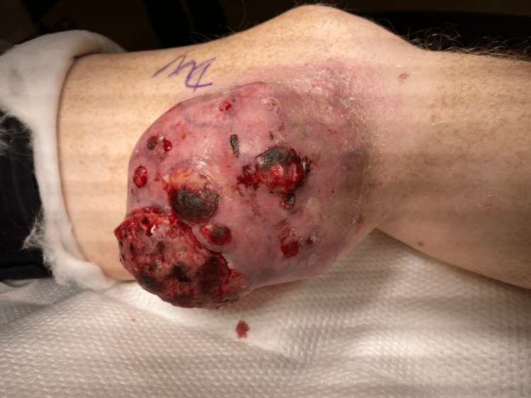
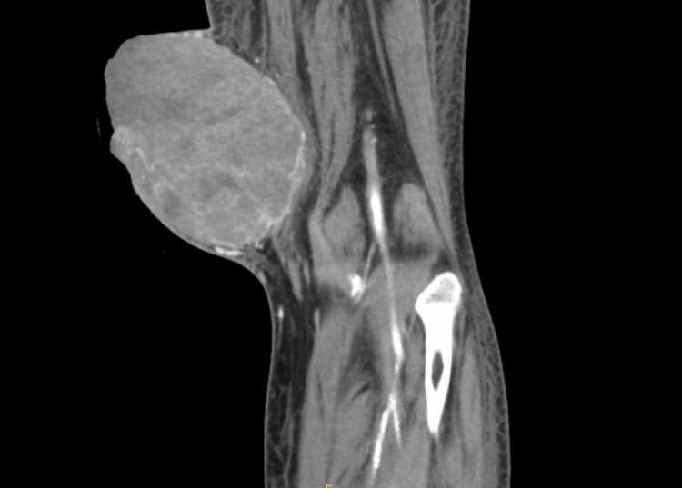
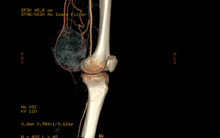
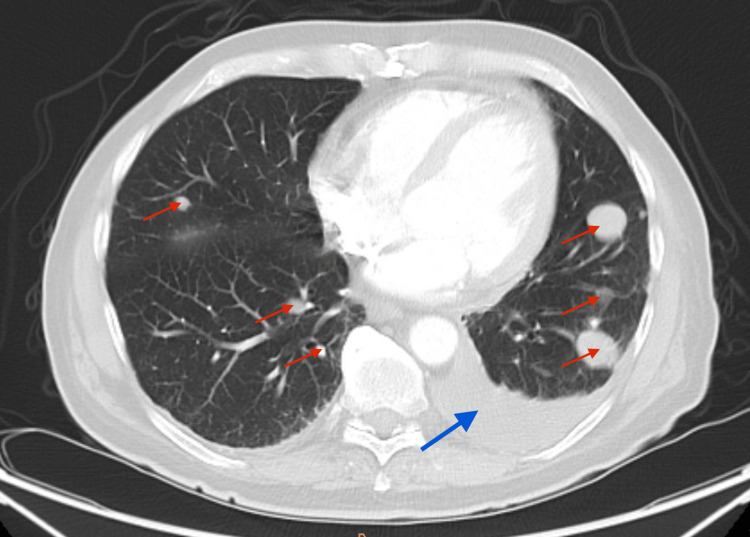
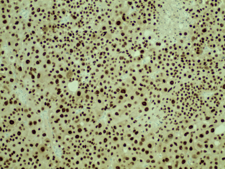
This case describes a unique presentation of a rare malignancy: giant melanoma. Due to the accessibility of healthcare in the United States, it is unusual for melanomas to grow to massive sizes without clinical intervention. In fact, an in-depth literature review elicited only a handful of similar cases. Giant malignant melanomas are typically defined by a cutoff size of no less than 10 cm in diameter. They often present with distant metastases and are highly invasive. Due to limited yet highly variable presentations, there is no standardized approach to treating this class of melanomas. We present a case with unique features not previously documented in similar cases that was ultimately treated with a novel approach.
Keywords: cancer immunotherapy; cutaneous malignancy; giant melanoma; malignant melanoma metastasis; oncology imaging.
Copyright © 2024, Al Zarkali et al.
Conflict of interest statement
The authors have declared that no competing interests exist.
Figures
Similar articles
-
Case Report and Literature Review of Giant Cutaneous Malignant Melanoma: What's Keeping Patients Away?Eplasty. 2022 Sep 23;22:e45. eCollection 2022. Eplasty. 2022. PMID: 36212603 Free PMC article.
-
Unusual Presentation of Giant Upper Limb Malignant Melanoma.Cureus. 2021 Aug 8;13(8):e16996. doi: 10.7759/cureus.16996. eCollection 2021 Aug. Cureus. 2021. PMID: 34540398 Free PMC article.
-
Two unique cases of metastatic malignant melanoma of the gastrointestinal tract.Int J Surg Case Rep. 2023 Feb;103:107907. doi: 10.1016/j.ijscr.2023.107907. Epub 2023 Feb 3. Int J Surg Case Rep. 2023. PMID: 36737869 Free PMC article.
-
Uncommon Subtypes of Malignant Melanomas: A Review Based on Clinical and Molecular Perspectives.Cancers (Basel). 2020 Aug 21;12(9):2362. doi: 10.3390/cancers12092362. Cancers (Basel). 2020. PMID: 32825562 Free PMC article. Review.
-
[Malignant melanoma in children--case presentation and statistical analysis].Nihon Hifuka Gakkai Zasshi. 1990 Apr;100(5):597-605. Nihon Hifuka Gakkai Zasshi. 1990. PMID: 2203921 Review. Japanese.
References
Publication types
LinkOut - more resources
Full Text Sources