. 2024 Jul;24(7):e2400111.
doi: 10.1002/mabi.202400111. Epub 2024 Apr 18.

Peptoid-Cross-Linked Hydrogel Stiffness Modulates Human Mesenchymal Stromal Cell Immunoregulatory Potential in the Presence of Interferon-Gamma

Affiliations

Peptoid-Cross-Linked Hydrogel Stiffness Modulates Human Mesenchymal Stromal Cell Immunoregulatory Potential in the Presence of Interferon-Gamma

David A Castilla-Casadiego et al. Macromol Biosci. 2024 Jul.

Abstract

Human mesenchymal stromal cell (hMSC) manufacturing requires the production of large numbers of therapeutically potent cells. Licensing with soluble cytokines improves hMSC therapeutic potency by enhancing secretion of immunoactive factors but typically decreases proliferative ability. Soft hydrogels, however, have shown promise for boosting immunomodulatory potential, which may compensate for decreased proliferation. Here, hydrogels are cross-linked with peptoids of different secondary structures to generate substrates of various bulk stiffnesses but fixed network connectivity. Secretions of interleukin 6, monocyte chemoattractive protein-1, macrophage colony-stimulating factor, and vascular endothelial growth factor are shown to depend on hydrogel stiffness in the presence of interferon gamma (IFN-γ) supplementation, with soft substrates further improving secretion. The immunological function of these secreted cytokines is then investigated via coculture of hMSCs seeded on hydrogels with primary peripheral blood mononuclear cells (PBMCs) in the presence and absence of IFN-γ. Cocultures with hMSCs seeded on softer hydrogels show decreased PBMC proliferation with IFN-γ. To probe possible signaling pathways, immunofluorescent studies probe the nuclear factor kappa B pathway and demonstrate that IFN-γ supplementation and softer hydrogel mechanics lead to higher activation of this pathway. Overall, these studies may allow for production of more efficacious therapeutic hMSCs in the presence of IFN-γ.

Keywords: human mesenchymal stromal cells; hydrogels; immunosuppression; secretome.


Conflict of interest statement

Declaration of Competing Interest

The authors declare that they have no known competing financial interests or personal relationships that could have appeared to influence the work reported in this paper.

Figures

Figure 1.
Figure 1.
FTIR analysis confirming the inclusion of the crosslinker and the maintenance of the hyaluronic acid peaks for each formulation. Chemical composition of hyaluronic acid, norbornene-functionalized hyaluronic acid, and peptoid crosslinked hydrogels. A) Chemical structure (i) HA, (ii) norbornene-functionalized HA, B) FTIR spectrums of HA and Norbornene-functionalized HA (Range: 3700–700 cm−1). C) Functional groups vs absorption location. FTIR spectrums of the peptoids and peptoid crosslinked hydrogels (Range: 3700–700 cm−1). D) Helical, E) Non-Helical, F) unstructured.
Figure 2.
Figure 2.
Crosslinker structure dictates the microstructure and compressive modulus of the resulting hydrogel. The resulting hydrogel morphology for each crosslinker, seeming to indicate a wide discrepancy in microstructures, particularly when comparing the unstructured peptoid to the other peptoid crosslinkers. A) Representative SEM pictures of each hydrogel formulation after lyophilization (n = 3) (High magnification 5000X). i) Helical peptoid crosslinked hydrogel, ii) Non-Helical peptoid crosslinked hydrogel, iii) Peptide crosslinked hydrogel, iv) Unstructured peptoid crosslinked hydrogel. Helical crosslinkers produce hydrogels with compressive moduli that are significantly higher than all other conditions, and a clear trend is present from helical to non-helical, and eventually to unstructured and peptide crosslinked hydrogels. B) Compression modulus of each hydrogel formulation. All bars represent n=4 hydrogels. * denotes p<0.05, ** p<0.01, *** p<0.001, **** p<0.0001 between conditions. All statistics were calculated by one-way ANOVA with post-hoc Tukey HSD test.
Figure 3.
Figure 3.
Softer hydrogels upregulate the production of immunoregulatory and regenerative cytokines in presence of IFN-γ. Cytokine levels are assessed by Luminex assay. Comparative hMSCs protein expression from donors 1 and 2 cultured on all hydrogel conditions and TCP with and without IFN-γ supplementation after 6 days of culture. A) IL-6, B) MCP-1, C) M-CSF, D) VEGF, E) Heatmap representing the concentration values of each cytokine released on each substrate, scaled to 100%. White; low expression, blue; high expression. All bars represent n=3 hydrogels. * denotes p<0.05, ** p<0.01, *** p<0.001, **** p<0.0001 between that condition with and without IFN-γ. # indicates p<0.05, $ indicates p<0.01, % indicates p<0.001, and & indicates p<0.0001 between the conditions marked. All statistics were calculated by one-way ANOVA with post-hoc Tukey HSD test.
Figure 4.
Figure 4.
Soft substrates with seeded hMSCs reduce the proliferation of cocultured PBMCs. A) Schematic of the experimental outline: Direct contact co-culture investigations of hMSCs and stimulated PBMCs. Quantification of proliferating PBMCs when exposed to hMSCs (2 donors) seeded on each condition with and without IFN-γ supplementation after 6 days of culture (PBMCs proliferation was assessed via EdU incorporation for 8 hours of culture). B) Donor 1, (i) with IFN-γ, (ii) without IFN-γ, C) Donor 2, (i) with IFN-γ, (ii) without IFN-γ, D) Representative fluorescence microscopy images of proliferating PBMCs stained for EdU, blue color represents the nucleus (DAPI), green represents EdU label, scale bar 100 μm. All bars represent n=4 hydrogels. * denotes p<0.05, ** p<0.01, *** p<0.001, **** p<0.0001 between conditions. $ indicates p<0.01 and & indicates p<0.0001 between the conditions marked with and without IFN-γ. All statistics were calculated by one-way ANOVA with post-hoc Tukey HSD test.
Figure 5.
Figure 5.
Soft substrates increase the nuclear localization of the NF-kB p65 activation in hMSCs in the presence and absence of IFN-γ. Representative fluorescent images of NF-kB activation in hMSCs with and without IFN-γ supplementation after 3 days of culture. A) without IFN-γ supplementation, B) with IFN-γ supplementation. Green represents NF-kB p65 staining and blue represents the nucleus (DAPI), scale bar 100 μm. Fluorescence intensity of the NF-kB immunostaining in cell nuclei and cytoplasm. C) without IFN-γ supplementation, D) with IFN-γ supplementation. All data presented are means ± standard deviations of n =20 cell measurements of two independent studies from two hMSCs donors across 4 hydrogel replicates. * denotes p<0.05, ** p<0.01, *** p<0.001 between conditions. $ indicates p<0.05 between the conditions marked with and without IFN-γ. All statistics were calculated by one-way ANOVA with post-hoc Tukey HSD test.
Figure 6.
Figure 6.
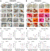
Peptoid crosslinked hydrogel did not negatively impact the differentiation capabilities of hMSCs with or without IFN-γ supplementation. A) Differentiated adipogenic hMSCs from donors 1 and 2 were stained with Oil Red and imaged after 10 days of culture in adipogenic medium with and without IFN-γ supplementation. B) Differentiated osteogenic hMSCs from donors 1 and 2 were stained with Alizarin Red and imaged after 10 days of culture in osteogenic medium with and without IFN-γ supplementation. Real-time RT-PCR data confirmed gene expression consistent with adipogenic and osteogenic differentiation of hMSCs of both donors seeded on our hydrogel formulations in the presence of soluble IFN-γ after differentiation induction for 10 days. C) Adipogenic differentiation: CEBPA gene (i) Donor 1 and (ii) donor 2, PPARG gene (iii) Donor 1 and (iv) donor 2. D) Osteogenic differentiation: ALPL gene (i) Donor 1 and (ii) donor 2, RUNX2 gene (iii) Donor 1 and (iv) donor 2. (Error bars shown are representative of RQ (relative quantity) minimum and maximum determined by technical replicates).
Scheme 1.
Scheme 1.
Peptoid Crosslinked hydrogels using sequence-defined secondary structure: helical (Hel), non-helical (N-hel), and unstructured (Unst).

References

    1. Trohatou O, Roubelakis MG, Cell. Reprogram 2017, 19, 217. - PubMed
    1. Mannino G, Russo C, Longo A, Anfuso CD, Lupo G, Lo Furno D, Giuffrida R, Giurdanella G, World J. Stem Cells 2021, 13, 632. - PMC - PubMed
    1. Rodríguez-Fuentes DE, Fernández-Garza LE, Samia-Meza JA, Barrera-Barrera SA, Caplan AI, Barrera-Saldaña HA, Arch. Med. Res 2021, 52, 93. - PubMed
    1. Hmadcha A, Martin-Montalvo A, Gauthier BR, Soria B, Capilla-Gonzalez V, Front. Bioeng. Biotechnol 2020, 8, 43. - PMC - PubMed
    1. Solis MA, Moreno Velásquez I, Correa R, Huang LLH, Diabetol. Metab. Syndr 2019, 11, 20. - PMC - PubMed

MeSH terms

LinkOut - more resources