. 2024 Apr 16;18(15):10439-10453.
doi: 10.1021/acsnano.3c11374. Epub 2024 Apr 3.

Self-Assembled STING-Activating Coordination Nanoparticles for Cancer Immunotherapy and Vaccine Applications

Affiliations

Self-Assembled STING-Activating Coordination Nanoparticles for Cancer Immunotherapy and Vaccine Applications

Xiaoqi Sun et al. ACS Nano. .

Abstract

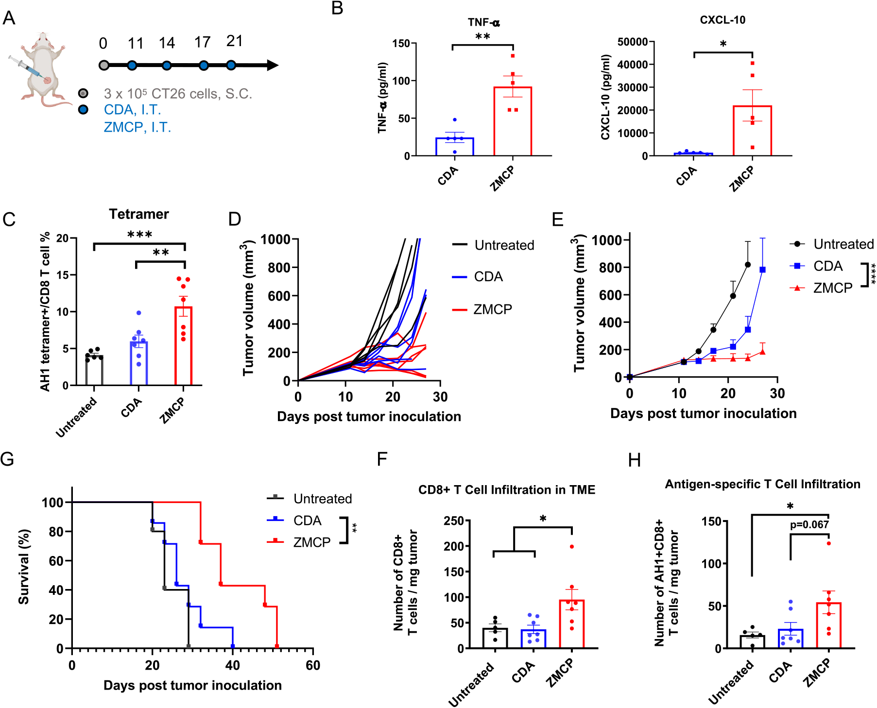
The cGAS-STING pathway plays a crucial role in innate immune activation against cancer and infections, and STING agonists based on cyclic dinucleotides (CDN) have garnered attention for their potential use in cancer immunotherapy and vaccines. However, the limited drug-like properties of CDN necessitate an efficient delivery system to the immune system. To address these challenges, we developed an immunostimulatory delivery system for STING agonists. Here, we have examined aqueous coordination interactions between CDN and metal ions and report that CDN mixed with Zn2+ and Mn2+ formed distinctive crystal structures. Further pharmaceutical engineering led to the development of a functional coordination nanoparticle, termed the Zinc-Mn-CDN Particle (ZMCP), produced by a simple aqueous one-pot synthesis. Local or systemic administration of ZMCP exerted robust antitumor efficacy in mice. Importantly, recombinant protein antigens from SARS-CoV-2 can be simply loaded during the aqueous one-pot synthesis. The resulting ZMCP antigens elicited strong cellular and humoral immune responses that neutralized SARS-CoV-2, highlighting ZMCP as a self-adjuvant vaccine platform against COVID-19 and other infectious pathogens. Overall, this work establishes a paradigm for developing translational coordination nanomedicine based on drug-metal ion coordination and broadens the applicability of coordination medicine for the delivery of proteins and other biologics.

Keywords: STING; cancer immunotherapy; coordination nanoparticle; cyclic dinucleotide; vaccine.


Figures

Scheme 1.
Scheme 1.. Aqueous One-Pot Assembly of Cyclic Dinucleotides Coordination Nanoparticle As a STING-Activating Platform for Cancer Immunotherapy and Vaccination against COVID-19a
a(A) Identification of unique macro-scale crystal structures of cyclic di-AMP (CDA)–Zn and CDA–Zn–Mn from screening of aqueous coordination interactions between CDA and metal ions. Further pharmaceutical engineering led to the development of a functional nanoparticle platform, termed Zinc–Mn–CDN Particle (ZMCP). (B) ZMCP enables systemic delivery of CDA for cancer immunotherapy and elicits robust anti-tumor efficacy. (C) ZMCP platform serves as a self-adjuvanted vaccine and ZMCP carrying SARS-CoV-2 antigens elicits strong anti-viral cellular and humoral immune responses. BioRender.com was used to create the scheme.
Figure 1.
Figure 1.
Development of aqueous one-pot-assembled cyclic dinucleotide (CDN) nanomedicine based on the coordination of CDN with metal ions. (A–D) Screening CDA–metal ion coordination polymorphs. ZnCl2, MgCl2, MnCl2, CaCl2, Al2(SO4)3, CuCl2, FeCl2, FeCl3, NiCl2, and CoCl2 solutions in water were added to CDA in 5 or 10 equivalents (n/n), followed by absorbance measurement at OD450 nm, OD500 nm, and OD600 nm (A). The loading efficacy of CDN in the coordination structure (B). Microscopy images of the CDN–metal coordination structures (C). Scale bar: 200 μm. (D) Titrating the ratio of CDA/Zn2+ (1:0.1 to 1:8) revealed ratio-dependent change of the crystal geometry, and doping Mn2+ into Zn2+/CDA induced needle-like crystals. Scale bar = 200 μm. (E,F) Aqueous one-pot assembly of CDN coordination nanoparticle. (E) CDA, H33-PEG20k, Zn2+, or/and Mn2+ were mixed in a one-pot synthesis. The resulting coordination nanoparticles, ZCP, ZMCP, and MCP, were visualized by TEM. Scale bar = 50 nm. (F) BMDCs were treated with ZMCP, ZCP, MCP, CDA+Mn2++Zn2+, CDA, or other controls, followed by flow cytometry analysis for the expression of CD80 and CD86. Data represent mean ± SEM, from a representative experiment of two independent experiments with n = 3 biological independent samples. *P < 0.05, **P < 0.01, ***P < 0.001, ****P < 0.0001, analyzed by one-way ANOVA (F) with Bonferroni’s multiple comparisons test.
Figure 2.
Figure 2.
ZMCP promotes cellular uptake, endosomal escape, and STING activation. (A,B) Hydrodynamic size (A) and zeta potential (B) of ZMCP. (C) Release kinetics of CDA, Mn, and Zn from ZMCP in PBS. (D) Cellular uptake of free CDG-Dy547 or CDG-Dy547@ZMCP by BMDCs over time was analyzed by flow cytometry. (E) As a surrogate marker for endosomal escape, hemolysis of murine red blood cells with ZMCP or soluble controls was measured at pH 7.4, 6, and 5. (F) BMDCs were incubated with ZMCP or CDA for 24 h, followed by ELISA-based measurement of IFN-β, TNF-α, CXCL-9, and CXCL-10 in the media. (G) Schematic mechanism of ZMCP-induced cellular uptake, endosomal escape, and STING activation. Data represent mean ± SEM, from a representative experiment of two independent experiments with n = 3 biological independent samples. *P < 0.05, **P < 0.01, ***P < 0.001, ****P < 0.0001, analyzed by two-way ANOVA (D, E, F) with Bonferroni’s multiple comparisons test. BioRender.com was used to create G.
Figure 3.
Figure 3.
Intratumoral injection of ZMCP induces antitumor immune responses and inhibits established tumors. (A) BALB/c mice were inoculated at S.C. flank with 3 × 10⁵ CT26 tumor cells. CDA or ZMCP containing 5 μg CDA were injected I.T. on days 11, 14, 17, and 21. (B) Serum cytokines/chemokines were measured by ELISA at 6 h post the second dose. (C) AH1 antigen-specific CD8+ T cell response was analyzed among PBMCs by flow cytometry on day 18. (D–F) ZMCP inhibited tumor growth (D,E) and prolonged animal survival (F). (G) Tumor-infiltrating total CD8+ T cells and (H) AH1 antigen-specific CD8+ T cells were analyzed in the tumor microenvironment by flow cytometry on day 27. Data represent mean ± SEM, from a representative experiment of two independent experiments with n = 5–8. *P <0.05, **P < 0.01, ***P < 0.001, ****P < 0.0001, analyzed by two-tailed Student’s t test (B), one-way ANOVA (C, G, H), or two-way ANOVA with Bonferroni’s multiple comparisons test or log-rank (Mantel-Cox) test (F).
Figure 4.
Figure 4.
Intravenous injection of ZMCP effectively induces an antitumor immune response and inhibits growth of established CT26 tumors. (A–G) BALB/c mice were inoculated at the S.C. flank with 3 × 10⁵ CT26 tumor cells. CDA or ZMCP containing 20 μg of CDA were injected I.V. on days 11, 14, 17, and 21 (A). Serum cytokines were measured by ELISA at 0, 4, 8, and 24 h post the second dose (B). Tumor growth (C–D) and animal survival (E) were monitored over time. (F–L) Mice were analyzed on day 27 by flow cytometry for immune cell profiles in PBMCs (F), tumor microenvironment (G–I) and spleen (J–L). Data represent mean ± SEM, from a representative experiment of two independent experiments with n = 5–8. *P < 0.05, **P < 0.01, ***P < 0.001, ****P < 0.0001, analyzed by one-way ANOVA (F–L) or two-way ANOVA (B, D) with Bonferroni’s multiple comparisons test or log-rank (Mantel-Cox) test (E).
Figure 5.
Figure 5.
Intravenous injection of ZMCP effectively induces an antitumor immune response and inhibits tumor growth in an established B16F10 melanoma model. (A) C57BL/6 mice were inoculated at the S.C. flank with 3 × 10⁵ B16F10 tumor cells. CDA or ZMCP containing 20 μg of CDA were injected I.V. on days 11, 14, 17, and 20. (B) Serum cytokines were measured by ELISA at 0, 4, 8, and 24 h post the second dose. (C–J) Mice were analyzed on day 26 by flow cytometry for immune cell profiles in spleens (C–F) and the tumor microenvironment (G–J). (K,L) Tumor growth (K) and animal survival (L) post-treatment were monitored over time. Data represent mean ± SEM, from a representative experiment of two independent experiments with n = 5–8. *P < 0.05, **P < 0.01, ***P < 0.001, ****P < 0.0001, analyzed by one-way ANOVA (C–J) or two-way ANOVA (B, K) with Bonferroni’s multiple comparisons test or log-rank (Mantel-Cox) test (L).
Figure 6.
Figure 6.
Delivery of the SARS-CoV2 spike protein receptor binding domain (RBD) by ZMCP as a self-adjuvanted COVID-19 vaccine against virus infection. (A,B) One-pot formulation of ZMCP-RBD. CDA, H33-PEG20k, Zn2++Mn2+, and RBD-H6 were mixed in a one-pot synthesis (A). Encapsulation of the RBD in nanoparticles was measured by SDS-PAGE (B). (C–E) Balb/c mice were vaccinated three times at the tail base with a two-week interval. Two weeks after each vaccination or 4 and 8 weeks after the last vaccination, sera were analyzed for anti-RBD antibody titer measurement by ELISA (C) and neutralizing activity using a spike pseudotyped virus (HEK293T-ACE2) (E). Draining LNs (dLNs) were harvested 1 week after the third vaccination for germinal center formation analysis (D). (F) C57BL mice were vaccinated twice at the tail base with a two-week interval. Spleens were harvested 1 week after the last vaccination for ELISPOT assay with RBD438–458 and RBD450–473. Data represent mean ± SEM, from a representative experiment of two independent experiments with n = 4–5. *P < 0.05, **P < 0.01, ***P < 0.001, ****P < 0.0001, analyzed by one-way ANOVA (D, F) or two-way ANOVA (C, E) with Bonferroni’s multiple comparisons test. BioRender.com was used to create A.
Figure 7.
Figure 7.
Delivery of SARS-CoV2 spike protein 1 (S1) by ZMCP as a self-adjuvanted COVID-19 vaccine against virus infectious. (A,B) Schematic for one-pot formulation of ZMCP-S1. CDA, H33-PEG20k, Zn2+, Mn2+, and S1–H6 were mixed in a one-pot synthesis (A). ZMCP-S1 and supernatant were separated by centrifugation, and S1 encapsulated in nanoparticles was measured by SDS-PAGE after particle lysis (B). (C–E) ZMCP-S1 induces an effective humoral immune response against SARS-CoV2. Balb/c mice were vaccinated three times at the tail base with a two-week interval. Sera samples were collected at the indicated time points for S1-specific antibody titer measurement (C) and a viral neutralization assay against SARS-CoV-2 infection in the Vero E6 culture assay (D). Draining LNs were harvested 1 week after the third vaccination and analyzed for germinal centers (E). Data represent mean ± SEM, from a representative experiment from two to three independent experiments with n = 4–5. *P < 0.05, **P < 0.01, ***P < 0.001, ****P < 0.0001, analyzed by one-way ANOVA (E) or two-way ANOVA (C, D) with Bonferroni’s multiple comparisons test. BioRender.com was used to create A.

References

    1. Wu J; Sun L; Chen X; Du F; Shi H; Chen C; Chen ZJ Cyclic GMP-AMP is an endogenous second messenger in innate immune signaling by cytosolic DNA. Science 2013, 339 (6121), 826–830. - PMC - PubMed
    1. Sun L; Wu J; Du F; Chen X; Chen ZJ Cyclic GMP-AMP synthase is a cytosolic DNA sensor that activates the type I interferon pathway. Science 2013, 339 (6121), 786–791. - PMC - PubMed
    1. Gajewski TF; Higgs EF Immunotherapy with a sting. Science 2020, 369 (6506), 921–922. - PubMed
    1. Ablasser A; Chen ZJ cGAS in action: Expanding roles in immunity and inflammation. Science 2019, 363 (6431). DOI: 10.1126/science.aat8657 - DOI - PubMed
    1. Ishikawa H; Ma Z; Barber GN STING regulates intracellular DNA-mediated, type I interferon-dependent innate immunity. Nature 2009, 461 (7265), 788–792. - PMC - PubMed

Publication types