. 2024 Sep-Oct;40(5):e3463.
doi: 10.1002/btpr.3463. Epub 2024 Apr 3.

Development of a pan-tau multivalent nanobody that binds tau aggregation motifs and recognizes pathological tau aggregates

Affiliations

Development of a pan-tau multivalent nanobody that binds tau aggregation motifs and recognizes pathological tau aggregates

Nikki McArthur et al. Biotechnol Prog. 2024 Sep-Oct.

Abstract

Alzheimer's disease and other tauopathies are characterized by the misfolding and aggregation of the tau protein into oligomeric and fibrillar structures. Antibodies against tau play an increasingly important role in studying these neurodegenerative diseases and the generation of tools to diagnose and treat them. The development of antibodies that recognize tau protein aggregates, however, is hindered by complex immunization and antibody selection strategies and limitations to antigen presentation. Here, we have taken a facile approach to identify single-domain antibodies, or nanobodies, that bind to many forms of tau by screening a synthetic yeast surface display nanobody library against monomeric tau and creating multivalent versions of our lead nanobody, MT3.1, to increase its avidity for tau aggregates. We demonstrate that MT3.1 binds to tau monomer, oligomers, and fibrils, as well as pathogenic tau from a tauopathy mouse model, despite being identified through screens against monomeric tau. Through epitope mapping, we discovered binding epitopes of MT3.1 contain the key motif VQIXXK which drives tau aggregation. We show that our bivalent and tetravalent versions of MT3.1 have greatly improved binding ability to tau oligomers and fibrils compared to monovalent MT3.1. Our results demonstrate the utility of our nanobody screening and multivalent design approach in developing nanobodies that bind amyloidogenic protein aggregates. This approach can be extended to the generation of multivalent nanobodies that target other amyloid proteins and has the potential to advance the research and treatment of neurodegenerative diseases.

Keywords: Alzheimer's disease; aggregate; amyloid; multivalency; nanobody; tau; yeast surface display.


Conflict of interest statement

Conflicts of Interest: No conflict of interest

Figures

Figure 1.
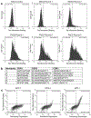
Figure 1.. Selection of tau-directed nanobodies.
a) A yeast surface display nanobody library was screened against tau monomer through two rounds of MACS and three rounds of FACS. After each sort, the selected yeast cells were incubated with 300 nM tau monomer and the extent of tau binding was assessed with flow cytometry. The fluorescence signal corresponding to tau binding to nanobodies displayed on yeast is plotted on the x-axis. Binding to tau above the level of the nanobody library appears on the right side of the vertical gate. b) CDR1, CDR2, and CDR3 sequences of the six nanobodies selected from sorts against tau monomer. c) Extent of tau binding to nanobodies MT2.1 (Nanobody 2), MT2.4 (Nanobody 4), and MT3.1(Nanobody 6) (y-axis) was assessed using flow cytometry. Yeast cells were incubated with 10 nM tau monomer. Expression levels of each nanobody on the surface of the yeast cells are shown on the x-axis.
Figure 2.
Figure 2.. MT3.1 binds to recombinant tau monomer, oligomers, and fibrils.
a) Dot blots with tau monomer (1), oligomers (2), and fibrils (3) reveal binding of bivalent MT3.1-Fc to all three forms of tau. An unprocessed image of this membrane is shown in Supplementary Figure 5a and Ponceau S staining of this membrane is shown in Supplementary Figure 4a. b) Characterization of the binding of MT3.1 to tau monomer (lane 1) and oligomers (lane 2) by western blot. An unprocessed image of this membrane is shown in Supplementary Figure 5b and a Coomassie blue stained SDS-PAGE gel with these proteins is shown in Supplementary Figure 4g. c) Characterization of the binding of MT3.1 to 100 nM tau monomer and oligomers (y-axis) by flow cytometry. Expression level of MT3.1 on the surface of the yeast cells is shown on the x-axis. d) Dot blot with tau fibrils (1), amyloid-β fibrils (2), α-synuclein fibrils (3), and BSA (4) show binding of bivalent MT3.1-Fc to tau fibrils and not other amyloid fibrils. An unprocessed image of this membrane is shown in Supplementary Figure 5c and Ponceau S staining of this membrane is shown in Supplementary Figure 4b.
Figure 3.
Figure 3.. Identification of MT3.1 binding epitope.
a) MT3.1 epitope mapping was performed with bivalent MT3.1-Fc and 108 15-mer peptides scanning the full length tau sequence. Peptides overlapped by 11 amino acids. Bivalent MT3.1-Fc bound to peptides 30–32, 68–69, 75–77, and 96–98 corresponding to tau residues 117–139, 269–287, 297–319, and 381–403. An unprocessed image of this membrane is shown in Supplementary Figure 5d. b) A second round of epitope mapping was performed. Bivalent MT3.1-Fc bound only to peptides containing the minimal binding epitopes 125ARMVSK130, 275VQIINK280, 306VQIVYK311, and 390AEIVYK395. An unprocessed image of this membrane is shown in Supplementary Figure 5e. c) Peptide sequences for the second round of epitope mapping are shown. The first three (row 1, 3, and 4) or two (row 2) peptides in each row are those that interacted with bivalent MT3.1-Fc in the first epitope mapping experiment. The following peptides in each row are glycine substitution mutants of these known binding peptides. The minimal binding epitopes identified are indicated in bold text.
Figure 4.
Figure 4.. Characterization of multivalent MT3.1.
a) Characterization of the purified monovalent and multivalent MT3.1 constructs by SDS-PAGE and stained by Coomassie blue dye. An unprocessed image of this gel is shown in Supplementary Figure 5f. b-c) Binding of MT3.1 constructs to tau oligomers (b) and fibrils (c) was evaluated by ELISA. Data points are averages of three repeats and error bars indicate standard deviation. d) EC50s for the binding of the MT3.1 constructs to tau oligomers or fibrils were calculated using a global nonlinear least-squares fit of the data presented in Figure 4b and 4c, respectively.
Figure 5.
Figure 5.. Western blotting of transgenic mice tissues.
Characterization of binding of AT8 (a) and bivalent MT3.1-Fc (b) and Tau5 and Tau13 (c) and bivalent MT3.1-Fc (d) to hippocampus (H) and somatosensory cortex (C) tissue lysate from wild-type (WT) and Tg4510 mice was performed with western blotting. Blotting revealed a prominent band at 59 kDa in the Tg4510 samples stained by both AT8 and bivalent MT3.1-Fc. Uncropped images of the membrane stained by AT8 and bivalent MT3.1-Fc are shown in Supplementary Figure 5g-i and Ponceau S staining of this membrane is shown in Supplementary Figure 4c. Uncropped images of the membrane stained by Tau5, Tau13, and bivalent MT3.1-Fc are shown in Supplementary Figure 5j-l and Ponceau S staining of this membrane is shown in Supplementary Figure 4d.

References

    1. Dhillon S Aducanumab: First Approval. Drugs. 2021;81(12):1437–1443. doi: 10.1007/S40265-021-01569-Z/METRICS - DOI - PubMed
    1. Mahase E Alzheimer’s disease: Lecanemab gets full FDA approval and black box safety warning. BMJ. 2023;382:p1580. doi: 10.1136/BMJ.P1580 - DOI - PubMed
    1. Karikari TK, Emeršič A, Vrillon A, et al. Head-to-head comparison of clinical performance of CSF phospho-tau T181 and T217 biomarkers for Alzheimer’s disease diagnosis. Alzheimer’s and Dementia. 2021;17(5):755–767. doi: 10.1002/ALZ.12236 - DOI - PMC - PubMed
    1. Horie K, Salvadó G, Barthélemy NR, et al. CSF MTBR-tau243 is a specific biomarker of tau tangle pathology in Alzheimer’s disease. Nature Medicine 2023 29:8. 2023;29(8):1954–1963. doi: 10.1038/s41591-023-02443-z - DOI - PMC - PubMed
    1. Breijyeh Z, Karaman R, Muñoz-Torrero D, Dembinski R. Comprehensive Review on Alzheimer’s Disease: Causes and Treatment. Molecules 2020, Vol 25, Page 5789. 2020;25(24):5789. doi: 10.3390/MOLECULES25245789 - DOI - PMC - PubMed

LinkOut - more resources