. 2024 May 21;90(5):e0011824.
doi: 10.1128/aem.00118-24. Epub 2024 Apr 3.

pH-dependent regulation of an acidophilic O-acetylhomoserine sulfhydrylase from Lactobacillus plantarum

Affiliations

pH-dependent regulation of an acidophilic O-acetylhomoserine sulfhydrylase from Lactobacillus plantarum

Yasuyuki Matoba et al. Appl Environ Microbiol. .

Abstract

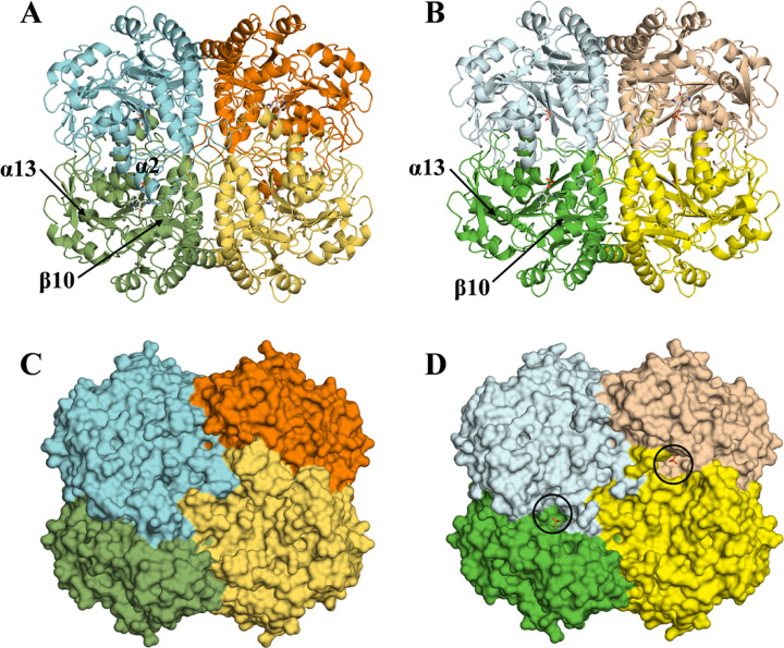
Bacteria have two routes for the l-methionine biosynthesis. In one route called the direct sulfuration pathway, acetylated l-homoserine is directly converted into l-homocysteine. The reaction using H2S as the second substrate is catalyzed by a pyridoxal 5'-phosphate-dependent enzyme, O-acetylhomoserine sulfhydrylase (OAHS). In the present study, we determined the enzymatic functions and the structures of OAHS from Lactobacillus plantarum (LpOAHS). The LpOAHS enzyme exhibited the highest catalytic activity under the weak acidic pH condition. In addition, crystallographic analysis revealed that the enzyme takes two distinct structures, open and closed forms. In the closed form, two acidic residues are sterically clustered. The proximity may cause the electrostatic repulsion, inhibiting the formation of the closed form under the neutral to the basic pH conditions. We concluded that the pH-dependent regulation mechanism using the two acidic residues contributes to the acidophilic feature of the enzyme.

Importance: In the present study, we can elucidate the pH-dependent regulation mechanism of the acidophilic OAHS. The acidophilic feature of the enzyme is caused by the introduction of an acidic residue to the neighborhood of the key acidic residue acting as a switch for the structural interconversion. The strategy may be useful in the field of protein engineering to change the optimal pH of the enzymes. In addition, this study may be useful for the development of antibacterial drugs because the l-methionine synthesis essential for bacteria is inhibited by the OAHS inhibitors. The compounds that can inhibit the interconversion between the open and closed forms of OAHS may become antibacterial drugs.

Keywords: Lactobacillus plantarum; O-acetylhomoserine sulfhydrylase; acidophilicity; l-methionine; pH-dependent regulation.


Conflict of interest statement

The authors declare no conflict of interest.

Figures

Fig 1
Fig 1
Bacterial biosynthetic pathways of l-methionine. In the forward transsulfuration pathway, activated l-homoserine and l-cysteine are linked by CGS, resulting in the generation of cystathionine. CBL cleaves the bond between the Cβ and the Sγ atoms and the bond between the amino nitrogen and the Cα atoms to generate l-homocysteine, pyruvate, and ammonium. The l-homocysteine is finally converted to l-methionine by another enzyme. The l-homocysteine is also generated by OAHS in the direct sulfuration pathway. The dotted lines in the figure indicate the covalent bonds to be cleaved by CBL.
Fig 2
Fig 2
Catalytic mechanism underlying the OAHS reaction. Details are described in the text.
Fig 3
Fig 3
pH profile of the catalytic activity of LpOAHS to generate l-homocysteine from l-OAH and H2S. The concentrations of l-OAH and Na2S were fixed at 50 mM and 4 mM, respectively. Filled squares and bars mean the averaged activity (v/Et) calculated from the data of at least three independent experiments and the standard deviation, respectively. Activities of the wild-type LpOAHS and the D119N and D272N mutants are shown in black, red, and blue, respectively.
Fig 4
Fig 4
Tetrameric structures of LpOAHS in the closed and open forms. Ribbon representations of the closed and open forms are shown in (A) and (B), respectively, while molecular surfaces of the closed and open forms are shown in (C) and (D), respectively. Subunits A, B, C, and D in the closed form are colored in dark green, cyan, orange, and luteofulvous, respectively, while subunits A, B, C, and D in the open form are colored in green, pale cyan, tan, and yellow, respectively. PLP molecules bond to the LpOAHS are shown in a stick model. Black circles in (D) indicate the substrate-binding pockets. The figures were drawn using PyMOL (18). Three twofold axes relate the subunits in the tetramer of LpOAHS. Two are horizontal and vertical lines passing through the center of the tetramer, and the other is a line orthogonal to the plane of the illustration.
Fig 5
Fig 5
Superposition between subunits in the closed and open forms. Subunit A in the closed form and the corresponding one in the open form are shown in yellow and blue colors, respectively.
Fig 6
Fig 6
Active site structures. Active site structures of LpOAHS in the closed and open forms are shown in (A) and (B), respectively, while that of T. maritima OAHS in the closed form is shown in (C). Carbon atoms from the residues in the subunits A and B of LpOAHS in the closed form, shown in (A), are colored in dark green and cyan, respectively, while those in the open form, shown in (B), are colored in green and pale cyan, respectively. In (C), carbon atoms from the residues in the subunits A and B of the intermediate-bound T. maritima OAHS (PDB ID: 7KB1) are colored in magenta and salmon pink, respectively. Carbon atoms of PLP covalently bound to LpOAHS in (A) and (B) and those of the PLP derivative (ketimine form of PLP complexed with vinylglycine) bound to the T. maritima OAHS in (C) are colored in white. Hydrogen bonds formed within the enzyme are indicated by black dashed lines, while those between the enzyme and the ligand are by gray dashed lines. The FoFc electron density maps for PLP-bound Lys211A are shown in (A) and (B), while that for the PLP derivative is shown in (C). To remove the model bias, a round of simulated annealing refinement was done during the calculation of the composite omit map. The map is contoured at 3.0 σ with gray color.
Fig 7
Fig 7
Structures around the active site in the closed and open forms of LpOAHS. Structures in the closed and open forms are shown in (A) and (B), respectively. Carbon atoms from the residues in the subunits A and B in the closed form are colored in dark green and cyan, respectively, while those in the subunits A and B in the open form are colored in green and pale cyan, respectively. Ribbon diagrams cover the residues His381A–Leu404A and Asp42B–Asn64B. The FoFc electron density maps for Asp119A, Arg271B, and Asp272B are shown in (A) and (B). The map is contoured at 3.0 σ with gray color.
Fig 8
Fig 8
Schematic representation of the catalytic mechanism of LpOAHS. By the binding of the first substrate l-OAH to the substrate-binding pocket, the open form with the internal aldimine bond (A) is converted into the closed form complexed with l-OAH-bound external aldimine (B). According to the conversion of the l-OAH-bound external aldimine to the β,γ-unsaturated ketimine intermediate and acetate, a partially disordered form is generated (C), which facilitates the release of acetate from the substrate-binding pocket and the binding of the second substrate HS- to the pocket. The binding of HS- regenerates the closed form (D), which enables the nucleophilic attack of the ion to the intermediate. After the formation of l-homocysteine-bound external aldimine, closed form is again converted to the partially disordered form (E). According to the regeneration of the internal aldimine bond, the partially disordered form is converted into the open form, which facilitates the release of l-homocysteine. For convenience, Gln389A residue is omitted from the figure.

Similar articles

Cited by

References

    1. Ferla MP, Patrick WM. 2014. Bacterial methionine biosynthesis. Microbiology (Reading) 160:1571–1584. doi:10.1099/mic.0.077826-0 - DOI - PubMed
    1. Ravanel S, Gakière B, Job D, Douce R. 1998. The specific features of methionine biosynthesis and metabolism in plants. Proc Natl Acad Sci U S A 95:7805–7812. doi:10.1073/pnas.95.13.7805 - DOI - PMC - PubMed
    1. Hesse H, Kreft O, Maimann S, Zeh M, Hoefgen R. 2004. Current understanding of the regulation of methionine biosynthesis in plants. J Exp Bot 55:1799–1808. doi:10.1093/jxb/erh139 - DOI - PubMed
    1. Clausen T, Huber R, Messerschmidt A, Pohlenz HD, Laber B. 1997. Slow-binding inhibition of Escherichia coli cystathionine β-lyase by l-aminoethoxyvinylglycine: a kinetic and X-ray study. Biochemistry 36:12633–12643. doi:10.1021/bi970630m - DOI - PubMed
    1. Ejim LJ, Blanchard JE, Koteva KP, Sumerfield R, Elowe NH, Chechetto JD, Brown ED, Junop MS, Wright GD. 2007. Inhibitors of bacterial cystathionine β-lyase: leads for new antimicrobial agents and probes of enzyme structure and function. J Med Chem 50:755–764. doi:10.1021/jm061132r - DOI - PubMed

MeSH terms

Substances

LinkOut - more resources