. 2024 Apr 11;187(8):2010-2028.e30.
doi: 10.1016/j.cell.2024.03.013. Epub 2024 Apr 2.

Charting the cellular biogeography in colitis reveals fibroblast trajectories and coordinated spatial remodeling

Affiliations

Charting the cellular biogeography in colitis reveals fibroblast trajectories and coordinated spatial remodeling

Paolo Cadinu et al. Cell. .

Abstract

Gut inflammation involves contributions from immune and non-immune cells, whose interactions are shaped by the spatial organization of the healthy gut and its remodeling during inflammation. The crosstalk between fibroblasts and immune cells is an important axis in this process, but our understanding has been challenged by incomplete cell-type definition and biogeography. To address this challenge, we used multiplexed error-robust fluorescence in situ hybridization (MERFISH) to profile the expression of 940 genes in 1.35 million cells imaged across the onset and recovery from a mouse colitis model. We identified diverse cell populations, charted their spatial organization, and revealed their polarization or recruitment in inflammation. We found a staged progression of inflammation-associated tissue neighborhoods defined, in part, by multiple inflammation-associated fibroblasts, with unique expression profiles, spatial localization, cell-cell interactions, and healthy fibroblast origins. Similar signatures in ulcerative colitis suggest conserved human processes. Broadly, we provide a framework for understanding inflammation-induced remodeling in the gut and other tissues.


Conflict of interest statement

Declaration of interests J.R.M. is a co-founder of, stakeholder in, and advisor for Vizgen, Inc. J.R.M. is an inventor on patents associated with MERFISH applied for on his behalf by Harvard University and Boston Children’s Hospital. J.R.M.’s interests were reviewed and are managed by Boston Children’s Hospital in accordance with their conflict-of-interest policies. R.N. is a paid consultant for Quris-AI. V.K.K. has an ownership interest in Tizona Therapeutics, Trishula Therapeutics, Celsius Therapeutics, Bicara Therapeutics, Larkspur Therapeutics. V.K.K. has financial interests in Biocon Biologic, Compass, Elpiscience Biopharmaceutical Ltd, Equilium Inc, PerkinElmer, and Syngene Intl. V.K.K. is a member of SABs for Cell Signaling Technology, Elpiscience Biopharmaceutical Ltd, GlaxoSmithKline, Larkspur, Novartis Sabatolimab, Tizona Therapeutics, Tr1X, and Werewolf. A.C.A. is a member of the SAB for Tizona Therapeutics, Trishula Therapeutics, Compass Therapeutics, Zumutor Biologics, ImmuneOncia, and Nekonal Sarl. A.C.A. is also a paid consultant for iTeos Therapeutics, Larkspur Biosciences, and Excepgen. R.N., V.K.K., and A.C.A.’s interests were reviewed and managed by Mass General Brigham in accordance with their conflict-of-interest policies.

Figures
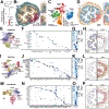
Figure 1:
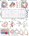
Figure 1:. Cellular remodeling in a mouse colitis model
(A) Dextran-sodium-sulfate administration (DSS; red) and tissue sampling (arrows) timing. (B) Spatial distribution of 7 of 940 RNAs measured with MERFISH in the mouse colon harvested at Day 0. Scale bar: 300 μm. Insets: Zoom-in on boxed regions. (C) UMAP of 1.35 million cells measured via MERFISH for mice at all days. Smooth muscle cells (SMC). Interstitial cells of Cajal (ICC). (D) Spatial distribution of major cell classes in the slice in (B). Color indicates cell class as in (C). Scale bar: 300 μm. Insets: Zoom-in on boxed regions. (E) UMAP representation of all epithelial cells labeled by population. (F) Key gene expression in epithelial populations. Average expression and fraction of expressing cells are indicated by circle color and diameter. (G) Normalized average per-slice abundance across different days. (H) Spatial distribution of cells in slices from Day 0 (left) and Day 9 (right). Epithelial cells colored as in (E) with other cells in gray. Scale bars: 200 μm. (I)-(L) As in (E)-(H) but for immune populations. (M)-(P) As in (E)-(H) but for fibroblast populations. See also Figures S1, S2, and S3, Tables S1 and S2, and Videos S1 and S2.
Figure 2:
Figure 2:. Cellular neighborhoods define the spatial remodeling of the colon during DSS-treatment
(A) Spatial distribution of all cells in slices from Day 0 and Day 9. Scale bars: 200 μm. (B) UMAP of cellular neighborhoods (STAR Methods) colored by neighborhood assignment. MU: mucosal; LUM: luminal; SM: submucosa; ME: muscularis externa; FOL: follicle; MES: mesentery. (C) As in (B) but colored by abundance of listed cell class (left) or cell cluster (right). (D) As in (A) but colored by neighborhood assignment as in (B). (E) Normalized fraction of cell populations within each cellular neighborhood. (F) Normalized average per-slice abundance across different days. (G) Spatial distribution of cells within a Day 0 slice colored by select neighborhoods (MU1, MU2, or MU3). Scale bar: 200 μm. (H) UMAP of epithelial, immune, and fibroblasts colored by select neighborhood assignment (MU1, MU2, or MU3). (I) Fraction of cells in mucosal (top) or non-mucosal (bottom) neighborhoods per slice, grouped by mouse of origin and disease stage. Circles represent weight loss (red) or colon length (yellow) at Day 9. (J) Spatial distribution of cells colored by neighborhood assignment. Scale bars: 200 μm. See also Table S3.
Figure 3:
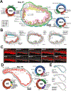
Figure 3:. Cellular neighborhoods evolve in a staged progression driven by polarization and recruitment of distinct cell populations
(A) Spatial distribution cells in a Day 0 and Day 3 slice colored by mucosal neighborhood. Pie charts represent neighborhood composition of cell classes (inner; fibroblast: green, epithelial: blue, immune: red, EC: purple, SMC: brown, ENS: yellow) or cell clusters (outer). Prominent populations are labeled. Scale bars: 200 μm. (B) Spatial distribution of cells that define MU4 in Day 0 or Day 3 slices. Inset: zoom-in on boxed region. Scale bars: 200 μm. (C) Spatial distribution of RNAs marking populations that define MU4. All RNAs are gray and the listed RNA is red. Scale bars: 200 μm. (D) As in (A) but for Day 9 mucosal neighborhoods. Scale bar: 200 μm. (E, F) As in (B) but for cells that define Day 9 neighborhoods (E) or IAFs in MU8 (F). Scale bars: 200 μm. (G) As in (C) for RNAs marking Day 9 neighborhood populations. Scale bar: 200 μm. (H) UMAP of fibroblasts (left) or IAF4 (right) colored by listed gene expression. (I) UMAP of neutrophils colored by cell population (top) or pseudo time (bottom). (J) As in (I) by with cells colored by listed gene expression. (K) Normalized gene expression sorted by neutrophil pseudo time as in (I). (L) Spatial distribution of neutrophils colored by pseudo time. Scale bar: 200 μm. (M) As in (A) but for non-mucosal neighborhoods in Day 0 and Day 9. Scale bars: 200 μm. (N) As in (B) but for cell populations that define Day 0 and Day 9 non-mucosal neighborhoods. Scale bars: 200 μm. (O) As in (C) for RNAs marking populations highlighted in (N). Scale bars: 200 μm. See also Figure S4 and Figure S5.
Figure 4:
Figure 4:. Cellular neighborhoods define the organization of repairing tissue
(A) Spatial distribution and cellular abundance (pie charts) of mucosal neighborhoods in a Day 21 slice. Scale bar: 200 μm. (B) Spatial distribution of cells that define Day 21 neighborhoods. Scale bars: 200 μm. (C) Spatial distribution of RNAs that highlight MU10 structure for boxed region in (B). (D) Spatial distribution of cells measured at Day 35 colored by tissue region: mucosa (MU), submucosa (SM), and muscularis externa (ME). Scale bar: 200 μm. (E) Spatial distribution of notable cells observed at Day 35. Scale bars: 200 μm.
Figure 5.
Figure 5.. Spatially prioritized receptor-ligand interactions suggest unique roles for IAFs in different cellular neighborhoods
(A, B, C) Predicted interactions between cell populations for Wnt-family interactions in MU1 (A) or chemokines (B, left) and interleukins (B, right) for MU3. Known interactions are highlighted. (C-E) Putative interactions to and from fibroblasts in MU4 (C), MU7 (D), and MU8 (E). Colored circles represent major cell populations (fibroblast: green, epithelial: blue, immune: red, EC: purple). Gray circles represent ligand-receptor complexes. Lines connecting cells to ligands or receptors are colored by the average expression of those genes in those cells, normalized to the maximum in any cell population. (F, G) Weight loss with 2.5% DSS (F) or 4% DSS (G) treatment, followed by rIL-11 or BSA administration. Markers: average; error bars: SEM (F: n=5, G: n=4–6). *p<0.05. (H,I) IL-1β and TNF concentration in colonic explant supernatants (H) or serum (I) from 4% DSS-treated mice on Day 14. Box: 1st and 3rd quartiles; line: median; and whiskers cover all measurements (markers). **p<0.01. ns: not-significant. See also Table S4.
Figure 6:
Figure 6:. Lineage tracing reveals distinct origins of IAF populations
(A) Immunofluorescence of Day 0 colon slices for CXCL12Lin, PDPN, PDGFRα, and DAPI (n=5). Right: zoom-in on boxed region. Scale bar: 20 μm. (B-D) As in (A) but for VCAM1 (B), CTGF (C), and SDC2 (D). Scale bars: 20 μm. (E) CXCL12Lin+ cell overlap with listed markers at Day 0. Box: 1st and 3rd quartiles; line: median; and whiskers extend to cover all measurements (markers). (F) Immunofluorescence of Day 9 colon slices sorted by mild or severe disease (n=5–6). Right: zoom-in on boxed regions. Scale bar: 200 μm. (G) CXCL12Lin cell density and area in ulcerated or non-ulcerated Day 9 sections. Bar: mean; error bars: SEM. ****p<0.0001. (H-K) Immunofluorescence images of Day 9 ulcerated regions for CXCL12Lin and IL-1β (H), PLAU (I), IL-11 (J), or GREM1 (K). Scale bars: 50 μm. (L) CXCL12Lin+ cell overlap with markers in (H-K). See also Figure S6.
Figure 7:
Figure 7:. Signatures of murine IAFs and inflammatory neighborhoods in human ulcerative colitis
(A) Key gene expression in fibroblasts in mouse (left) or human UC (right). The average expression and the fraction of expressing cells are indicated by circle color and diameter. Red box: IAF in mouse or human. Colored lines: markers for IAF1 (yellow), IAF2 (green), IAF3 (blue), and IAF4 (red). (B) Pearson correlation coefficients for gene expression in human IAF sorted by similarity (top tree). Boxes: groups of co-varying genes. Colored lines: human homologs of mouse markers for IAFs as in (A). (C) Normalized average gene expression in each neighborhood for mouse homologs of human treatment-resistant biomarkers. (D) Expression score for genes in (C) colored by associated disease stage. (E) UMAP of immune (left) and fibroblast (right) cells colored by population (top) or expression of listed genes (bottom). (F) Key gene expression as in (A) for cells observed in MU8 or LUM. See also Figure S7.

Update of

References

    1. Mowat AM, and Agace WW (2014). Regional specialization within the intestinal immune system. Nat. Rev. Immunol 14, 667–685. - PubMed
    1. Beumer J, and Clevers H (2021). Cell fate specification and differentiation in the adult mammalian intestine. Nat. Rev. Mol. Cell Biol 22, 39–53. - PubMed
    1. Nowarski R, Jackson R, and Flavell RA (2017). The stromal intervention: Regulation of immunity and inflammation at the epithelial-mesenchymal barrier. Cell 168, 362–375. - PubMed
    1. Abraham C, and Cho JH (2009). Inflammatory Bowel Disease. New England Journal of Medicine; 361, 2066–2078. 10.1056/nejmra0804647. - DOI - PMC - PubMed
    1. Friedrich M, Pohin M, and Powrie F (2019). Cytokine networks in the pathophysiology of inflammatory bowel disease. Immunity 50, 992–1006. - PubMed

LinkOut - more resources