

Novel Endoscopic Findings of Lesions with a Short White Hair-like Appearance in the Lower Esophagus
- PMID: 38569905
- PMCID: PMC11671187
- DOI: 10.2169/internalmedicine.3396-23
Novel Endoscopic Findings of Lesions with a Short White Hair-like Appearance in the Lower Esophagus
Abstract
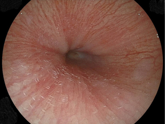
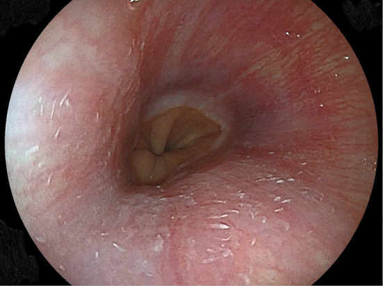
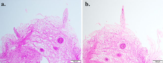
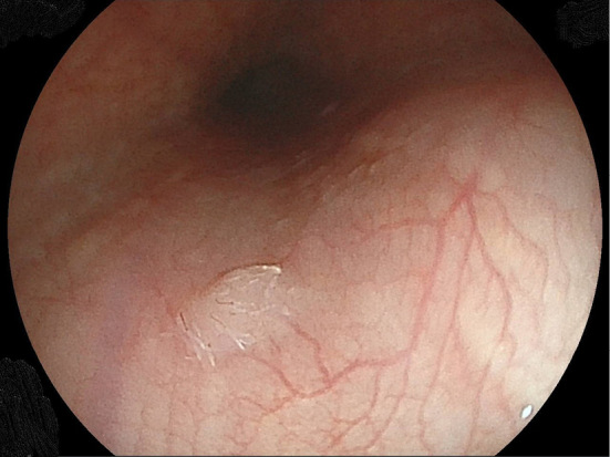
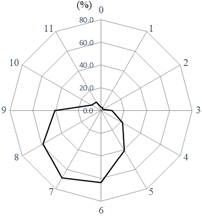
Objective The presence of a short white hair-like appearance in the lower esophagus has recently been noted during esophagogastroduodenoscopy (EGD) at our institution. Histological findings showed that this formation was a spiked protrusion of the esophageal papilla. The results regarding the prevalence of such lesions in individuals who underwent EGD examinations as part of medical checkup procedures are herein presented. Methods The EGD results of 14,338 individuals (9,225 males, 5,113 females; mean age 54.0±9.8 years) were examined. The findings related to the presence of multiple lesions with a short white hair-like appearance in the lower esophagus of patients with reflux esophagitis, esophageal squamous papilloma, or gastric mucosal atrophy (GMA), as well as the hiatal hernia width, were investigated. Results Endoscopic findings indicating short white hair-like appendages in the lower esophagus were noted in 167 patients, with a prevalence rate of 1.2%. A female sex, younger age, lower body mass index, lower percentages of habitual smoking and drinking, and the presence of esophageal squamous papilloma were characteristic features of cases with such findings. In addition, a significantly lower prevalence of reflux esophagitis and a smaller diaphragmatic hiatus size were observed. A multiple logistic regression analysis indicated that a female sex, absence of reflux esophagitis, presence of esophageal squamous papilloma, and a smaller diaphragmatic hiatus were factors significantly related to the presence of these short white hair-like appendages. An analysis of circumferential localization revealed the main location to be the left-posterior wall. Conclusion This study is the first to report the prevalence of multiple short white hair-like appendages in the lower esophagus. The occurrence of such lesions is inversely associated with the presence of reflux esophagitis.
Keywords: esophageal papilla; esophageal papilloma; esophagus; female-predominance; protrusion; reflux esophagitis.
Conflict of interest statement
The authors state that they have no Conflict of Interest (COI).
Figures
Similar articles
-
Hiatal hernia size is the dominant determinant of esophagitis presence and severity in gastroesophageal reflux disease.Am J Gastroenterol. 2001 Jun;96(6):1711-7. doi: 10.1111/j.1572-0241.2001.03926.x. Am J Gastroenterol. 2001. PMID: 11419819
-
Role of hiatus hernia and gastric mucosal atrophy in the development of reflux esophagitis in the elderly.J Gastroenterol Hepatol. 2001 Feb;16(2):132-6. doi: 10.1046/j.1440-1746.2001.02408.x. J Gastroenterol Hepatol. 2001. PMID: 11207891
-
[The influence of Barrett's esophagus on the clinical signs and postoperative results of GERD].Zentralbl Chir. 2004 Apr;129(2):99-103. doi: 10.1055/s-2004-816278. Zentralbl Chir. 2004. PMID: 15106039 German.
-
Severe reflux esophagitis.Gastrointest Endosc Clin N Am. 1994 Oct;4(4):677-98. Gastrointest Endosc Clin N Am. 1994. PMID: 7812641 Review.
-
[Diagnosis of gastroesophageal reflux and Barrett esophagus].Zentralbl Chir. 2000;125(5):414-23. Zentralbl Chir. 2000. PMID: 10929625 Review. German.
References
-
- Adachi K, Mishiro T, Tanaka S, Hanada K, Kinoshita Y. Gender differences in the time-course changes of reflux esophagitis in Japanese patients. Intern Med 54: 863-873, 2015. - PubMed
-
- Adachi K, Mishiro T, Tanaka S, Kinoshita Y. Suitable biopsy site for detection of esophageal eosinophilia in eosinophilic esophagitis suspected cases. Dig Endosc 28: 139-144, 2016. - PubMed