Deep Learning for Automated Measurement of Total Cardiac Volume for Heart Transplantation Size Matching
- PMID: 38570368
- PMCID: PMC11842492
- DOI: 10.1007/s00246-024-03470-4
Deep Learning for Automated Measurement of Total Cardiac Volume for Heart Transplantation Size Matching
Abstract
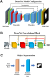
Total Cardiac Volume (TCV)-based size matching using Computed Tomography (CT) is a novel technique to compare donor and recipient heart size in pediatric heart transplant that may increase overall utilization of available grafts. TCV requires manual segmentation, which limits its widespread use due to time and specialized software and training needed for segmentation. This study aims to determine the accuracy of a Deep Learning (DL) approach using 3-dimensional Convolutional Neural Networks (3D-CNN) to calculate TCV, with the clinical aim of enabling fast and accurate TCV use at all transplant centers. Ground truth TCV was segmented on CT scans of subjects aged 0-30 years, identified retrospectively. Ground truth segmentation masks were used to train and test a custom 3D-CNN model consisting of a DenseNet architecture in combination with residual blocks of ResNet architecture. The model was trained on a cohort of 270 subjects and a validation cohort of 44 subjects (36 normal, 8 heart disease retained for model testing). The average Dice similarity coefficient of the validation cohort was 0.94 ± 0.03 (range 0.84-0.97). The mean absolute percent error of TCV estimation was 5.5%. There is no significant association between model accuracy and subject age, weight, or height. DL-TCV was on average more accurate for normal hearts than those listed for transplant (mean absolute percent error 4.5 ± 3.9 vs. 10.5 ± 8.5, p = 0.08). A deep learning-based 3D-CNN model can provide accurate automatic measurement of TCV from CT images. This initial study is limited as a single-center study, though future multicenter studies may enable generalizable and more accurate TCV measurement by inclusion of more diverse cardiac pathology and increasing the training data.
Keywords: Artificial intelligence; Congenital heart disease; Deep learning; Heart transplant; Imaging; Size matching.
© 2024. The Author(s).
Conflict of interest statement
Declarations. Conflict of Interest: Dr. Morales reports contributions from Cormatrix, Inc., personal fees from Syncardia, Inc., personal fees and other contributions from Abbott Medical Inc., personal fees from Xeltis, Inc., personal fees from Azyio, Inc. all outside the submitted work. All other authors have no financial conflicts of interest to disclose. Dr. Zafar is Vice President of Cardiothoracic Clinical Development at Transmedics, Inc, his employment is outside the submitted work. All other authors report no conflicts of interest.
Figures


Update of
-
Deep Learning for Automated Measurement of Total Cardiac Volume for Heart Transplantation Size Matching.Res Sq [Preprint]. 2023 Dec 28:rs.3.rs-3788726. doi: 10.21203/rs.3.rs-3788726/v1. Res Sq. 2023. Update in: Pediatr Cardiol. 2025 Mar;46(3):590-598. doi: 10.1007/s00246-024-03470-4. PMID: 38234758 Free PMC article. Updated. Preprint.
References
-
- Kransdorf E et al (2017) Predicted Heart Mass Is the Optimal Metric for Size Matching in Heart Transplantation. J Heart Lung Transplant 36(4):S113–S113 - PubMed
-
- Riggs KW et al (2019) Time for evidence-based, standardized donor size matching for pediatric heart transplantation. J Thorac Cardiovasc Surg 158(6):1652–1660 - PubMed
MeSH terms
Grants and funding
LinkOut - more resources
Full Text Sources
Medical

