Review
. 2024 Apr 3;9(1):83.
doi: 10.1038/s41392-024-01779-3.

Progression from ductal carcinoma in situ to invasive breast cancer: molecular features and clinical significance

Affiliations
Review

Progression from ductal carcinoma in situ to invasive breast cancer: molecular features and clinical significance

Jing Wang et al. Signal Transduct Target Ther. .

Abstract

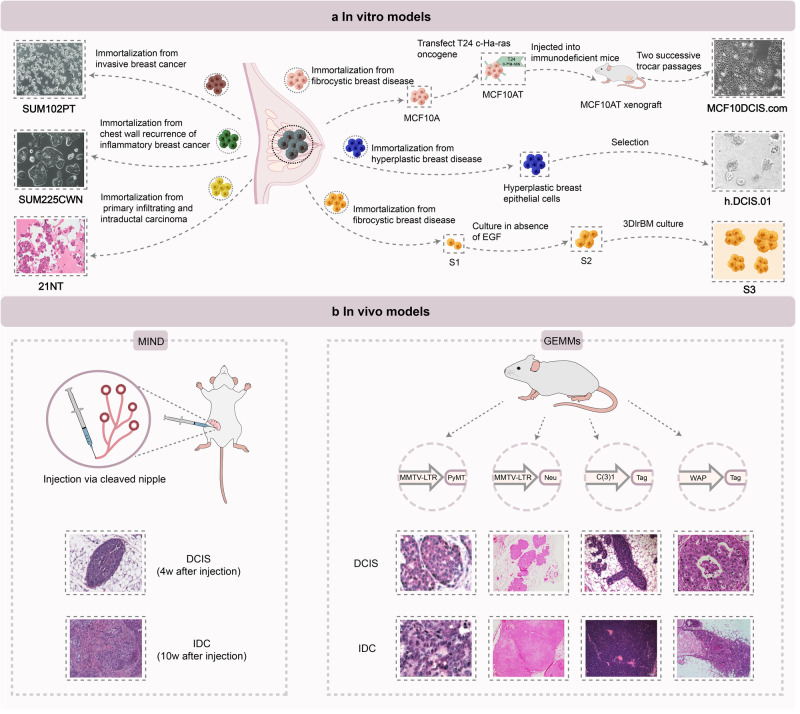
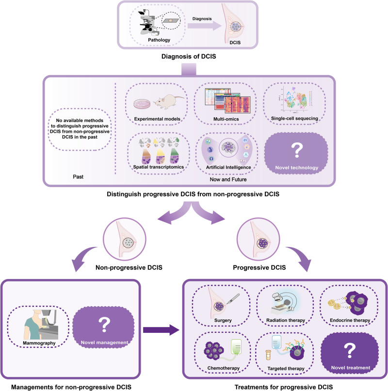
Ductal carcinoma in situ (DCIS) represents pre-invasive breast carcinoma. In untreated cases, 25-60% DCIS progress to invasive ductal carcinoma (IDC). The challenge lies in distinguishing between non-progressive and progressive DCIS, often resulting in over- or under-treatment in many cases. With increasing screen-detected DCIS in these years, the nature of DCIS has aroused worldwide attention. A deeper understanding of the biological nature of DCIS and the molecular journey of the DCIS-IDC transition is crucial for more effective clinical management. Here, we reviewed the key signaling pathways in breast cancer that may contribute to DCIS initiation and progression. We also explored the molecular features of DCIS and IDC, shedding light on the progression of DCIS through both inherent changes within tumor cells and alterations in the tumor microenvironment. In addition, valuable research tools utilized in studying DCIS including preclinical models and newer advanced technologies such as single-cell sequencing, spatial transcriptomics and artificial intelligence, have been systematically summarized. Further, we thoroughly discussed the clinical advancements in DCIS and IDC, including prognostic biomarkers and clinical managements, with the aim of facilitating more personalized treatment strategies in the future. Research on DCIS has already yielded significant insights into breast carcinogenesis and will continue to pave the way for practical clinical applications.


Conflict of interest statement

The authors declare no competing interests.

Figures

Fig. 1
Fig. 1
Highlights of DCIS-IDC research. Since DCIS was termed as the precursor of IDC in 1973, research on the transition from DCIS to IDC has rapidly advanced. This progress has been fueled by the establishment of in vitro DCIS cell lines and in vivo animal models. In recent years, technological advancements have offered deeper insights into the changes occurring in both tumor cells and the microenvironment during the transition from DCIS to IDC
Fig. 2
Fig. 2
Key signaling pathways in breast cancer. Breast cancer is a heterogeneous disease characterized by diverse subtypes. The development and progression of breast cancer result from the influence of subtype-specific and shared signaling pathways, as well as the intricate crosstalk between them. Inhibitors that target these key signaling pathways have led to improvement in the prognosis of breast cancer. E2 estradiol, ER estrogen receptor, ERE estrogen response element, GPER G protein-coupled ER, HER2 human epidermal growth factor receptor 2, TNBC triple-negative breast cancer, EGFR epidermal growth factor receptor, NICD Notch intracellular domain, SERM selective ER modulator, AI aromatase inhibitor, CDK4/6i cyclin-dependent kinase 4/6 inhibitor, mAb monoclonal antibody, TKI tyrosine kinase inhibitor, ADC antibody–drug conjugates, PARPi Poly (ADP‑ribose) polymerase inhibitor
Fig. 3
Fig. 3
Proposed models of DCIS progression. a Independent lineage model, which presumes that DCIS and IDC derive from two distinct normal epithelial cells which share no overlapping CNAs or mutations. b Evolutionary bottleneck model, which presumes that a specific clone within DCIS is selected and it evolves into IDC. c Multiclonal invasion model, which presumes that multiple clones escape and co-migrate to invasive regions to generate IDC. d Convergent phenotype model, which presumes that subclones of different genotypes within DCIS can all give rise to an invasive phenotype to establish IDC
Fig. 4
Fig. 4
Role of microenvironment in DCIS progression. Tumor-microenvironment crosstalk may facilitate DCIS progression. The environment consists of a range of components including MECs, immune cells, fibroblasts, blood vessels, basement membrane and ECM. a MECs: Upregulation of αvβ6 integrin expressed in MECs activates TGFβ signaling, resulting in upregulation of MMP9 and MMP13 and ECM remodeling. Moreover, increased secretion of CXCL12 and CXCL14 from MECs promotes DCIS tumor cell invasion. b Immune cells: DCIS cell-derived CCL2 recruit macrophages into the tumor, driving increased Wnt-1 secretion from macrophages, contributing to myoepithelium disruption and the breakdown of E-cadherin junctions. c Fibroblasts: Increased secretion of CXCL1 and IL-6 from CAFs drives activation of NF-κB and COX-2 in DCIS cells, which induced and upregulation of MMP9 and MMP14 from DCIS cells, resulting in ECM remodeling and basement membrane degradation. DCIS ductal carcinoma in situ, MECs myoepithelial cells, ECM extracellular matrix, TGFβ transforming growth factor-beta, MMP9 matrix metalloproteinase 9, CXCL12 C-X-C motif chemokine ligand 12, CCL2 C-C motif ligand 2, IL6 interleukin-6, CAFs tumor-associated fibroblasts, NF-κB nuclear factor-kappaB, COX-2 cyclooxygenase 2
Fig. 5
Fig. 5
In vitro and in vivo models for DCIS progression research. a In vitro models, in which DCIS cell lines that include the MCF10 series, HMT-3522 series, 21Tseries, SUM225CWN, SUM102PT, and h.DCIS.01 authentically mimic human DCIS progression for 2D and 3D culture in vitro. b In vivo models, in which MIND models and GEMMs including MMTV-PyMT, MMTV-Neu, C3(1)/Tag, and WAP-T mouse are available for studying the biology of DCIS in vivo
Fig. 6
Fig. 6
Future management of non-progressive and progressive DCIS. Historically, distinguishing between progressive and non-progressive DCIS in women was challenging, resulting in overtreatment with limited benefits. It is crucial to find a solution to this problem. Advanced technologies, such as experimental models, multi-omics, single-cell sequencing, spatial transcriptomics, artificial intelligence and other emerging technologies, can facilitate the development of a risk stratification system for personalized treatment. Tailoring treatments for non-progressive DCIS differently from progressive DCIS is essential to alleviate the unnecessary burdens associated with overtreatment

References

    1. DeSantis CE, et al. Breast cancer statistics, 2019. CA Cancer J. Clin. 2019;69:438–451. doi: 10.3322/caac.21583. - DOI - PubMed
    1. Giaquinto AN, et al. Breast cancer statistics, 2022. CA Cancer J. Clin. 2022;72:524–541. doi: 10.3322/caac.21754. - DOI - PubMed
    1. Badve SS, Gökmen-Polar Y. Ductal carcinoma in situ of breast: update 2019. Pathology. 2019;51:563–569. doi: 10.1016/j.pathol.2019.07.005. - DOI - PMC - PubMed
    1. Shaaban AM, et al. Pathological features of 11,337 patients with primary ductal carcinoma in situ (DCIS) and subsequent events: results from the UK Sloane Project. Br. J. Cancer. 2021;124:1009–1017. doi: 10.1038/s41416-020-01152-5. - DOI - PMC - PubMed
    1. Groen EJ, et al. Prognostic value of histopathological DCIS features in a large-scale international interrater reliability study. Breast Cancer Res. Treat. 2020;183:759–770. doi: 10.1007/s10549-020-05816-x. - DOI - PMC - PubMed

Publication types

MeSH terms