. 2024 Apr 3;14(1):7889.
doi: 10.1038/s41598-024-55614-4.

Nobiletin alleviates cisplatin-induced ototoxicity via activating autophagy and inhibiting NRF2/GPX4-mediated ferroptosis

Affiliations

Nobiletin alleviates cisplatin-induced ototoxicity via activating autophagy and inhibiting NRF2/GPX4-mediated ferroptosis

Wenao Song et al. Sci Rep. .

Abstract

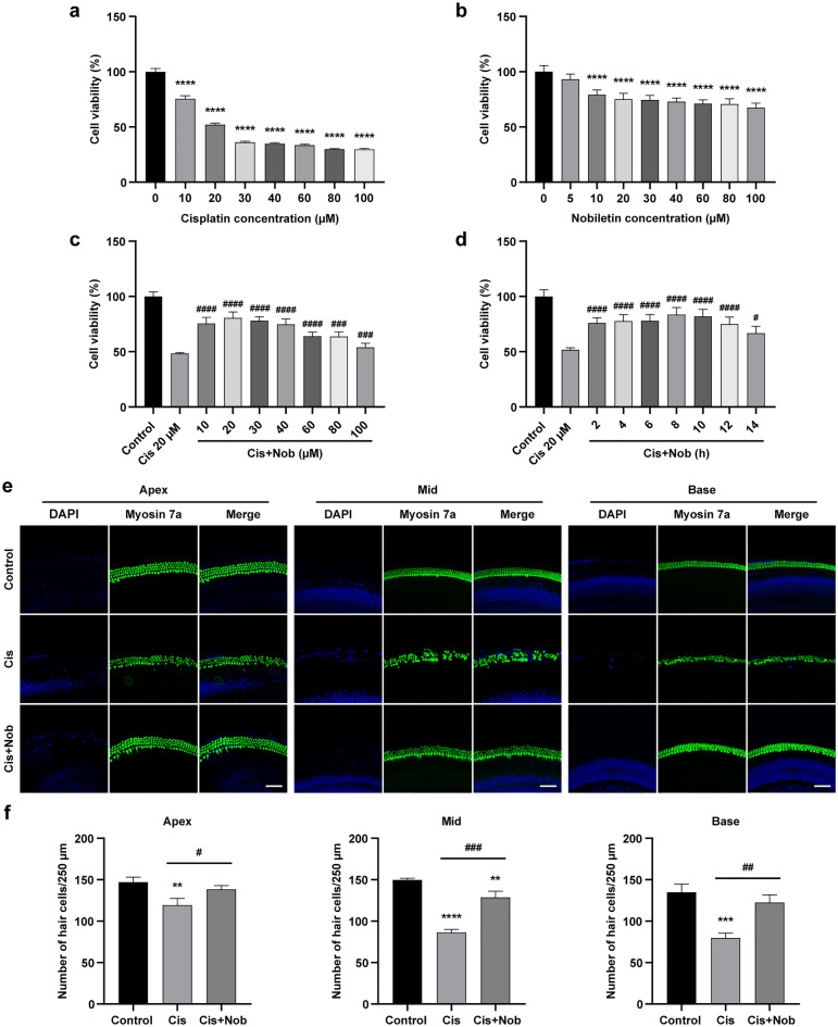
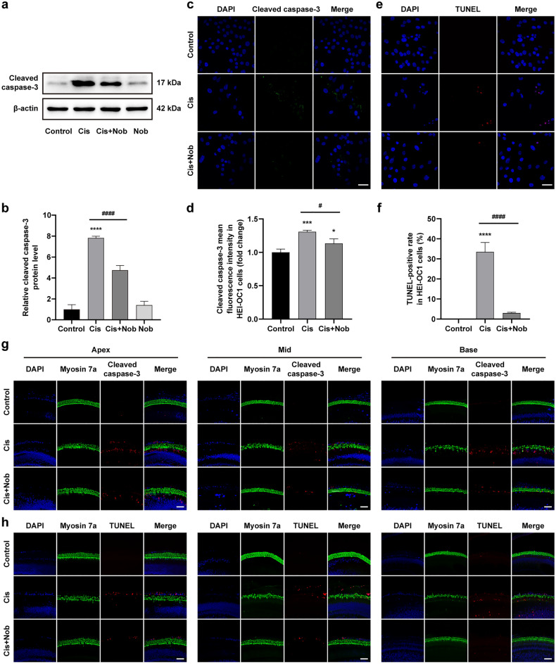
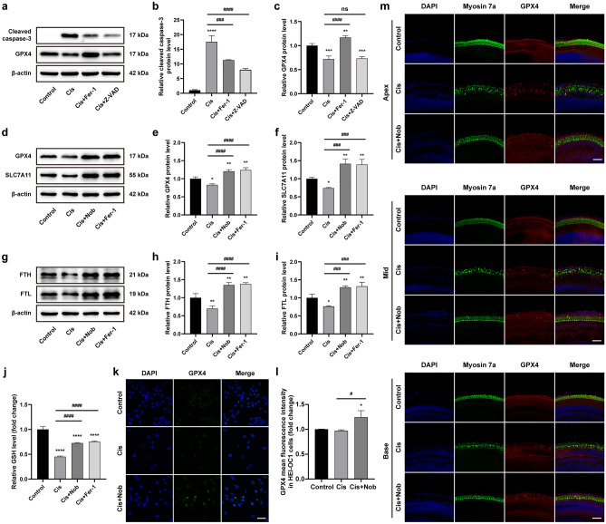
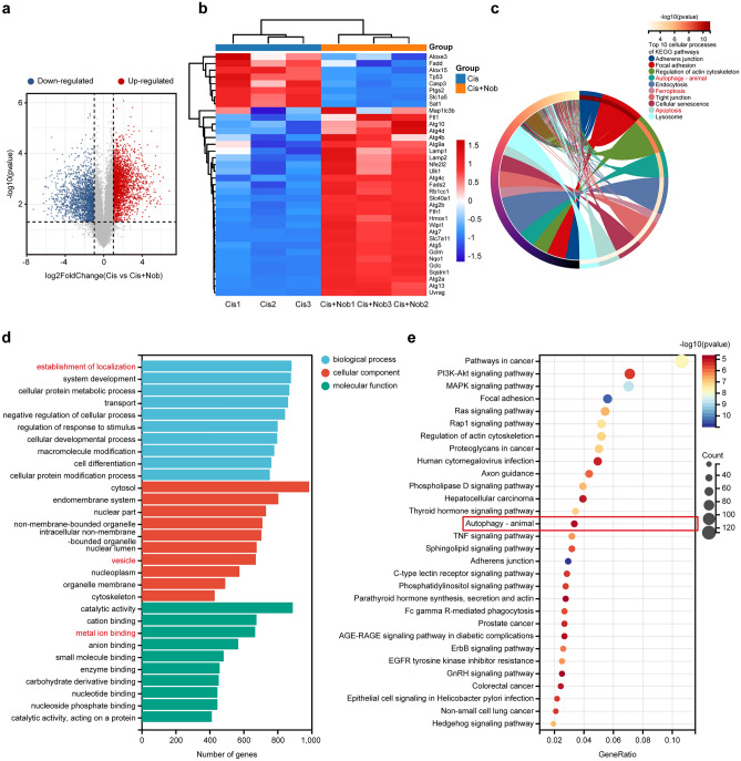
Nobiletin, a citrus polymethoxy flavonoid with antiapoptotic and antioxidative properties, could safeguard against cisplatin-induced nephrotoxicity and neurotoxicity. Cisplatin, as the pioneer of anti-cancer drug, the severe ototoxicity limits its clinical applications, while the effect of nobiletin on cisplatin-induced ototoxicity has not been identified. The current study investigated the alleviating effect of nobiletin on cisplatin-induced ototoxicity and the underlying mechanisms. Apoptosis and ROS formation were evaluated using the CCK-8 assay, Western blotting, and immunofluorescence, indicating that nobiletin attenuated cisplatin-induced apoptosis and oxidative stress. LC3B and SQSTM1/p62 were determined by Western blotting, qPCR, and immunofluorescence, indicating that nobiletin significantly activated autophagy. Nobiletin promoted the nuclear translocation of NRF2 and the transcription of its target genes, including Hmox1, Nqo1, and ferroptosis markers (Gpx4, Slc7a11, Fth, and Ftl), thereby inhibiting ferroptosis. Furthermore, RNA sequencing analysis verified that autophagy, ferroptosis, and the NRF2 signaling pathway served as crucial points for the protection of nobiletin against ototoxicity caused by cisplatin. Collectively, these results indicated, for the first time, that nobiletin alleviated cisplatin-elicited ototoxicity through suppressing apoptosis and oxidative stress, which were attributed to the activation of autophagy and the inhibition of NRF2/GPX4-mediated ferroptosis. Our study suggested that nobiletin could be a prospective agent for preventing cisplatin-induced hearing loss.


Conflict of interest statement

The authors declare no competing interests.

Figures

Figure 1
Figure 1
Nobiletin protects HEI-OC1 cells and cochlear explants against cisplatin-induced cytotoxicity. (a) Cell viability of HEI-OC1 cells treated with the designated doses of cisplatin for 24 h based on CCK-8 assay (n = 5). (b) Cell viability of HEI-OC1 cells treated with the designated doses of nobiletin for 24 h based on CCK-8 assay (n = 5). (c) Cell viability of HEI-OC1 cells after the pretreatment with incremental doses of nobiletin for 3 h and co-treated with 20 µM cisplatin for 24 h based on CCK-8 assay (n = 4). (d) Cell viability of HEI-OC1 cells after the pretreatment with 20 µM nobiletin for incremental times and co-treated with 20 µM cisplatin for 24 h based on CCK-8 assay (n = 4). (e) Representative images of Myosin 7a and DAPI immunofluorescence staining of hair cells in different cochlear regions under various treatments. (f) Quantification of hair cells in different cochlear regions (n = 3). Scale bars = 50 µm. *P < 0.05, **P < 0.01, ***P < 0.001, and ****P < 0.0001 vs. control group; # P < 0.05, ## P < 0.01, and ### P < 0.001 vs. cisplatin group.
Figure 2
Figure 2
Nobiletin suppresses cisplatin-induced apoptosis in HEI-OC1 cells and cochlear explants. (a, b) Protein levels of cleaved caspase-3 in HEI-OC1 cells assessed by Western blotting (n = 3). The original blots are presented in Supplementary Fig. S1. (c) Representative images of cleaved caspase-3 immunofluorescence staining of HEI-OC1 cells under various treatments. (d) Quantitation of intensity levels of cleaved caspase-3 immunofluorescence shown in (c) (n = 3). (e) Representative images of TUNEL staining of HEI-OC1 cells under various treatments. (f) Quantification of TUNEL-positive cells shown in (e) (n = 3). (g, h) Representative images of cleaved caspase-3 immunofluorescence staining and TUNEL staining of hair cells in different cochlear regions under various treatments. Scale bar = 50 µm. *P < 0.05, **P < 0.01, ***P < 0.001, and **** P < 0.0001 vs. control group; # P < 0.05, ## P < 0.01, ### P < 0.001, and #### P < 0.0001 vs. cisplatin group.
Figure 3
Figure 3
Nobiletin relieves cisplatin-induced oxidative damage in HEI-OC1 cells and cochlear explants. (a) Representative images of DCFH-DA and Hoechst 33,258 immunofluorescence staining of HEI-OC1 cells under various treatments. (b) Quantitation of intensity levels of DCFH-DA immunofluorescence shown in (a) (n = 3). (c) Representative images of DCFH-DA and Myosin 7a immunofluorescence staining of cochlear middle regions under various treatments. (d) Quantitation of intensity levels of DCFH-DA immunofluorescence shown in (c) (n = 3). (e–h) Protein levels of 4-HNE and SOD1 in HEI-OC1 cells based on Western blotting (n = 3). The original blots are presented in Supplementary Fig. S1. (i) The mRNA levels of gene encoding Sod1 in HEI-OC1 cells based on qPCR (n = 3). Scale bar = 50 µm. *P < 0.05, **P < 0.01, ***P < 0.001, and ****P < 0.0001 vs. control group; #P < 0.05, ##P < 0.01, ###P < 0.001, and ####P < 0.0001 vs. cisplatin group.
Figure 4
Figure 4
Nobiletin alleviates cisplatin-induced damage via activating autophagy. (a–e) Protein levels of LC3B and SQSTM1 in HEI-OC1 cells based on Western blotting (n = 3). The original blots are presented in Supplementary Fig. S1. (f, g) The mRNA levels of genes encoding Lc3b and Sqstm1 in HEI-OC1 cells based on qPCR (n = 3). (h) Representative images of LC3B immunofluorescence staining of HEI-OC1 cells under various treatments. (i) Quantitation of intensity levels of LC3B immunofluorescence shown in (h) (n = 3). (j) Representative images of LC3B and Myosin 7a immunofluorescence staining of hair cells in different cochlear regions under various treatments. Scale bar = 50 µm. *P < 0.05, **P < 0.01, ***P < 0.001, and ****P < 0.0001 vs. control group; #P < 0.05, ##P < 0.01, ###P < 0.001, and ####P < 0.0001 vs. cisplatin group.
Figure 5
Figure 5
Nobiletin activates NRF2-mediated pathway in HEI-OC1 cells and cochlear explants. (a–f) Protein levels of nuclear NRF2 and cytoplasmic NRF2, HMOX1, and NQO1 in HEI-OC1 cells based on Western blotting (n = 3). The original blots are presented in Supplementary Fig. S2. (g–i) The mRNA levels of genes encoding Nrf2, Hmox1, and Nqo1 in HEI-OC1 cells based on qPCR (n = 3). (j) Representative images of NRF2 immunofluorescence staining of HEI-OC1 cells under various treatments. (k) Quantitation of intensity levels of NRF2 immunofluorescence shown in (j) (n = 3). (l) Representative images of NRF2 and Myosin 7a immunofluorescence staining of hair cells in different cochlear regions under various treatments. Scale bar = 50 µm. *P < 0.05, **P < 0.01, ***P < 0.001, and ****P < 0.0001 vs. control group; #P < 0.05, ##P < 0.01, ###P < 0.001, and ####P < 0.0001 vs. cisplatin group.
Figure 6
Figure 6
Nobiletin ameliorates cisplatin-induced injury via inhibiting ferroptosis. (a–c) Protein levels of cleaved caspase-3 and GPX4 in HEI-OC1 cells treated with cisplatin after pretreatment with Fer-1 or Z-VAD-FMK based on Western blotting (n = 3). The original blots are presented in Supplementary Fig. S3. (d–i) Protein levels of GPX4, SLC7A11, FTH, and FTL in HEI-OC1 cells treated with cisplatin after pretreatment with nobiletin or ferrostatin-1 based on Western blotting (n = 3). The original blots are presented in Supplementary Fig. S4. (j) Levels of GSH measured in HEI-OC1 cells under various treatments (n = 3). (k) Representative images of GPX4 immunofluorescence staining of HEI-OC1 cells under various treatments. (l) Quantitation of intensity levels of GPX4 immunofluorescence shown in (k) (n = 3). (m) Representative images of GPX4 and Myosin 7a immunofluorescence staining of hair cells in different cochlear regions under various treatments. Scale bar = 50 µm. *P < 0.05, **P < 0.01, ***P < 0.001, and ****P < 0.0001 vs. control group; #P < 0.05, ##P < 0.01, ###P < 0.001, and ####P < 0.0001 vs. cisplatin group.
Figure 7
Figure 7
Validation of targets and signaling pathways associated with the protection of nobiletin against cisplatin-induced ototoxicity using RNA sequencing. (a) Volcanic map of differentially expressed genes (DEGs). (b) Heatmap of DEGs related to autophagy, ferroptosis, and NRF2 signaling pathway. (c) The top 10 cellular processes based on KEGG enrichment analysis. (d) GO annotation analysis based on the DEGs. (e) The top 30 pathways based on KEGG enrichment analysis.

Similar articles

Cited by

References

    1. Muller U, Barr-Gillespie PG. New treatment options for hearing loss. Nat. Rev. Drug Discov. 2015;14:346–U384. doi: 10.1038/nrd4533. - DOI - PubMed
    1. Tang Q, et al. Cisplatin-induced ototoxicity: Updates on molecular mechanisms and otoprotective strategies. Eur. J. Pharm. Biopharm. 2021;163:60–71. doi: 10.1016/j.ejpb.2021.03.008. - DOI - PubMed
    1. Smoorenburg, G. F., De Groot, J., Hamers, F. P. T. & Klis, S. F. L. in Ototoxicity: Basic Science and Clinical Applications Vol. 884 Annals of the New York Academy of Sciences (eds D. Henderson et al.) 192–210 (New York Acad Sciences, 1999).
    1. Rezaee R, Momtazi AA, Monemi A, Sahebkar A. Curcumin: A potentially powerful tool to reverse cisplatin-induced toxicity. Pharmacol. Res. 2017;117:218–227. doi: 10.1016/j.phrs.2016.12.037. - DOI - PubMed
    1. Gentilin E, Simoni E, Candito M, Cazzador D, Astolfi L. Cisplatin-induced ototoxicity: updates on molecular targets. Trends Mol. Med. 2019;25:1123–1132. doi: 10.1016/j.molmed.2019.08.002. - DOI - PubMed