Review
. 2024 Apr 3;23(1):98.
doi: 10.1186/s12944-024-02062-8.

The novel molecular mechanism of pulmonary fibrosis: insight into lipid metabolism from reanalysis of single-cell RNA-seq databases

Affiliations
Review

The novel molecular mechanism of pulmonary fibrosis: insight into lipid metabolism from reanalysis of single-cell RNA-seq databases

Xiangguang Shi et al. Lipids Health Dis. .

Abstract

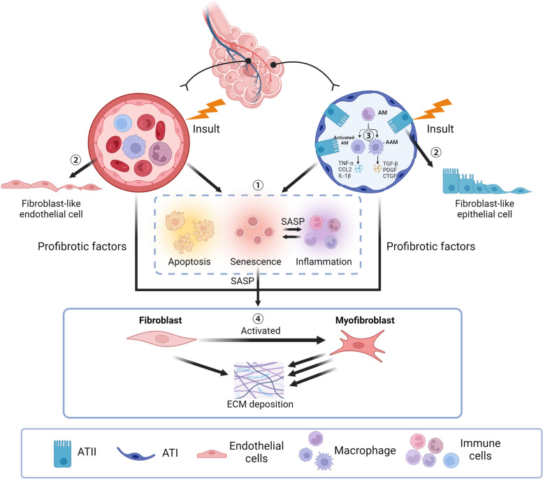
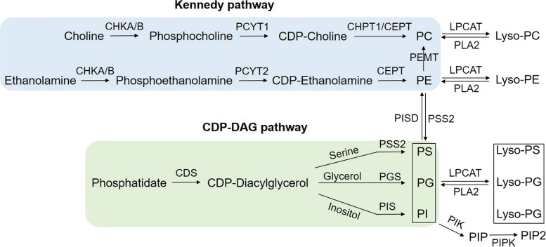
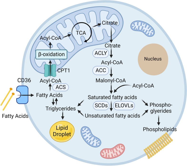
Pulmonary fibrosis (PF) is a severe pulmonary disease with limited available therapeutic choices. Recent evidence increasingly points to abnormal lipid metabolism as a critical factor in PF pathogenesis. Our latest research identifies the dysregulation of low-density lipoprotein (LDL) is a new risk factor for PF, contributing to alveolar epithelial and endothelial cell damage, and fibroblast activation. In this study, we first integrative summarize the published literature about lipid metabolite changes found in PF, including phospholipids, glycolipids, steroids, fatty acids, triglycerides, and lipoproteins. We then reanalyze two single-cell RNA-sequencing (scRNA-seq) datasets of PF, and the corresponding lipid metabolomic genes responsible for these lipids' biosynthesis, catabolism, transport, and modification processes are uncovered. Intriguingly, we found that macrophage is the most active cell type in lipid metabolism, with almost all lipid metabolic genes being altered in macrophages of PF. In type 2 alveolar epithelial cells, lipid metabolic differentially expressed genes (DEGs) are primarily associated with the cytidine diphosphate diacylglycerol pathway, cholesterol metabolism, and triglyceride synthesis. Endothelial cells are partly responsible for sphingomyelin, phosphatidylcholine, and phosphatidylethanolamines reprogramming as their metabolic genes are dysregulated in PF. Fibroblasts may contribute to abnormal cholesterol, phosphatidylcholine, and phosphatidylethanolamine metabolism in PF. Therefore, the reprogrammed lipid profiles in PF may be attributed to the aberrant expression of lipid metabolic genes in different cell types. Taken together, these insights underscore the potential of targeting lipid metabolism in developing innovative therapeutic strategies, potentially leading to extended overall survival in individuals affected by PF.

Keywords: Lipid metabolism; Lipid metabolomic gene, Single-cell RNA-sequencing reanalysis; Pulmonary fibrosis.


Conflict of interest statement

The authors declare no competing interests.

Figures

Fig. 1
Fig. 1
The mechanism of PF. Excessive apoptosis of endothelial and epithelial cells leads to lung injury and releases various pro-inflammatory and profibrotic factors. Injured EC and alveolar epithelial cells also undergo senescence, inducing an SASP phenotype, which further enhances the pro-inflammatory and profibrotic effects. The apoptosis-resistant endothelial and epithelial cells undergo an activated process to obtain fibroblast-like properties. The polarisation of macrophages confers AM and IM cells to differentiate into activated macrophages, which produce abundant pro-inflammatory and profibrotic factors and further promote PF. Over-proliferation and hyperactivation of fibroblast lead to ECM deposition and fibrosis formation
Fig. 2
Fig. 2
The metabolites and genes involved in the Kennedy pathway and CDP-DAG pathway. The synthesis of PC and PE via the Kennedy pathway, and the synthesis of PS, PG and PI through the CDP-DAG pathway
Fig. 3
Fig. 3
The metabolites and genes involved in SM metabolism. ASAHs catalyze ceramide to sphingosine, which is phosphorylated by SPHK to produce S1P. S1P can bind to S1PRs on the cell surface to regulate cell function or be degraded by SGPL1
Fig. 4
Fig. 4
Regulation of Lipid Droplet Formation by TG. The left panel of this figure depicts the multi-step synthesis of triglycerides (TAGs). Initially, glycerol-3-phosphate acyltransferase (GPAT) catalyzes the biosynthesis of lysophosphatidic acid (LPA) with a preference for saturated fatty acids and glycerol-3-phosphate (G3P) as substrates. GPATs exist in two forms, the mitochondrial isoform (GPAT1/2) and the endoplasmic isoform (GPAT3/4). Next, 1-acylglycerol-3-phosphate O-acyltransferases (AGPATs) convert LPA to phosphatidic acid (PA). Following this, the enzyme lipin, a magnesium-ion-dependent phosphatidic acid phosphohydrolase, dephosphorylates PA to yield diacylglycerol (DAG). Diacylglycerol O-acyltransferases (DGAT1/2) catalyze DAG and fatty acyl-CoA to TAG. The biogenesis of lipid droplets commences with TG synthesis, which accumulates between the ER membrane’s two leaflets. Proteins bound to the lipid droplet surface, such as perilipins (PLINs), localize to the phospholipid monolayer, while the neutral lipid core comprises triacylglycerols and sterol esters. The right panel illustrates the hydrolysis of TG. Adipose triglyceride lipase (ATGL), encoded by the PNPLA2 gene, initiates TAG degradation to produce DAG, which is subsequently hydrolyzed to monoacylglycerol (MAG) by hormone-sensitive lipase (LIPE). LIPE also participates in steroid hormone synthesis by converting cholesteryl esters to free cholesterol. Finally, monoglyceride lipase (MGLL) hydrolyzes MAG to free fatty acids and glycerol
Fig. 5
Fig. 5
Regulation of FA Metabolism Pathways. ACLY utilizes cytoplasmic citrate to produce cytosolic acetyl-CoA, which is then used as a substrate by ACC to produce malonyl-CoA. FASN could convert acetyl-CoA and malonyl-CoA into long-chain saturated fatty acids for palmitate synthesis. SCD plays a critical role in the synthesis of unsaturated FAs, particularly oleic acid. The ELOVLs are involved in fatty acid elongation. In FA catabolism, acyl-CoA synthetases (ACSLs) first degrade long-chain FAs (LCFAs) to fatty acyl-CoA esters. Subsequently, mitochondrial carnitine palmitoyltransferases (CPT1/II), in conjunction with a carnitine-acylcarnitine translocase, induce the oxidation of LCFAs, which are ultimately broken down to acetyl-CoA via the β-oxidation pathway in the mitochondria

References

    1. Jee AS, Sahhar J, Youssef P, Bleasel J, Adelstein S, Nguyen M, Corte TJ. Review: serum biomarkers in idiopathic pulmonary fibrosis and systemic sclerosis associated interstitial lung disease - frontiers and horizons. Pharmacol Ther. 2019;202:40–52. doi: 10.1016/j.pharmthera.2019.05.014. - DOI - PubMed
    1. Hutchinson J, Fogarty A, Hubbard R, McKeever T. Global incidence and mortality of idiopathic pulmonary fibrosis: a systematic review. Eur Respir J. 2015;46:795–806. doi: 10.1183/09031936.00185114. - DOI - PubMed
    1. Khanna D, Tashkin DP, Denton CP, Renzoni EA, Desai SR, Varga J. Etiology, risk factors, and biomarkers in systemic sclerosis with interstitial lung disease. Am J Respir Crit Care Med. 2020;201:650–660. doi: 10.1164/rccm.201903-0563CI. - DOI - PMC - PubMed
    1. Liu X, Jiang Q, Wu P, Han L, Zhou P. Global incidence, prevalence and disease burden of silicosis: 30 years' overview and forecasted trends. BMC Public Health. 2023;23:1366. doi: 10.1186/s12889-023-16295-2. - DOI - PMC - PubMed
    1. Alysandratos KD, Russo SJ, Petcherski A, Taddeo EP, Acin-Perez R, Villacorta-Martin C, Jean JC, Mulugeta S, Rodriguez LR, Blum BC, et al. Patient-specific iPSCs carrying an SFTPC mutation reveal the intrinsic alveolar epithelial dysfunction at the inception of interstitial lung disease. Cell Rep. 2021;36:109636. doi: 10.1016/j.celrep.2021.109636. - DOI - PMC - PubMed

MeSH terms