. 2024 Apr 3;19(1):25.
doi: 10.1186/s13062-024-00466-1.

Circ_0002669 promotes osteosarcoma tumorigenesis through directly binding to MYCBP and sponging miR-889-3p

Affiliations

Circ_0002669 promotes osteosarcoma tumorigenesis through directly binding to MYCBP and sponging miR-889-3p

Ying Zhang et al. Biol Direct. .

Abstract

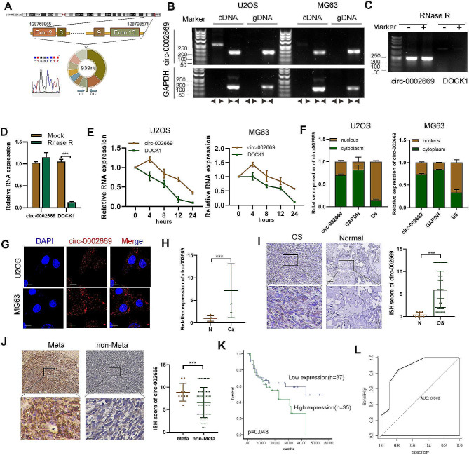
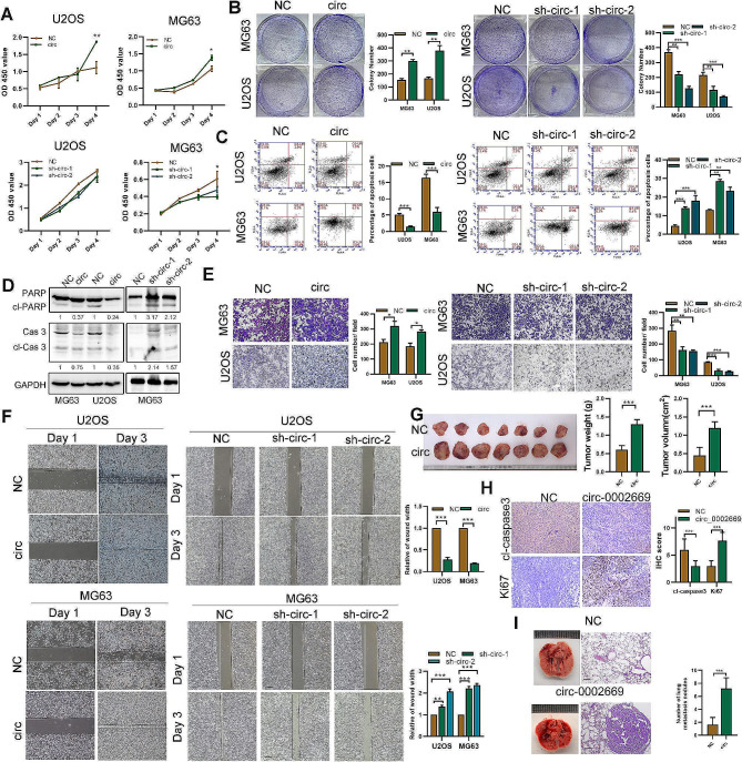
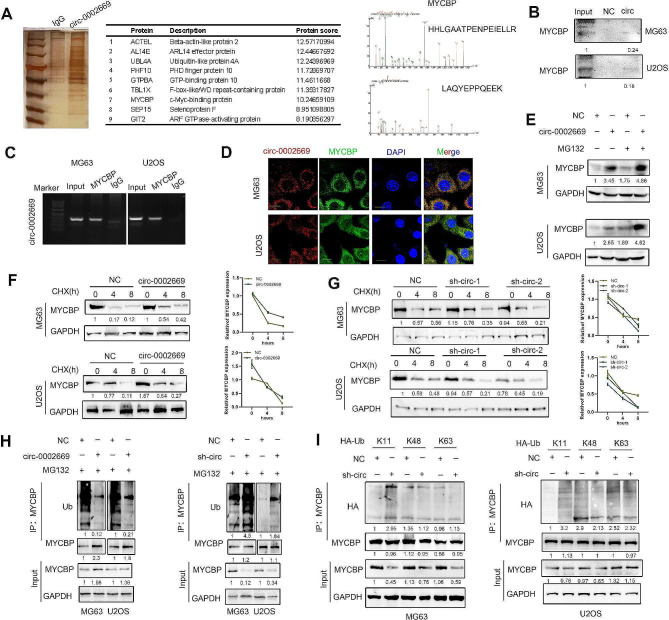
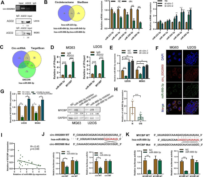
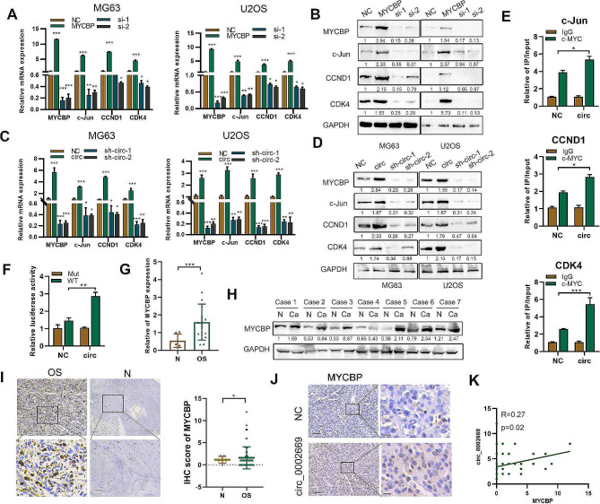
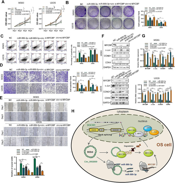
Circular RNAs (circRNAs) are a class of highly multifunctional single-stranded RNAs that play crucial roles in cancer progression, including osteosarcoma (OS). Circ_0002669, generated from the dedicator of cytokinesis (DOCK) gene, was highly expressed in OS tissues, and negatively correlated with OS patient survival. Elevated circ_0002669 promoted OS cell growth and invasion in vivo and in vitro. By biotin pulldown and mass spectroscopy, we found that circ_0002669 directly bound to MYCBP, a positive regulator of c-myc, to prevent MYCBP from ubiquitin-mediated proteasome degradation. In addition, circ_0002669 interacted with miR-889-3p and served as a miRNA sponge to increase the expression of MYCBP, as determined by luciferase assays and RNA immunoprecipitation. Functional rescue experiments indicated MYCBP acted as a key factor for circ_0002669- and miR-889-3p-regulated OS cell proliferation and migration. Increased expression of c-myc-associated genes, such as CCND1, c-Jun and CDK4, were found in circ_0002669- and MYCBP-overexpressing OS cells. Our data thus provide evidence that circ_0002669 promotes OS malignancy by protecting MYCBP from protein ubiquitination and degradation and blocking miR-889-3p-mediated inhibition of MYCBP expression.

Keywords: Circ_0002669; MYCBP; Osteosarcoma; miR-889-3p.


Conflict of interest statement

The authors declare no competing interests.

Figures

Fig. 1
Fig. 1
Verification of the structure and location of circ_0002669 and its clinical significance. A Circ_0002669 was generated from the DOCK1 gene located on chromosome 10. Sanger sequencing was used to identify the back-splice junction. B The closed loop structure of circ_0002669 was verified by PCR using convergent and divergent primers. C, D Upon RNase R treatment, expression of circ_0002669 and DOCK1 mRNA were detected by PCR and qRT-PCR in U2OS cells. E Circ_0002669 and DOCK1 mRNA expression were detected by qRT-PCR in OS cells treated with actinomycin D (1 μm) at the indicated time point. F Expression level of circ_0002669 in the nucleus and cytoplasm of cells was measured by qRT-PCR. G FISH was performed to identify the cellular location of circ_0002669 in OS cells (scale bar, 20 μm). H Circ_0002669 expression in 12 OS tissues and normal tissues (n = 5) was analyzed by qRT-PCR. (I) Circ_0002669 expression was detected in OS tissues and normal tissues by ISH (scale bar, 50 μm). J Circ_0002669 expression was detected in metastatic lesions and primary OS tissues (scale bar, 50 μm). K Survival analysis of 72 OS patients based on circ_0002669 ISH scores. L ROC curve based on the expression of circ_0002669 in OS tissues. Data shown are from three independent experiments, *p < 0.05, **p < 0.01, ***p < 0.001
Fig. 2
Fig. 2
Circ_0002669 enhances OS cell proliferation and migration both in vitro and in vivo. A Cell proliferation was quantitated by CCK-8 assay. B Cell colony forming ability was measured. C Apoptosis of OS cells quantitated by flow cytometry via Annexin V-FITC/PI staining. D Expression of apoptosis-related proteins was detected using western blotting in OS cells. E Cell migration of OS cells determined by transwell assays (scale bar, 100 μm). F Migration of OS cells determined by wound healing assay. G Images of xenograft tumors in control and circ_0002669 groups (n = 7). H IHC staining of KI-67 and cleaved caspase-3 in control and circ_0002669 groups (scale bar, 50 μm). I Lung metastasis following tail vein injection of stable circ_0002669-overexpressing or control cells into nude mice. The mice were euthanized at 35 days. HE staining of lungs displayed metastatic nodules (scale bar, 50 μm). Data shown are from three independent experiments, *p < 0.05, **p < 0.01, ***p < 0.001
Fig. 3
Fig. 3
Circ_0002669 binds to MYCBP to upregulate MYCBP expression. A RNA pull-down was performed using the circ_0002669 biotin probe in U2OS cell lysates. After silver staining, MYCBP was identified as a candidate protein binding with circ_0002669 by mass spectrometry and western blot. B RNA pull-down was performed using biotin circ_0002669, and the relative expression of MYCBP was quantitated by western blotting. C RIP was performed on U2OS cell lysates using anti-MYCBP or anti-IgG, then the enrichment of circ_0002669 was detected by RT-PCR. D Circ_0002669 was co-localized with MYCBP in OS cells by FISH-IF (scale bar, 25 μm). E Stable circ_0002669-overexpressing or control OS cells were treated with MG132 (10 µM) and immunoblotted and probed with MYCBP antibody. FG Stable circ_0002669-overexpressing, -knockdown or control OS cells were treated with cycloheximide (CHX, 10 µM) and collected at different time points. Cell lysates were immunoblotted and probed with MYCBP antibody. H Ubiquitination of MYCBP in control, circ_002669-overexpressing or -knockdown OS cells were determined by immunoprecipitation with anti-MYCBP and then immunoblotting with the indicated antibodies. I Ubiquitination of MYCBP in OS cells, transfected with plasmids encoding different ubiquitin chains (K11, K48 and K63), was determined by immunoprecipitation with anti-MYCBP and then immunoblotting with the indicated antibodies. Data shown are from three independent experiments, *p < 0.05, **p < 0.01, ***p < 0.001
Fig. 4
Fig. 4
Circ_0002669 functions as a sponge for miR-889-3p. B RIP and RT-PCR was performed using U2OS cells to determine the enrichment of circ_0002669. RNA pull-down was performed, followed by western blotting to determine AGO2 expression. B The potential miRNAs binding to circ_0002669, as predicted by StarBase and CircInteractome. Expression of the miRNAs was determined in circ_0002669-overexpressing or -knockdown OS cells. C Identification of miR-889-3p by using TargetScan and ENCORI to screen for upstream miRNAs of MYCBP. D RIP was performed to detect the binding between miR-889-3p or MYCBP mRNA and AGO2 protein. E MiR-889-3p expression was determined using qRT-PCR in circ_0002669-overexpressing or -knockdown OS cells. F FISH was performed to determine the intracellular location of circ_0002669 (red) and miR-889-3p (green) (scale bar, 20 μm). G Expression of MYCBP was determined in miR-889-3p mimic- or inhibitor-transfected OS cells, as determined by qRT-PCR and western blotting. H Relative expression of miR-889-3p in OS tissues (n = 12) and non-tumor tissues (n = 5) was determined by qRT-PCR. I Correlation between miR-889-3p and MYCBP expression in OS tissues. J Schematic illustration of circ_002669-wildtype (WT) and circ_002669-Mut (MT) luciferase reporter vectors. Relative luciferase activities were determined in the indicated transfected OS cells. K Schematic illustration of MYCBP-WT and MYCBP-MT luciferase reporter vectors. The relative luciferase activities were quantitated in the indicated transfected OS cells. Data shown are from three independent experiments, *p < 0.05, **p < 0.01, ***p < 0.001
Fig. 5
Fig. 5
Circ_0002669 increases c-MYC transcriptional activity. A, B Relative expression of MYCBP and c-MYC downstream genes was determined, by qRT-PCR and western blotting, in OS cells transfected with MYCBP plasmid or siRNAs. C, D Relative expression of MYCBP and c-MYC downstream genes was determined in circ_0002669-overexpressing or -knockdown OS cells by qRT-PCR and western blotting. E CHIP-qPCR showing circ_002669 enhanced c-MYC occupancy on the c-Jun, CCND1 and CDK4 promoters in U2OS cells. F Circ_002669 in U2OS cells increased transcription of the c-Jun promoter. G MYCBP expression in OS (n = 12) and normal tissues (n = 5) was determined by qRT-PCR. H MYCBP expression in OS and normal tissues (n = 7) was determined by western blotting. I MYCBP expression in OS tissues (n = 72) and normal tissues (n = 6) was determined by IHC. J MYCBP expression in control or circ_002669-overexpressing samples derived from xenografts (scale bar, 100 μm). K Pearson correlation analysis showing the correlation between expression of MYCBP and circ_002669 in OS tissues. Data shown are from three independent experiments, *p < 0.05, **p < 0.01, ***p < 0.001
Fig. 6
Fig. 6
Circ_0002669 promotes OS malignancy through a miR-889-3p/MYCBP axis. A, B Cell proliferation and colony formation was determined by CCK-8 and colony formation assays in control, miR-889-3p, miR-889-3p + circ_0002669, si-MYCBP and si-MYCBP + circ_0002669 OS cell subgroups. C Apoptosis was measured by flow cytometry following annexin V-FITC/PI staining in control, miR-889-3p, miR-889-3p + circ_0002669, si-MYCBP and si-MYCBP + circ_0002669 OS cell groups. D, E Cell migration was determined by transwell and wound healing assays in control, miR-889-3p, miR-889-3p + circ_0002669, si-MYCBP and si-MYCBP + circ_0002669 OS cell groups (scale bar, 100 μm). F, G Relative expression of MYCBP, c-Jun, CCND1 and CDK4 mRNA and protein levels was determined by qRT-PCR and western blotting in control, miR-889-3p, miR-889-3p + circ_0002669, si-MYCBP and si-MYCBP + circ_0002669 OS cell groups. H Schematic diagram of this paper. Data shown are from three independent experiments, *p < 0.05, **p < 0.01, ***p < 0.001

Similar articles

Cited by

References

    1. Whelan JS, Davis LE. Osteosarcoma, Chondrosarcoma, and Chordoma. J Clin Oncology: Official J Am Soc Clin Oncol. 2018;36(2):188–93. doi: 10.1200/JCO.2017.75.1743. - DOI - PubMed
    1. Gill J, Gorlick R. Advancing therapy for osteosarcoma. Nat Reviews Clin Oncol. 2021;18(10):609–24. doi: 10.1038/s41571-021-00519-8. - DOI - PubMed
    1. Kristensen LS, Andersen MS, Stagsted LVW, Ebbesen KK, Hansen TB, Kjems J. The biogenesis, biology and characterization of circular RNAs. Nat Rev Genet. 2019;20(11):675–91. doi: 10.1038/s41576-019-0158-7. - DOI - PubMed
    1. Li Z, Li X, Xu D, Chen X, Li S, Zhang L, Chan MTV, Wu WKK. An update on the roles of circular RNAs in osteosarcoma. Cell Prolif. 2021;54(1):e12936. doi: 10.1111/cpr.12936. - DOI - PMC - PubMed
    1. Yang B, Li L, Tong G, Zeng Z, Tan J, Su Z, Liu Z, Lin J, Gao W, Chen J, et al. Circular RNA circ_001422 promotes the progression and metastasis of osteosarcoma via the miR-195-5p/FGF2/PI3K/Akt axis. J Experimental Clin cancer Research: CR. 2021;40(1):235. doi: 10.1186/s13046-021-02027-0. - DOI - PMC - PubMed

Publication types