

Suppression of the growth and metastasis of mouse melanoma by Taenia crassiceps and Mesocestoides corti tapeworms
- PMID: 38571957
- PMCID: PMC10987685
- DOI: 10.3389/fimmu.2024.1376907
Suppression of the growth and metastasis of mouse melanoma by Taenia crassiceps and Mesocestoides corti tapeworms
Abstract
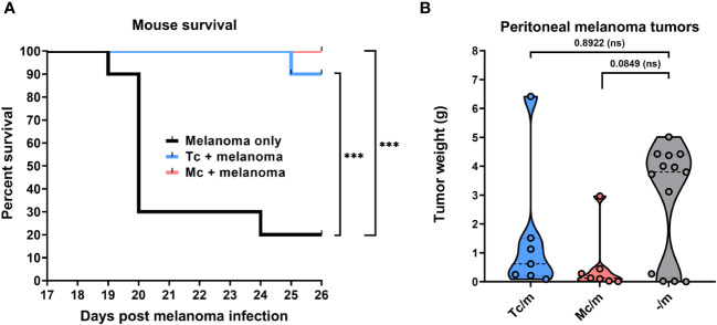
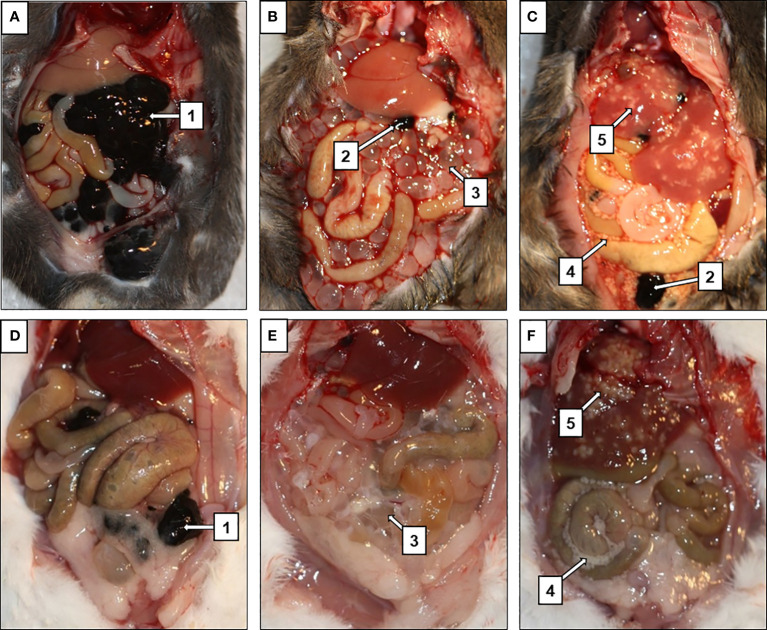
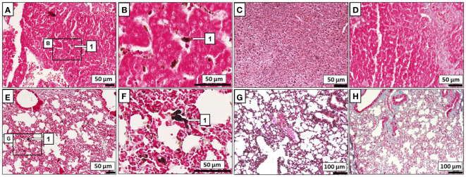
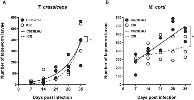
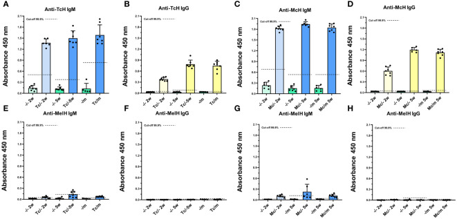
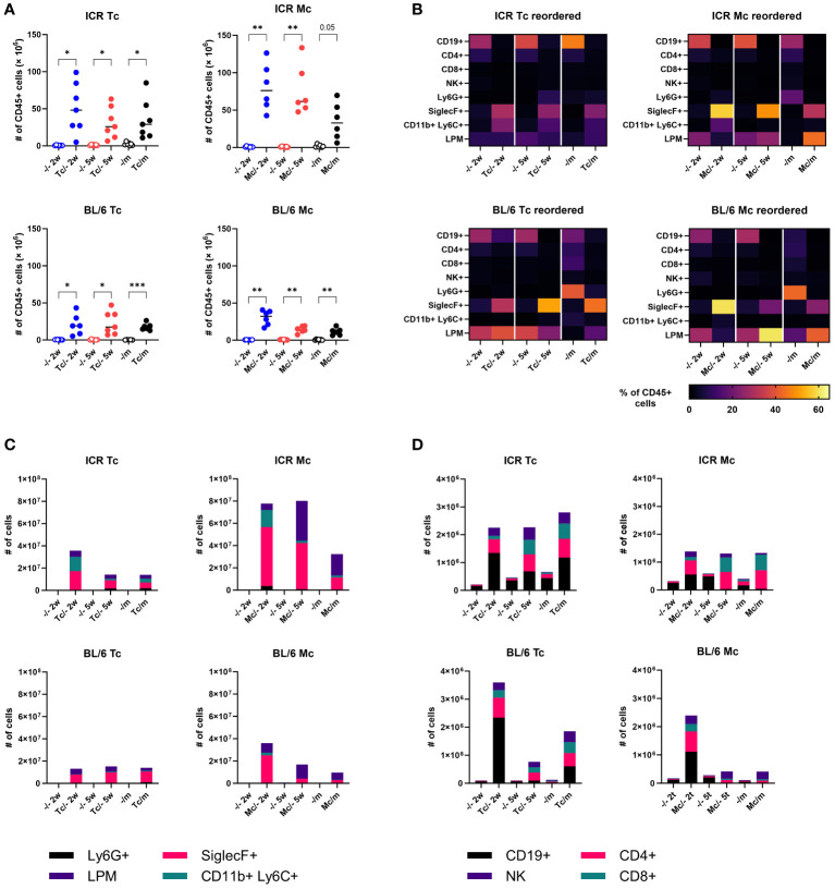
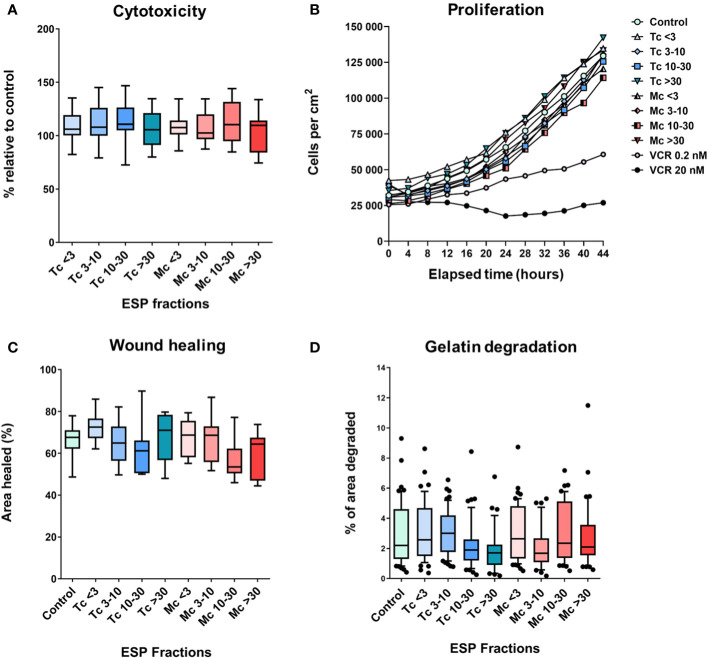
Cancer is still one of the leading causes of death, with an estimated 19.3 million new cases every year. Our paper presents the tumor-suppressing effect of Taenia crassiceps and Mesocestoides corti on B16F10 melanoma, the intraperitoneal application of which followed the experimental infection with these tapeworms, resulting in varying degrees of effectiveness in two strains of mice. In the case of M. corti-infected ICR mice, a strong tumor growth suppression occurred, which was accompanied by a significant reduction in the formation of distant metastases in the liver and lung. Tapeworm-infected C57BL/6J mice also showed a suppression of tumor growth and, in addition, the overall survival of infected C57BL/6J mice was significantly improved. Experiments with potential cross-reaction of melanoma and tapeworm antigens with respective specific antibodies, restimulation of spleen T cells, or the direct effect of tapeworm excretory-secretory products on melanoma cells in vitro could not explain the phenomenon. However, infections with T. crassiceps and M. corti increased the number of leukocytes possibly involved in anti-tumor immunity in the peritoneal cavity of both ICR and C57BL/6J mice. This study unveils the complex interplay between tapeworm infections, immune responses, and melanoma progression, emphasizing the need for further exploration of the mechanisms driving observed tumor-suppressive effects.
Keywords: Mesocestoides; Taenia; cancer; melanoma; metastasis; suppression; tapeworm.
Copyright © 2024 Schreiber, Macháček, Vajs, Šmídová, Majer, Hrdý, Tolde, Brábek, Rösel and Horák.
Conflict of interest statement
The authors declare that the research was conducted in the absence of any commercial or financial relationships that could be construed as a potential conflict of interest.
Figures
Similar articles
-
Cestode larvae excite host neuronal circuits via glutamatergic signalling.Elife. 2025 Jul 4;12:RP88174. doi: 10.7554/eLife.88174. Elife. 2025. PMID: 40613653 Free PMC article.
-
Suppression of Taenia crassiceps during concurrent infections with Mesocestoides corti in mice.Parasitology. 1986 Feb;92 ( Pt 1):199-207. doi: 10.1017/s0031182000063551. Parasitology. 1986. PMID: 3754325
-
Systemic treatments for metastatic cutaneous melanoma.Cochrane Database Syst Rev. 2018 Feb 6;2(2):CD011123. doi: 10.1002/14651858.CD011123.pub2. Cochrane Database Syst Rev. 2018. PMID: 29405038 Free PMC article.
-
Sentinel lymph node biopsy followed by lymph node dissection for localised primary cutaneous melanoma.Cochrane Database Syst Rev. 2015 May 16;2015(5):CD010307. doi: 10.1002/14651858.CD010307.pub2. Cochrane Database Syst Rev. 2015. PMID: 25978975 Free PMC article.
-
Comparison of Two Modern Survival Prediction Tools, SORG-MLA and METSSS, in Patients With Symptomatic Long-bone Metastases Who Underwent Local Treatment With Surgery Followed by Radiotherapy and With Radiotherapy Alone.Clin Orthop Relat Res. 2024 Dec 1;482(12):2193-2208. doi: 10.1097/CORR.0000000000003185. Epub 2024 Jul 23. Clin Orthop Relat Res. 2024. PMID: 39051924
Cited by
-
How tapeworms interact with cancers: a mini-review.PeerJ. 2024 Mar 29;12:e17196. doi: 10.7717/peerj.17196. eCollection 2024. PeerJ. 2024. PMID: 38563013 Free PMC article. Review.
-
Impact of a previous infection with Taenia crassiceps cysticerci on the susceptibility to Leishmania (L.) major or L. (V.) braziliensis.World J Microbiol Biotechnol. 2025 Mar 28;41(4):115. doi: 10.1007/s11274-025-04327-5. World J Microbiol Biotechnol. 2025. PMID: 40148716
References
-
- NCI-SEER Database . Melanoma of the skin — Cancer stat facts(2023). Available online at: https://seer.cancer.gov/statfacts/html/melan.html (Accessed August 31, 2023).
-
- Elder DE, Bastian BC, Cree IA, Chb MB, Massi D, Scolyer RA. The 2018 World Health Organization classification of cutaneous, mucosal, and uveal melanoma detailed analysis of 9 distinct subtypes defined by their evolutionary pathway. Arch Pathol Lab Med. (2020) 144:500–22. doi: 10.5858/arpa.2019-0561-RA - DOI - PubMed
Publication types
MeSH terms
LinkOut - more resources
Full Text Sources
Medical