

The Arabidopsis leaf quantitative atlas: a cellular and subcellular mapping through unified data integration
- PMID: 38572078
- PMCID: PMC10988163
- DOI: 10.1017/qpb.2024.1
The Arabidopsis leaf quantitative atlas: a cellular and subcellular mapping through unified data integration
Abstract
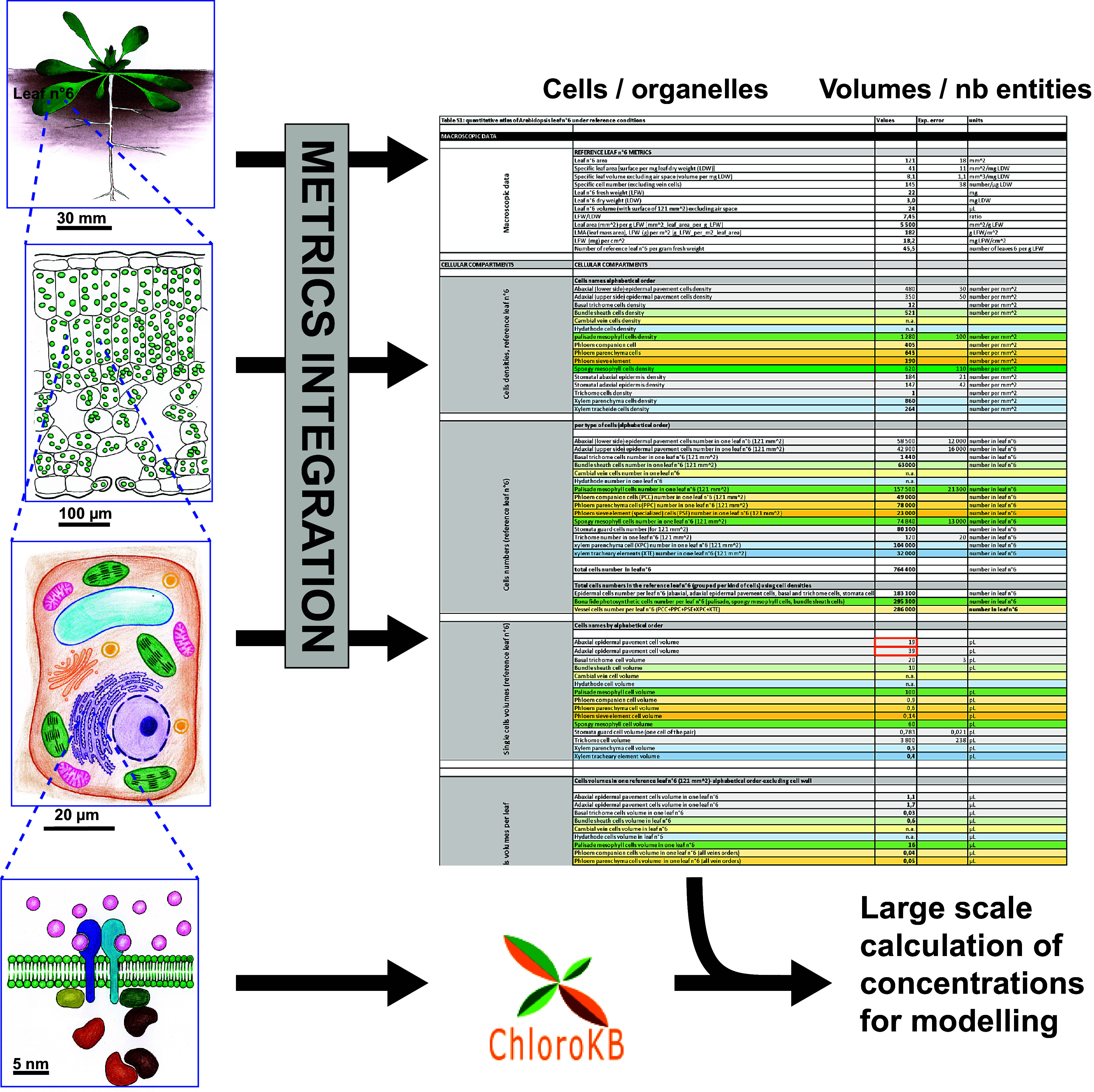
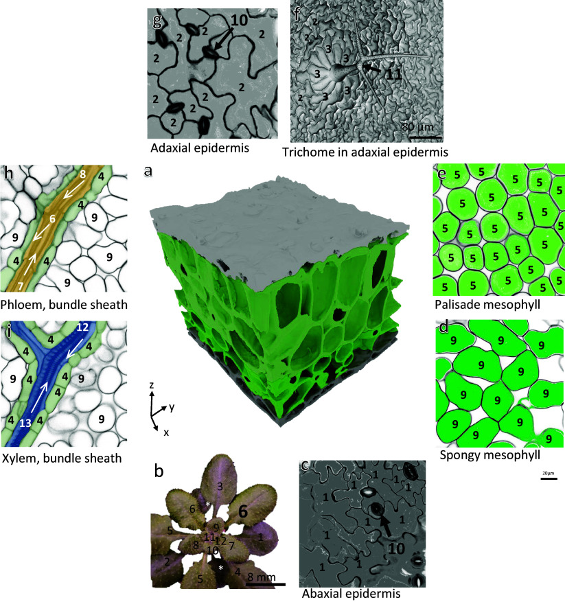
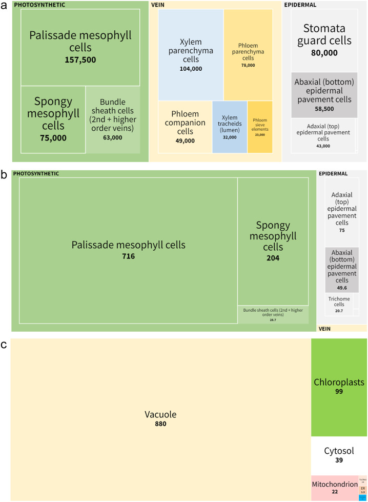
Quantitative analyses and models are required to connect a plant's cellular organisation with its metabolism. However, quantitative data are often scattered over multiple studies, and finding such data and converting them into useful information is time-consuming. Consequently, there is a need to centralise the available data and to highlight the remaining knowledge gaps. Here, we present a step-by-step approach to manually extract quantitative data from various information sources, and to unify the data format. First, data from Arabidopsis leaf were collated, checked for consistency and correctness and curated by cross-checking sources. Second, quantitative data were combined by applying calculation rules. They were then integrated into a unique comprehensive, referenced, modifiable and reusable data compendium representing an Arabidopsis reference leaf. This atlas contains the metrics of the 15 cell types found in leaves at the cellular and subcellular levels.
Keywords: Arabidopsis; curation; leaf anatomy; modelling; organelle.
© The Author(s) 2024.
Conflict of interest statement
The authors declare they have no competing interests.
Figures
Similar articles
-
The leaf ionome as a multivariable system to detect a plant's physiological status.Proc Natl Acad Sci U S A. 2008 Aug 19;105(33):12081-6. doi: 10.1073/pnas.0804175105. Epub 2008 Aug 12. Proc Natl Acad Sci U S A. 2008. PMID: 18697928 Free PMC article.
-
A topological map of the compartmentalized Arabidopsis thaliana leaf metabolome.PLoS One. 2011 Mar 15;6(3):e17806. doi: 10.1371/journal.pone.0017806. PLoS One. 2011. PMID: 21423574 Free PMC article.
-
LEAFDATA: a literature-curated database for Arabidopsis leaf development.Plant Methods. 2016 Feb 15;12:15. doi: 10.1186/s13007-016-0115-9. eCollection 2016. Plant Methods. 2016. PMID: 26884807 Free PMC article.
-
New insights into the regulation of leaf senescence in Arabidopsis.J Exp Bot. 2018 Feb 12;69(4):787-799. doi: 10.1093/jxb/erx287. J Exp Bot. 2018. PMID: 28992051 Review.
-
Preliminary Planning for Mars Sample Return (MSR) Curation Activities in a Sample Receiving Facility (SRF).Astrobiology. 2022 Jun;22(S1):S57-S80. doi: 10.1089/AST.2021.0105. Epub 2022 May 19. Astrobiology. 2022. PMID: 34904890 Review.
Cited by
-
Subcellular plant carbohydrate metabolism under elevated temperature.Plant Physiol. 2025 Jul 3;198(3):kiaf117. doi: 10.1093/plphys/kiaf117. Plant Physiol. 2025. PMID: 40238939 Free PMC article.
References
-
- Aguirrezabal, L. , Bouchier-Combaud, S. , Radziejwoski, A. , Dauzat, M. , Cookson, S. J. , & Granier, C. (2006). Plasticity to soil water deficit in Arabidopsis thaliana: Dissection of leaf development into underlying growth dynamic and cellular variables reveals invisible phenotypes. Plant Cell and Environment, 29, 2216–2227. - PubMed
-
- Ahuja, I. , Kissen, R. , Hoang, L. , Sporsheim, B. , Halle, K. K. , Wolff, S. A. , Ahmad, S. J. N. , Ahmad, J. N. , & Bones, A. M. (2021). The imaging of guard cells of thioglucosidase (tgg) mutants of Arabidopsis further links plant chemical defence systems with physical defence barriers. Cell, 10(2), Article 227. - PMC - PubMed
-
- Armstrong, A. F. , Logan, D. C. , Tobin, A. K. , O'Toole, P. , & Atkin, O. K. (2006). Heterogeneity of plant mitochondrial responses underpinning respiratory acclimation to the cold in Arabidopsis thaliana leaves. Plant Cell and Environment, 29, 940–949. - PubMed
-
- Baerenfaller, K. , Massonnet, C. , Walsh, S. , Baginsky, S. , Buhlmann, P. , Hennig, L. , Hirsch-Hoffmann, M. , Howell, K. A. , Kahlau, S. , Radziejwoski, A. , Russenberger, D. , Rutishauser, D. , Small, I. , Stekhoven, D. , Sulpice, R. , Svozil, J. , Wuyts, N. , Stitt, M. , Hilson, P. , … Gruissem, W. (2012). Systems-based analysis of Arabidopsis leaf growth reveals adaptation to water deficit. Molecular Systems Biology, 8, Article 606. - PMC - PubMed
-
- Baerenfaller, K. , Shu, H. , Hirsch-Hoffmann, M. , Futterer, J. , Opitz, L. , Rehrauer, H. , Hennig, L. , & Gruissem, W. (2016). Diurnal changes in the histone H3 signature H3K9ac|H3K27ac|H3S28p are associated with diurnal gene expression in Arabidopsis. Plant Cell and Environment, 39(11), 2557–2569. - PubMed
LinkOut - more resources
Full Text Sources