. 2024 Apr;14(4):1624-1643.
doi: 10.1016/j.apsb.2024.01.006. Epub 2024 Jan 17.

Blockade of the deubiquitinating enzyme USP48 degrades oncogenic HMGA2 and inhibits colorectal cancer invasion and metastasis

Affiliations

Blockade of the deubiquitinating enzyme USP48 degrades oncogenic HMGA2 and inhibits colorectal cancer invasion and metastasis

Can Cheng et al. Acta Pharm Sin B. 2024 Apr.

Abstract

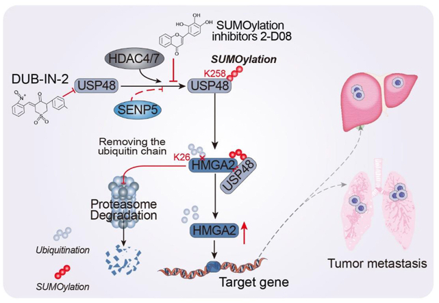
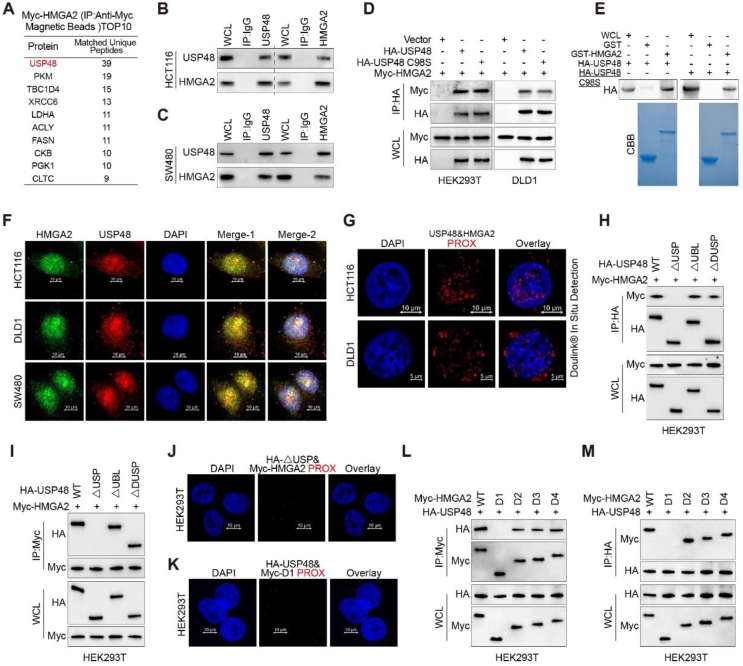
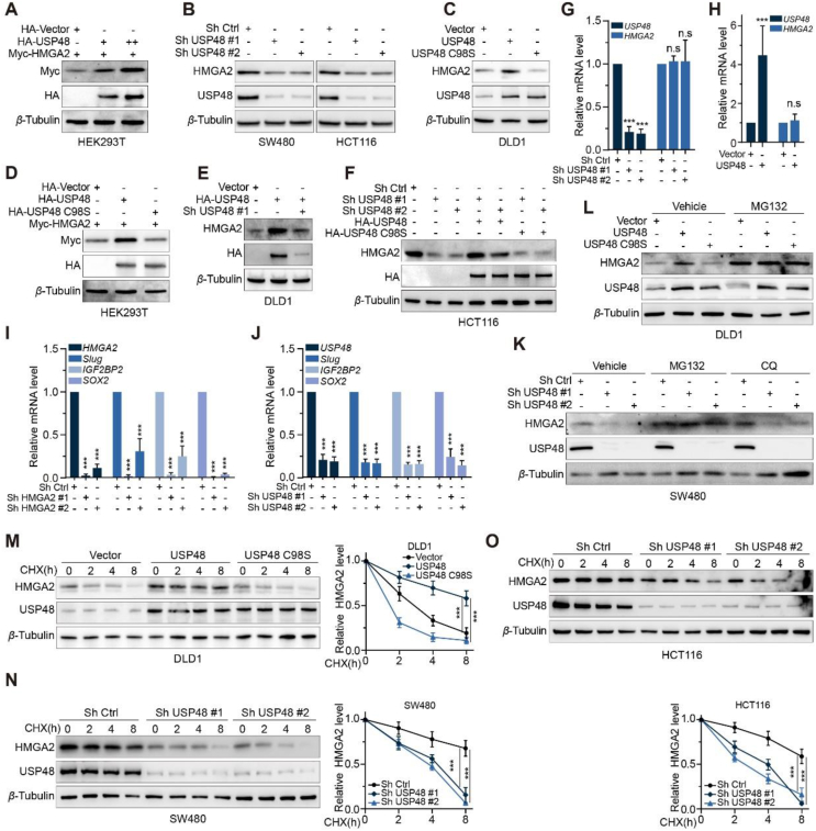
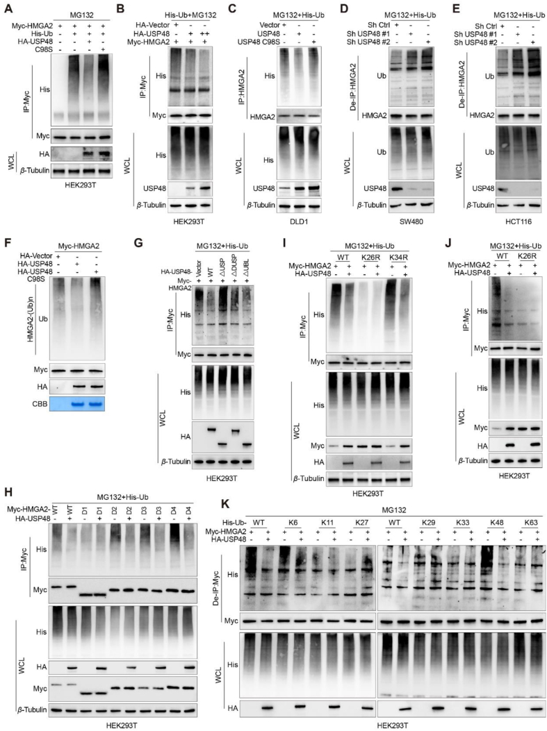
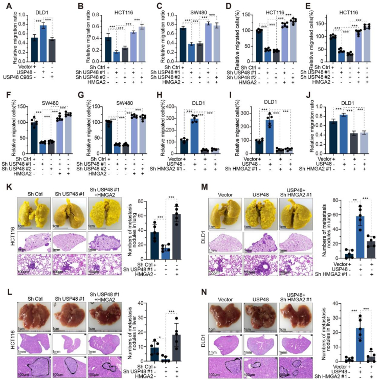
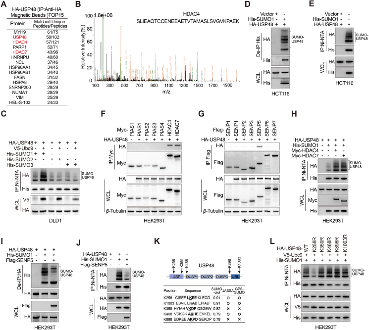
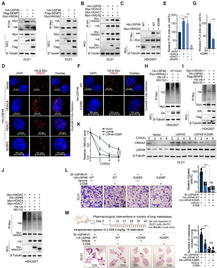
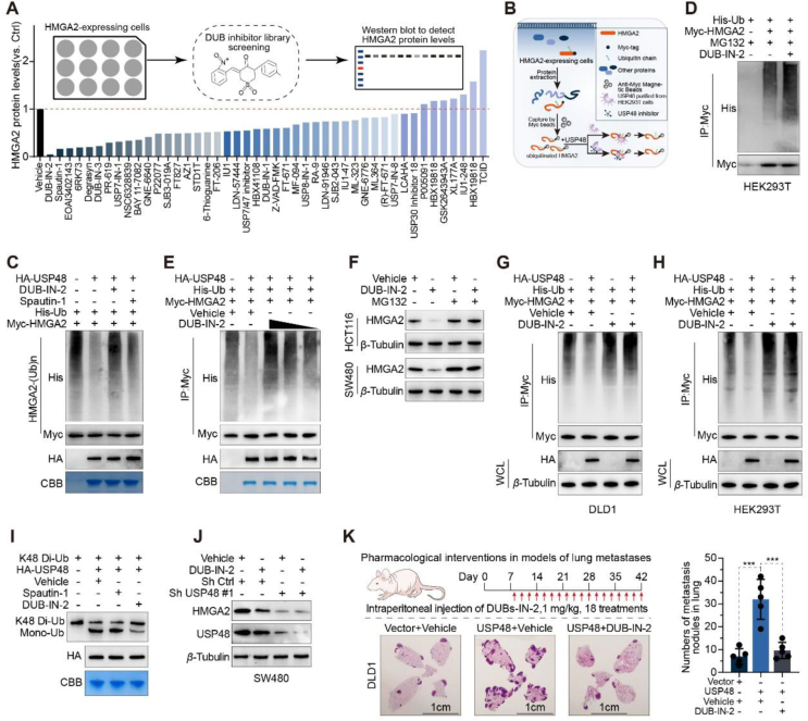
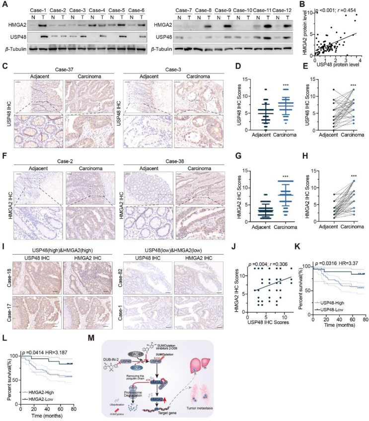
HMGA2, a pivotal transcription factor, functions as a versatile regulator implicated in the progression of diverse aggressive malignancies. In this study, mass spectrometry was employed to identify ubiquitin-specific proteases that potentially interact with HMGA2, and USP48 was identified as a deubiquitinating enzyme of HMGA2. The enforced expression of USP48 significantly increased HMGA2 protein levels by inhibiting its degradation, while the deprivation of USP48 promoted HMGA2 degradation, thereby suppressing tumor invasion and metastasis. We discovered that USP48 undergoes SUMOylation at lysine 258, which enhances its binding affinity to HMGA2. Through subsequent phenotypic screening of small molecules, we identified DUB-IN-2 as a remarkably potent pharmacological inhibitor of USP48. Interestingly, the small-molecule inhibitor targeting USP48 induces destabilization of HMGA2. Clinically, upregulation of USP48 or HMGA2 in cancerous tissues is indicative of poor prognosis for patients with colorectal cancer (CRC). Collectively, our study not only elucidates the regulatory mechanism of DUBs involved in HMGA2 stability and validates USP48 as a potential therapeutic target for CRC, but also identifies DUB-IN-2 as a potent inhibitor of USP48 and a promising candidate for CRC treatment.

Keywords: Colorectal cancer; HMGA2; Invasion and metastasis; Post-translational modification; SUMOylation; Specific inhibitors; USP48; Ubiquitination.


Conflict of interest statement

The authors have declared that no conflict of interest exists.

Figures

Image 1
Graphical abstract
Figure 1
Figure 1
USP48 interacts with HMGA2. (A) HMGA2-interacting proteins were detected by mass spectrometry using anti-Myc affinity magnetic beads, following transfection of Myc-HMGA2 into HCT116 cells for 36 h. (B–C) IP assays were performed on HCT116 and SW480 cell lysates using control IgG, anti-USP48, or anti-HMGA2 antibodies, followed by detection of resulting immunoprecipitates with appropriate antibodies. (D) HEK293T and DLD1 cells were transfected with Myc-HMGA2 alone or in combination with HA-tagged USP48-WT or USP48 CS, and cell lysates were analyzed by IP with anti-HA affinity magnetic beads followed by IB with antibodies against Myc and HA. (E) Purified HA-USP48 or HA-USP48 C98S from HEK293T cells was incubated with purified recombinant GST-HMGA2 or GST, respectively. The retained USP48 or USP48 C98S on sepharose was detected using the HA antibody. (F) HCT116, DLD1 and SW480 cells were stained with USP48 antibody (red) and HMGA2 antibody (green), then analyzed using confocal microscopy. Nuclei were counterstained with DAPI (blue). Scale bar, 10 μm. (G) In situ PLA was used to detect the interaction between endogenous USP48 and HMGA2 in HCT116 and DLD1 cells. Representative merged PLA and nuclei (DAPI) images from the experiments are shown, with upper panel scaled at 10 μm and lower panel at 5 μm. (H) HEK293T cells were co-transfected with Myc-HMGA2 and either full-length HA-tagged USP48 or its deletion mutants, followed by IP using anti-HA magnetic beads and IB with antibodies against HA and Myc. (I) HEK293T cells were co-transfected with Myc-HMGA2 and either full-length HA-tagged USP48 or its deletion mutants, followed by IP using anti-Myc magnetic beads and IB with antibodies against HA and Myc. (J) In situ PLA was employed to investigate the interaction between exogenous Myc-HMGA2 WT and HA-USP48 △USP in HEK293T cells, with a scale bar of 10 μm. (K) In situ PLA was employed to investigate the interaction between exogenous HA-USP48 WT and Myc-HMGA2 D1 in HEK293T cells, with a scale bar of 10 μm. (L) HEK293T cells were co-transfected with HA-USP48 and either full-length Myc-tagged HMGA2 or its deletion mutants, followed by IP using anti-Myc magnetic beads and IB with antibodies against HA and Myc. (M) HEK293T cells were co-transfected with HA-USP48 and either full-length Myc-tagged HMGA2 or its deletion mutants, followed by IP using anti-HA magnetic beads and IB with antibodies against HA and Myc. The presented panels depict representative outcomes from three independent experiments. The panels presented illustrate representative results obtained from three independent experiments.
Figure 2
Figure 2
USP48 maintains HMGA2 stability. (A) Increasing amounts of HA tagged USP48 were transfected into HEK293T cells along with equal amounts of Myc-HMGA2. Cell lysates were analyzed using IB and antibodies against Myc. “+” denotes 2.5 μg plasmid transfection, while “++” indicates 5 μg. (B) Two independent shRNAs were employed to silence USP48 expression in SW480 and HCT116 cells, followed by IB analysis of HMGA2 protein levels. (C) USP48-WT or USP48–C98S was overexpressed in DLD1 cells, followed by analysis of HMGA2 protein levels. (D) Equal amounts of Myc-HMGA2 and either USP48-WT or USP48–C98S were co-transfected into HEK293T cells, followed by IB analysis using Myc antibodies. (E) DLD1 cells were transduced with HA-tagged USP48-WT alone or in combination with USP48 shRNA, and HMGA2 levels were analyzed by IB analysis. (F) HCT116 cells were transduced with USP48 shRNA and either HA-tagged USP48-WT or USP48–C98S, followed by IB analysis to analyze HMGA2 levels. (G) qRT-PCR analysis was conducted to assess the expression of HMGA2 mRNA in SW480 cells with depleted endogenous USP48 via two independent shRNA. (H) The expression of HMGA2 mRNA was assessed by qRT-PCR in DLD1 cells following transduction with either vector control or HA-USP48. (I) qRT-PCR was performed to evaluate the expression levels of Slug, IGF2BP2 and SOX2 in SW480 cells transduced with two distinct HMGA2 shRNA constructs. (J) The expression levels of Slug, IGF2BP2 and SOX2 were quantified by qRT-PCR in SW480 cells that had been transduced with USP48 shRNA. (K) SW480 cells transfected with two independent USP48 shRNA were treated with DMSO, MG132 (10 μmol/L), or CQ (20 mmol/L) for 6 h, followed by IB analysis of USP48 and HMGA2. (L) DLD1 cells transfected with vector control, or HA-USP48 were treated with DMSO or MG132 (10 μmol/L) for 6 h, and then USP48 and HMGA2 were analyzed. (M) DLD1 cells were transfected with vector control, HA-USP48 or USP48-C98S, treated with 50 μg/mL of CHX, collected at the indicated times, and then subjected to IB analysis with antibodies against USP48 and HMGA2. Quantification of HMGA2 levels relative to β-Tubulin is shown. (N) SW480 cells with stable expression of either control shRNA or USP48 shRNA were treated with 50 μg/mL CHX, harvested at indicated time points, and analyzed by IB using antibodies against HMGA2 and USP48. Quantification of HMGA2 levels relative to β-Tubulin is presented as mean ± SD. (O) HCT116 stably expressing control shRNA or USP48 shRNA were treated with 50 μg/mL CHX, harvested at the indicated times, and then subjected to IB with antibodies against HMGA2 and USP48. Quantification of HMGA2 levels relative to β-Tubulin is shown. One-way ANOVA test (G, I, J, M‒O). Unpaired two-tailed Student's t-test (H). All experiments were performed independently at least three times. ∗∗∗P < 0.001, ns indicates no statistical significance.
Figure 3
Figure 3
USP48 deubiquitinates HMGA2. (A) HEK293T cells were co-transfected with Myc-HMGA2, His-Ub, and either HA-tagged USP48-WT or USP48-C98S. Cell lysates were then subjected to IP using anti-Myc magnetic beads followed by IB analysis with antibodies against His, HA, and Myc. Prior to harvesting, the cells were treated with 10 μmol/L MG132 for 6 h. (B) Increasing amounts of HA-tagged USP48 and equal amounts of Myc-HMGA2 were transfected into the cells, followed by IP with Myc antibodies and IB analysis with anti-His antibodies to detect ubiquitinated HMGA2 after treatment with MG132 for 6 h. (C) DLD1 cells were transfected with USP48-WT or USP48-C98S. After 48 h, the cells were treated with MG132 for 6 h. The whole-cell lysates were subjected to IP with HMGA2 antibodies and IB analysis with anti-Ub antibodies to detect ubiquitinated HMGA2. (D) SW480 cells were transfected with shRNA as indicated, and lysates were subjected to IP using HMGA2 antibody, followed by IB analysis with antibodies against Ub and HMGA2. (E) HCT116 cells were transfected with shRNA as indicated, and lysates were subjected to IP using HMGA2 antibody, followed by IB analysis with antibodies against Ub and HMGA2. Cells were treated with 10 μmol/L MG132 for 6 h prior to harvesting. Before IP, lysates were denatured at 95 °C for 5 min in the presence of 1% SDS, then diluted tenfold with lysis buffer and sonicated. (F) The ubiquitinated Myc-HMGA2 protein was subjected to in vitro deubiquitination assay using USP48-WT or USP48-C98S, and the reaction mixes were analyzed by IB analysis. (G) Myc-HMGA2 was co-transfected with either USP48-WT or USP48 deletion mutants into HEK293T cells. Whole-cell lysates were subjected to IP using Myc antibody, followed by IB analysis with anti-His antibodies after treatment with MG132 for 6 h to detect ubiquitinated HMGA2. (H) De-ubiquitination assay of HMGA2 WT and mutant domains (D1‒D4) was performed in HEK293T cells co-transfected with His-Ub and HA-USP48, followed by treatment with 10 μmol/L MG132 for 6 h. (I) De-ubiquitination assay of HMGA2 in HEK293T cells co-transfected with His-Ub, HA-USP48, Myc-HMGA2-K26R, and Myc-HMGA2-K34R and treated with 10 μmol/L MG132 for 6 h. (J) De-ubiquitination assay of HMGA2 in HEK293T cells co-transfected with His-Ub, HA-USP48, Myc-HMGA2-K26R and treated with 10 μmol/L MG132 for 6 h. (K) Myc-HMGA2, HA-USP48 and His-Ub plasmids with different ubiquitin linkages (K6, K11, K27, K29, K33, K48 or K63) were co-transfected into HEK293T cells to analyze HMGA2 ubiquitination. The data shown in all panels are representative of three independent experiments.
Figure 4
Figure 4
Ablation of USP48 expression impairs the metastatic potential of CRC cells. (A) Wound-healing assay of DLD1 cells transfected with USP48-WT, USP48-C98S, or vector control. Migration rates in each group were quantified by measuring four different wound areas. (B–G) USP48 knockdown was performed in HCT116 and SW480 cells, with or without HMGA2 overexpression, followed by quantification of migration rates through measurement of five distinct wound areas (B, C). The relative number of migrated cells in each group was determined by counting the cells in five random fields of view (D–G). (H–J) USP48 was upregulated in DLD1 cells with or without HMGA2 knockdown. The relative number of migrated cells was quantified by counting the cells in five random fields of view (H, I), while migration rates were determined by measuring five different wound areas (J). (K–L) USP48 knockdown was conducted in HCT116 cells, with or without HMGA2 overexpression. Representative images of lung tissue and HE-stained sections are presented in K, while the number of metastatic nodules in each group was calculated (K). Representative images of liver tissue and HE-stained sections are shown in (L), along with the calculation of metastatic nodules for each group (L). (M–N) USP48 was upregulated in DLD1 cells with or without HMGA2 knockdown. Representative images of the lung and HE-staining sections are shown in (M), and the metastasis nodules in each group were calculated (M). Representative images of the liver and HE-staining sections are shown in N, and the metastasis nodules in each group were calculated (N). The Scale bars were shown in (K–N). Data are presented as mean ± SD. One-way ANOVA test (A–N). All in vitro experiments were performed independently at least three times. ∗∗∗P < 0.001.
Figure 5
Figure 5
USP48 is SUMOylated by SUMO1 at Lys258. (A) Mass spectrometry detection of USP48-interacting proteins (obtained from anti-HA affinity magnetic beads) after HCT116 cells were transfected with HA-USP48 for 36 h. (B) Mass-spectrometry analysis of a HDAC4 peptide in HA-USP48 precipitate. (C) DLD1 cells were co-transfected with His-SUMO1, His-SUMO2, or His-SUMO3, with the HA-USP48 in the presence and absence of ubiquitin-conjugating enzyme 9 (V5-Ubc9), and cell lysates were subjected to Ni-NTA magnetic beads pulldown followed by IB with antibodies against His, HA, and V5. (D–E) IB analysis of total lysates, anti-His immunoprecipitates (D) and Ni-NTA magnetic beads pulldown products (E) of HCT116 cells transfected with control vector or His-SUMO1 as indicated. The intensity of total USP48 band was quantitated and shown. (F) IB analysis of total lysates and anti-HA immunoprecipitates of HEK293T cells transfected with HA-USP48 and Myc-tagged PIAS1, PIAS2, PIAS3, PIAS4, HDAC4, or HDAC7 constructs, as indicated. (G) IB analysis of total lysates and anti-HA immunoprecipitates of HEK293T cells transfected with HA-USP48 and flag-tagged SENP1, SENP2, SENP3, SENP5, SENP6, or SENP7 constructs, as indicated. (H) IB analysis of total lysates and Ni-NTA magnetic beads pulldown products of HEK293T cells transfected with HA-USP48, His-SUMO1 and Myc-HDAC4 or Myc-HDAC7 constructs, as indicated. (I–J) IB analysis of total lysates, anti-HA immunoprecipitates (I) and Ni-NTA magnetic beads pulldown products (J) of HEK293T cells transfected with HA-USP48, His-SUMO1 and Flag-SENP5 constructs, as indicated. (K) A schematic representation of the five predicted SUMO-conjugation motifs (ψKXE) in the USP48 protein. Down-hand side, bioinformatics analysis of USP48 using three independent computational programmes to detect SUMOylation sites (SUMOplotTM Analysis Programme (Abgent), JASSA and GPS-SUMO). (L) IB analysis of total lysates and Ni-NTA magnetic beads pulldown products of HEK293T cells transfected with V5-Ubc9, His-SUMO1, and HA-USP48 WT or USP48 SUMO mutants (USP48 K258R, USP48 K359R, USP48 K468R, USP48 K898R, and USP48 K1003R), as indicated.
Figure 6
Figure 6
The function of USP48 on HMGA2 is significantly enhanced by SUMOylation at Lys258. (A) IB analysis of total lysates and anti-HA (left panel) or anti-Myc immunoprecipitates (right panel) from DLD1 cells transfected with control vector or Flag-SENP5. (B) IB analysis of total lysates and anti-HA immunoprecipitates derived from DLD1 cells transfected with the control vector, Myc-HDAC4, or Myc-HDAC7, as indicated. (C) IB analysis of total lysates and anti-Myc immunoprecipitates derived from HEK293T cells transfected with HA-USP48 WT or K258R and Myc-HMGA2, as indicated. (D–E) Representative images are shown with merged PLA and DAPI channels from PLA experiments. Scale bar, 10 μm. Each red dot represents the detection of the USP48–HMGA2 interaction complex, and the graphs representing mean ± SD are shown in (E). (F–G) HEK293T cells were transfected with HA-USP48 WT or K258R. After fixation, in situ PLA for USP48-HMGA2 was performed with anti-HA and anti-Myc antibodies. Scale bar, 10 μm. Each red dot represents the detection of the USP48-HMGA2 interaction complex, and the graphs representing mean ± SD are shown in (G). (H) IB analysis of total lysates and anti-Myc immunoprecipitates of HEK293T cells transfected with His-Ub, HA-USP48 WT/K258R and Myc-HMGA2 and treated with MG132 (10 μmol/L for 6 h), as indicated. (I–J) IB analysis of total lysates and anti-Myc immunoprecipitates of HEK293T cells transfected with Myc-HMGA2, His-Ub, HA-USP48, and control vector, Flag-SENP5 (I), Myc-HDAC4 (J), or Myc-HDAC7 (J) and treated with MG132 (10 μmol/L for 6 h), as indicated. (K) DLD1 cells were transfected with specific plasmids, as indicated and treated with CHX (50 μg/mL) for the indicated time points. Quantification of HMGA2 levels relative to β-Tubulin is shown. (L) DLD1 cells stably expressing USP48 shRNA were transfected with indicated plasmids and treated with 200 μmol/L 2-D08 or DMSO for 24 h. Graphic representation of the migration ability from cells described above was examined by transwell migration assay. Scale bar, 200 μm. (M) Representative images of lung sections were stained with HE, and metastatic nodules were calculated in each group. Four-week-old BALB/c nude mice were injected intravenously with 2 × 106 cells as in (I). 2-D08 (4 mg/kg) or vehicle was injected intraperitoneally on Day 8 and every other day for 5 weeks. Scale bar, 1 cm. Data are presented as mean ± SD. One-way ANOVA test (E). Unpaired two-tailed Student's t-test (G). All experiments were performed independently at least three times. ∗∗∗P < 0.001, ns indicates no statistical significance.
Figure 7
Figure 7
DUB-IN-2 hinders the deubiquitinating activity of USP48 and induces the degradation of HMGA2. (A) Relative protein level of HMGA2 in the screening of DUBs inhibitors. The SW480 cells were treated with the library of 45 reported DUB inhibitors at a concentration of 2.5 μmol/L for 12 h as indicated, and then, the protein levels of HMGA2 were assessed by Western blotting. (B) Schematic diagram of USP48 activity detection after treatment with compounds in vitro. (C) Ubiquitinated HMGA2 was immunoprecipitated from HEK293T cells transfected with Myc-HMGA2 and His-Ub plasmids with after 10 μmol/L MG132 treatment. Purified USP48 from HEK293T cells overexpressing HA-USP48 was pretreated with DUB inhibitors for 1 h and subsequently incubated with ubiquitinated HMGA2 for 6 h. Then, the ubiquitylation level of HMGA2 was evaluated. (D) HEK293T cells were co-transfected with MYC-HMGA2 and His-ubiquitin, and then treated with 2.5 μmol/L DUB-IN-2 24 h. After pretreatment with MG132 (10 μmol/L) for 6 h, anti-Myc beads were used for IP, and anti-Myc and His antibodies were used for IB analysis. (E) The deubiquitinating effect of USP48 on HMGA2 upon treatment with different concentrations of DUB-IN-2. HA-USP48 was exposed to 5, 2.5 and 1 μmol/L DUB-IN-2 and then incubated with ubiquitinated HMGA2. The ubiquitylation level of HMGA2 was measured. (F) IB analysis of HMGA2 and β-Tubulin in HCT116 and SW480 cells pretreated with 2.5 μmol/L DUB-IN-2 24 h and then treated with MG132 (10 μmol/L) for 6 h. (G, H) DLD1 and HEK293T cells were co-transfected with MYC-HMGA2, HA-USP48 and His-ubiquitin, and then treated with 2.5 μmol/L DUB-IN-2 24 h. After pretreatment with MG132 (10 μmol/L) for 6 h, anti-Myc beads were used for IP, and anti-Myc, anti-HA and His antibodies were used for IB analysis. (I) Purified HA-USP48 was pretreated with Spautin-1 (2.5 μmol/L) or DUB-IN-2 (2.5 μmol/L) for 10 min and then incubated with K48-linked Di-ubiquitin in the presence of the compound at 37 °C for 1.5 h. Samples were then analyzed by IB analysis with a ubiquitin-specific antibody. (J) IB analysis of HMGA2 and β-Tubulin in control and USP48-knockdown SW480 cells treated with 2.5 μmol/L DUB-IN-2 24 h. (K) Representative images of lung sections were stained with HE, and metastatic nodules were calculated in each group. Four-week-old BALB/c nude mice were injected intravenously with 2 × 106 DLD1 cells with high USP48 expression. DUB-IN-2 (1 mg/kg) or vehicle was injected intraperitoneally on Day 8 and every other day for 5 weeks. Scale bar, 1 cm ∗∗∗P < 0.001.
Figure 8
Figure 8
Elevated levels of USP48 are positively associated with the upregulation of HMGA2 and serve as a predictor for an unfavorable prognosis in patients with CRC. (A) Cell lysates from 42 pairs CRC tissues and matched adjacent tissues were blotted with USP48 and HMGA2 antibodies. (B) Correlation analysis of USP48 and HMGA2 in CRC tissues and matched adjacent tissues. Statistical analyses were performed using the chi-square test. Pearson r indicates the correlation coefficient. (C–E) Representative images of USP48 highly expressed in CRC tissues and IHC score of USP48 in CRC and matched adjacent tissue, respectively. Data are presented as mean ± SD. The comparison between cancer tissues and adjacent or matched adjacent tissues can be conducted using either a Wilcoxon unpaired test or paired t-test. (F–H) Representative images of HMGA2 highly expressed in CRC tissues and IHC score of HMGA2 in CRC and matched adjacent tissue, respectively. Data are presented as mean ± SD. The comparison between cancer tissues and adjacent or matched adjacent tissues can be conducted using either a Wilcoxon unpaired test or paired t-test. (I) Representative images showing the simultaneous high or low expression of both USP48 and HMGA2 in CRC tissues with a scale bar of 100 μm. (J) USP48 protein scores (X axis) in CRC positively correlate with HMGA2 protein scores (Y axis) in individual patients. The P-value was calculated from a linear regression analysis. r is the correlation coefficient. (K–L) Kaplan–Meier curves of the overall survival of CRC patients (90 patients), stratified by USP48 (K) or HMGA2 (L) protein levels. The P-value was calculated from a log-rank test. HR, hazard ratio. (M) Working model of SUMOylated USP48-mediated HMGA2 deubiquitination. ∗∗∗P < 0.001.

References

    1. Keum N., Giovannucci E. Global burden of colorectal cancer: emerging trends, risk factors and prevention strategies. Nat Rev Gastroenterol Hepatol. 2019;16:713–732. - PubMed
    1. Ferlay J., Soerjomataram I., Dikshit R., Eser S., Mathers C., Rebelo M., et al. Cancer incidence and mortality worldwide: sources, methods and major patterns in globocan 2012. Int J Cancer. 2015;136:E359–E386. - PubMed
    1. Harrigan J.A., Jacq X., Martin N.M., Jackson S.P. Deubiquitylating enzymes and drug discovery: emerging opportunities. Nat Rev Drug Discov. 2018;17:57–78. - PMC - PubMed
    1. Hoeller D., Dikic I. Targeting the ubiquitin system in cancer therapy. Nature. 2009;458:438–444. - PubMed
    1. Senft D., Qi J., Ronai Z.A. Ubiquitin ligases in oncogenic transformation and cancer therapy. Nat Rev Cancer. 2018;18:69–88. - PMC - PubMed

LinkOut - more resources