. 2024 Apr 4;134(11):e171002.
doi: 10.1172/JCI171002.

Neutrophil-mediated innate immune resistance to bacterial pneumonia is dependent on Tet2 function

Affiliations

Neutrophil-mediated innate immune resistance to bacterial pneumonia is dependent on Tet2 function

Candice Quin et al. J Clin Invest. .

Abstract

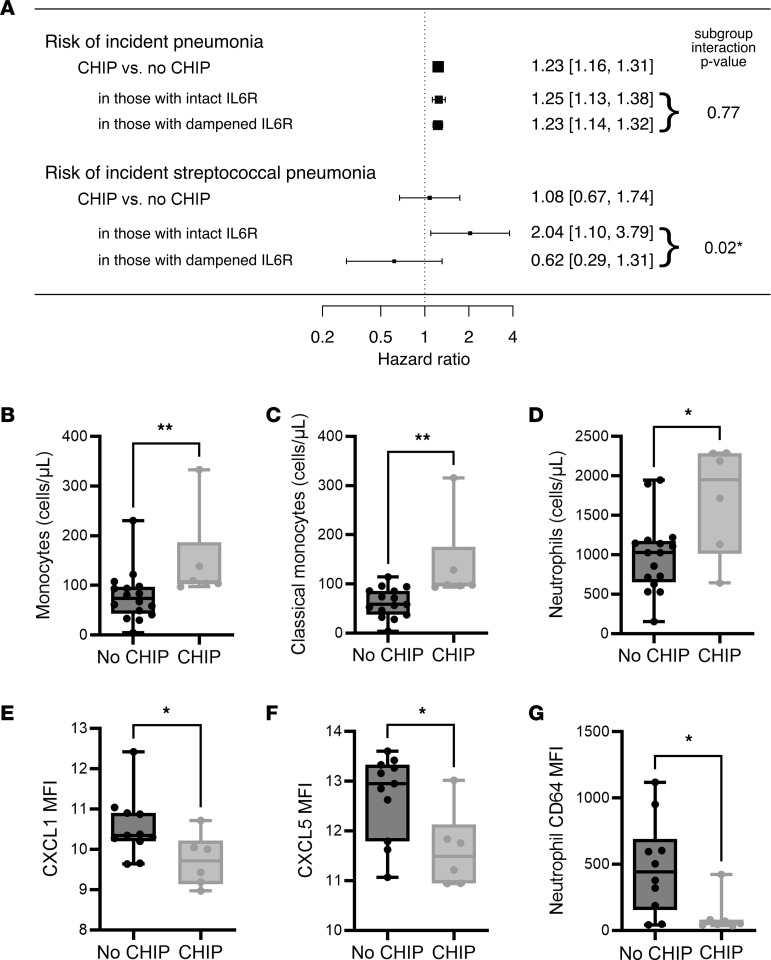
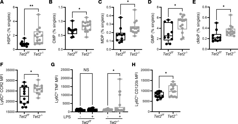
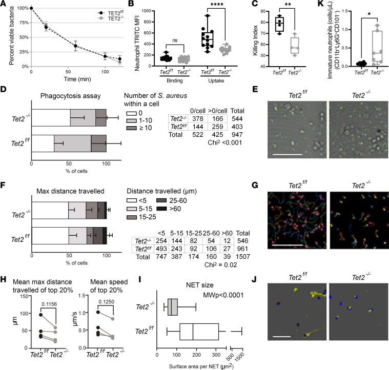
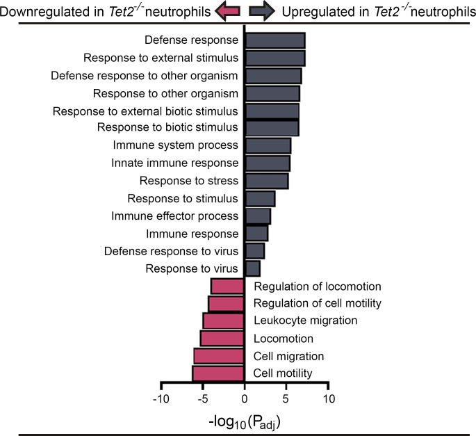
Individuals with clonal hematopoiesis of indeterminate potential (CHIP) are at increased risk of aging related health conditions and all-cause mortality, but whether CHIP affects risk of infection is much less clear. Using UK Biobank data, we revealed a positive association between CHIP and incident pneumonia in 438,421 individuals. We show that inflammation enhanced pneumonia risk, as CHIP carriers with a hypomorphic IL6 receptor polymorphism were protected. To better characterize the pathways of susceptibility, we challenged hematopoietic Tet Methylcytosine Dioxygenase 2-knockout (Tet2-/-) and floxed control mice (Tet2fl/fl) with Streptococcus pneumoniae. As with human CHIP carriers, Tet2-/- mice had hematopoietic abnormalities resulting in the expansion of inflammatory monocytes and neutrophils in peripheral blood. Yet, these cells were insufficient in defending against S. pneumoniae and resulted in increased pathology, impaired bacterial clearance, and higher mortality in Tet2-/- mice. We delineated the transcriptional landscape of Tet2-/- neutrophils and found that, while inflammation-related pathways were upregulated in Tet2-/- neutrophils, migration and motility pathways were compromised. Using live-imaging techniques, we demonstrated impairments in motility, pathogen uptake, and neutrophil extracellular trap (NET) formation by Tet2-/- neutrophils. Collectively, we show that CHIP is a risk factor for bacterial pneumonia related to innate immune impairments.

Keywords: Immunology; Infectious disease; Innate immunity.


Conflict of interest statement

Conflict of interest: The authors have declared that no conflict of interest exists.

Figures

Figure 1
Figure 1. CHIP is positively associated with incident pneumonia caused by Streptococcus pneumoniae.
(A) CHIP is positively associated with incident all-cause pneumonia among 438,421 individuals in the UK Biobank without a history of pneumonia in a Cox proportional hazards regression model adjusted for age, age2, sex, smoking history, history of chronic inflammatory lung disease (COPD), and 10 principal components of genetic ancestry. In a model adding an interaction term for a common SNP in the IL6 receptor associated with lower IL6 signaling (rs2228415), CHIP is associated with incident confirmed S. pneumoniae pneumonia. The CHIP × rs2228415 interaction term is significantly below 0, suggesting that lower IL6R signaling mitigates the effects of CHIP on pneumonia risk. (BG) Leukocyte populations were quantified in whole blood from CHIP carriers and noncarriers in a local cohort using flow cytometry. Compared with noncarriers, the numbers of (B) peripheral blood monocytes (No CHIP [78.4 ± 12.9]; CHIP [147.6 ± 37.5]), (C) classical monocytes (No CHIP [62.4 ± 7.7]; CHIP [138.3 ± 35.9]), and (D) neutrophils (No CHIP [990 ± 117]; CHIP [1709 ± 281]), were increased in CHIP carriers. Chemokines (E) CXCL1 (No CHIP [10.5 ± 0.23]; CHIP [9.7 ± 0.26]), and (F) CXCL5 (No CHIP [12.7 ± 0.25]; CHIP [11.6 ± 0.32]), were decreased in the sera of CHIP carriers. (G) Surface expression of CD64 was decreased on circulating blood neutrophils in CHIP carriers (No CHIP [474.9 ± 113.5]; CHIP [106.6 ± 53.0]). Data are presented as box and whisker plots, minimum to maximum, where the center line represents the median and each dot is a participant. Sample size: 16 No CHIP, 6 CHIP participants. Significant outliers removed using ROUT method. MFI, Geometric Mean Fluorescence Intensity. Significance was assessed by Mann-Whitney test. *P ≤ 0.05, **P ≤ 0.01.
Figure 2
Figure 2. Expansion of inflammatory monocytes in peripheral blood following TET2 loss.
(A) Flow cytometric analysis of peripheral blood revealed an increase in the number (Tet2fl/fl [111 ± 13.07], n = 21; Tet2–/– [157.5 ± 15], n = 24) and proportion (Tet2fl/fl [9.85 ± 0.89]; Tet2–/– [15.43 ± 1.66]) of monocytes in Tet2–/– mice. (B) There was a tendency toward increase in the number (Tet2fl/fl [135 ± 12.7], n = 21; Tet2–/– [177.4 ± 18.58], n = 24) and relative proportion (Tet2fl/fl [12.3 ± 0.78]; Tet2–/– [14.4 ± 1.34]) of circulating neutrophils in Tet2–/– mice. (C) The proportion of Ly6Chi inflammatory monocytes, as a proportion of total CD45+ leukocytes, increased in the circulation of Tet2–/– mice (6.63 ± 0.81) compared with Tet2fl/fl mice (4.60 ± 0.39). (D) This corresponded with a decrease in the surface expression of CX3CR1 (Tet2fl/fl [20,657 ± 945.6]; Tet2–/– [17,057 ± 1355]). (E) Intracellular staining of TNF revealed higher TNF expression in the peripheral blood of Tet2–/– mice (4,050 ± 662) following 4-hour stimulation with LPS, compared with Tet2fl/fl mice (2,202 ± 566.4) (F) Results from a multiplex-ELISA showed that whole blood from Tet2–/– mice had a significant increase in TNF (Tet2fl/fl [147 ± 24.3]; Tet2–/– [357 ± 71.2]), and (G) IL6 (Tet2fl/fl [225 ± 26.3]; Tet2–/– [553 ± 119]) compared with Tet2fl/fl mice, following 4 hour stimulation with LPS ex vivo. (H) There was a significant induction of IL1-β in whole blood from Tet2–/– mice following LPS stimulation (4.48 ± 1.3) that was not observed in blood stimulated from Tet2fl/fl mice (2.3 ± 0.7). Data are presented as box and whisker plots, minimum to maximum, where the center line represents the median and each dot is a mouse. MFI, Geometric Mean Fluorescence Intensity. Significance was assessed by Kruskal-Wallis test. *P ≤ 0.05, **P ≤ 0.01.
Figure 3
Figure 3. Mutations in Tet2 increase the proportion of myeloid progenitor cells in the bone marrow.
Flow cytometry analysis of the hematopoietic compartment showed an increase in the relative proportion of (A) hematopoietic stem and progenitor cells (HSPC) (Tet2fl/fl [0.83 ± 0.10], n = 10; Tet2–/– [1.95 ± 0.30], n = 14), (B) common myeloid progenitor (CMP) (Tet2fl/fl [0.66 ± 0.05]; Tet2–/– [0.82 ± 0.04]), (C) monocyte-dendritic progenitor (MDP) (Tet2fl/fl [0.17 ± 0.02]; Tet2–/– [0.25 ± 0.02]), (D) granulocyte-monocyte progenitors (GMP) (Tet2fl/fl [3.06 ± 0.54]; Tet2–/– [4.82 ± 0.40]), (E) and common monocyte progenitors (cMoP) (Tet2fl/fl [0.24 ± 0.03]; Tet2–/– [0.35 ± 0.02]) in Tet2–/– mice compared with Tet2fl/fl mice. (F) Inflammatory Ly6Chi monocytes within the bone marrow of Tet2–/– mice had higher expression of the surface C-C chemokine receptor type 2 (CCR2) (Tet2fl/fl [22695 ± 1222]; Tet2–/– [25971 ± 656]), compared with Tet2fl/fl mice. (G) These monocytes were hyper-responsive to ex vivo stimulation with LPS and had a significant induction of intracellular TNF expression, whereas monocytes from Tet2fl/fl did not. (H) Expression of the cell surface TNF receptor, CD120b, was increased on mutant-TET2 Ly6Chi monocytes (Tet2fl/fl [8055 ± 412]; Tet2–/– [10013 ± 746]) in the bone marrow. Data are presented as box and whisker plots, minimum to maximum, where the center line represents the median and each dot is a mouse. MFI, Geometric Mean Fluorescence Intensity. Significance was assessed by Mann-Whitney test. *P ≤ 0.05, **P ≤ 0.01.
Figure 4
Figure 4. Pneumococcal pneumonia–induced sepsis and accompanying inflammatory responses are exacerbated in Tet2–/– mice.
(A) Experimental timeline of infection and concurrent survival (n = 13 Tet2–/–; n = 12 Tet2fl/fl). (B) Representative H&E-stained lung sections of mice at 10 days p.i. with S. pneumoniae. Original magnification 20-fold. (C) Histopathological analysis of lung H&E tissue sections [as shown in (B)] attained by 2 blinded scorers. (D) Counts and representative IHC staining of mononuclear phagocytes (F4/80+) and neutrophils (Ly6G+) on lung sections at 10 days p.i. Original magnification 200-fold. Tet–/– mice had increased numbers of mononuclear phagocytes in the lungs (Tet2fl/fl [27 ± 1]; Tet2–/– [35 ± 3.6]). In contrast, neutrophils were decreased (Tet2fl/fl [49 ± 2.3]; Tet2–/– [36 ± 4.2]). (E) Relative frequency, as a percentage of 100 cells counted via LeukoSpins, of neutrophils and monocytes in circulation 10 days p.i. showed an increase in neutrophils (Tet2fl/fl [20.9 ± 2.1]; Tet2–/– [46.9 ± 10.5]) and monocytes (Tet2fl/fl [4.9 ± 0.7]; Tet2–/– [7.6 ± 2.2]) in Tet2–/– mice. Representative images are shown. (F) Surface expression of CCR2 is decreased on peripheral blood neutrophils in Tet2–/– mice (Tet2fl/fl [4328 ± 449, n = 19]; Tet2–/– [2875 ± 407], n = 18), at steady-state. (G) Relative expression of CCL2 is lower in the lungs of Tet2–/– at steady-state (Tet2fl/fl [1.96 ± 0.35], n = 8; Tet2–/– [0.87 ± 0.17], n = 16). (H) Enumeration of CFUs in lungs and complete nasal turbinate (CNT) 10 days p.i. showed increased pathogen burden in the CNT of Tet2–/– mice (Tet2fl/fl [3.1 ± 0.1]; Tet2–/– [3.6 ± 0.22]). (I) Results from a simple linear regression between CFUs in the CNT at 10 days p.i. and whole blood inflammatory mediators showed a positive association between inflammation and pathogen burden, with a greater relationship in Tet2–/– mice. Shaded area represents 95% confidence intervals. * P < 0.05
Figure 5
Figure 5. Loss of Tet2 impairs bactericidal capacity of neutrophils.
(A) There were no differences in bacterial killing between Tet2fl/fl and Tet2–/– BM-derived macrophages. (B) Bacterial binding and uptake, measured with pHrodo-Red-labeled Streptococcus pneumoniae, showed decreased pathogen uptake in Tet2–/– neutrophils (Tet2fl/fl [500 ± 14.6]; Tet2–/– [296 ± 14.1]) compared with Tet2fl/fl neutrophils. (C) Intracellular killing of engulfed S. pneumoniae was reduced in Tet2–/– neutrophils (Tet2fl/fl [78.5 ± 3.5]; Tet2–/– [58.3 ± 4.1]). (DJ) Functional assays comparing Tet2fl/fl versus Tet2–/– neutrophils over 30 minutes coculture with Staphylococcus aureus. n = 4 independent experiments each. (D) Phagocytosis of S. aureus was impaired in Tet2–/– neutrophils compared with Tet2fl/fl neutrophils. Mean ± SD of percentages of cells with certain counts of internalized bacteria. Significance tested with χ2 test. (E) Representative image showing GFP-labeled bacteria (green) at end of 30 minutes coincubation with neutrophils. Scale bar: 100 μm. (FH) Migration qualities of Tet2–/– neutrophils in response to S. aureus were impaired compared with Tet2fl/fl neutrophils. (F) Mean ± SD of percentages of cells travelling certain max distances. Significance tested with χ2 test. (G) Representative tracks of cells over 30 minutes coincubation with S. aureus. Scale bar: 100 μm. (H) Mean maximum distances travelled (left) and mean speeds of top 20% of cells (right). Significance tested with paired t test and Wilcoxon, respectively. (I) Neutrophil extracellular traps (NETs) were less expansive in Tet2–/– (72 μm2 [range 54–106]) versus. Tet2fl/fl neutrophils (183 μm2 [range 115–312]). Boxes represent median [IQR] of individual NETs quantified, with minimum to maximum whiskers; Mann-Whitney test. (J) Representative image of NETs stained for dsDNA (Alexa568, yellow), nuclear DNA, (DAPI, blue), mitochondria (MitoTracker, red), S. aureus (GFP, green). Scale bar: 100 μm. (K) Immature neutrophil counts were higher in the circulation of Tet2–/– mice (0.45 ± 0.16; n = 7) versus Tet2fl/fl mice (0.07 ± 0.01; n = 7).
Figure 6
Figure 6. RNA-Seq of neutrophils isolated from Tet2–/– mice.
Gene pathways related to immunity and defense were upregulated in neutrophils isolated from Tet2–/– mice, whereas pathways related to motility and locomotion were downregulated.

Comment in

  • CHIPing away at immunity: the role of clonal hematopoiesis of indeterminate potential in bacterial pneumonia

References

    1. Groarke EM, Young NS. Aging and hematopoiesis. Clin Geriatr Med. 2019;35(3):285–293. doi: 10.1016/j.cger.2019.03.001. - DOI - PMC - PubMed
    1. Cooper JN, Young NS. Clonality in context: hematopoietic clones in their marrow environment. Blood. 2017;130(22):2363–2372. doi: 10.1182/blood-2017-07-794362. - DOI - PMC - PubMed
    1. Steensma DP, et al. Clonal hematopoiesis of indeterminate potential and its distinction from myelodysplastic syndromes. Blood. 2015;126(1):9–16. doi: 10.1182/blood-2015-03-631747. - DOI - PMC - PubMed
    1. Vlasschaert C, et al. Association of clonal hematopoiesis of indeterminate potential with worse kidney function and anemia in two cohorts of patients with advanced chronic kidney disease. J Am Soc Nephrol. 2022;33(5):985–995. doi: 10.1681/ASN.2021060774. - DOI - PMC - PubMed
    1. Jaiswal S, Ebert BL. Clonal hematopoiesis in human aging and disease. Science. 2019;366(6465):eaan4673. doi: 10.1126/science.aan4673. - DOI - PMC - PubMed

MeSH terms