. 2024 Apr 4;19(4):e0301175.
doi: 10.1371/journal.pone.0301175. eCollection 2024.

Selection, engineering, and in vivo testing of a human leukocyte antigen-independent T-cell receptor recognizing human mesothelin

Affiliations

Selection, engineering, and in vivo testing of a human leukocyte antigen-independent T-cell receptor recognizing human mesothelin

Martyn J Hiscox et al. PLoS One. .

Abstract

Background: Canonical α/β T-cell receptors (TCRs) bind to human leukocyte antigen (HLA) displaying antigenic peptides to elicit T cell-mediated cytotoxicity. TCR-engineered T-cell immunotherapies targeting cancer-specific peptide-HLA complexes (pHLA) are generating exciting clinical responses, but owing to HLA restriction they are only able to target a subset of antigen-positive patients. More recently, evidence has been published indicating that naturally occurring α/β TCRs can target cell surface proteins other than pHLA, which would address the challenges of HLA restriction. In this proof-of-concept study, we sought to identify and engineer so-called HLA-independent TCRs (HiTs) against the tumor-associated antigen mesothelin.

Methods: Using phage display, we identified a HiT that bound well to mesothelin, which when expressed in primary T cells, caused activation and cytotoxicity. We subsequently engineered this HiT to modulate the T-cell response to varying levels of mesothelin on the cell surface.

Results: The isolated HiT shows cytotoxic activity and demonstrates killing of both mesothelin-expressing cell lines and patient-derived xenograft models. Additionally, we demonstrated that HiT-transduced T cells do not require CD4 or CD8 co-receptors and, unlike a TCR fusion construct, are not inhibited by soluble mesothelin. Finally, we showed that HiT-transduced T cells are highly efficacious in vivo, completely eradicating xenografted human solid tumors.

Conclusion: HiTs can be isolated from fully human TCR-displaying phage libraries against cell surface-expressed antigens. HiTs are able to fully activate primary T cells both in vivo and in vitro. HiTs may enable the efficacy seen with pHLA-targeting TCRs in solid tumors to be translated to cell surface antigens.


Conflict of interest statement

This study was funded by Adaptimmune. Adaptimmune contributed to study design, data analysis, decision to publish, and preparation of the manuscript. Writing and editorial support was funded by Adaptimmune. All authors are employees of Adaptimmune or were at the time of the study. Authors may hold stock in Adaptimmune. This does not alter our adherence to PLOS ONE policies on sharing data and materials

Figures

Fig 1
Fig 1. Immunomodulatory molecules that can be added to T cells for adoptive T-cell therapies.
CAR T cells use an antibody fragment (such as a single-chain variable fragment as shown here) as the binding domain—which is fused to CD3ζ and often contains additional co-stimulatory domains—that targets a cell surface protein. TRuC T cells also use an antibody fragment (such as a single-domain antibody as shown here) that targets a cell surface protein; however, this antibody fragment is fused to another part of the TCR:CD3 complex—shown here, it is fused to the CD3ε, using natural T-cell signaling pathways. FvTCR T cells replace the variable domains of the TCR with the light and heavy chains of the antibody. HiT T cells use TCR binding domains to target cell surface proteins directly and therefore use natural cell signaling pathways. TCR T cells use a TCR binding domain to specifically target a peptide presented by the HLA molecule. CAR, chimeric antigen receptor; FvTCR, variable fragment T-cell receptor; HiT, human leukocyte antigen–independent T-cell receptor; HLA, human leukocyte antigen; TCR, T-cell receptor; TRuC, T-cell receptor fusion construct.
Fig 2
Fig 2. Initial characterization of a HiT against human mesothelin.
(A) Representative example of surface plasmon resonance data. Equilibrium binding with a fit to a 1:1 Langmuir equation (one of four independent experiments is shown). (B) Epitope analysis of T-cell receptor binding to full-length (M7) and truncated (M1–6, MN1–4) mesothelin. Reduced binding is defined as a reduction in affinity (greater than five-fold increase in KD) or reduction in maximal binding response (maximal binding response less than 10% of the theoretical maximum based on the amount of immobilized mesothelin). (C) ntd 2B9 Jurkat T cells (left column) and mesothelin HiT–transduced Jurkat T cells (right column) were analyzed for CD3 expression, demonstrating successful transduction. (D) Jurkat T cells were cultured alone or co-cultured with antigen-positive (HCT116, K562.MSLN) or antigen-negative (K562) cell lines. CD69 surface expression was monitored by flow cytometry. HiT, human leukocyte antigen–independent T-cell receptor; KD, equilibrium dissociation constant; ntd, non-transduced; T1/2, dissociation half-life.
Fig 3
Fig 3. Primary T-cell activity of a HiT targeting human mesothelin.
(A) IFN-γ and (B) granzyme B released by ntd T cells (gray) or mesothelin HiT T cells (red) from a representative donor are shown in triplicate against a panel of target cell lines (shown in descending order of mesothelin quantitative polymerase chain reaction score, from left to right). (C–F) Cytotoxic activity of the mesothelin HiT against target cell lines with varying mesothelin expression. The number of apoptotic target cells per millimeter squared over time is shown for (C) SNG-M, (D) HCT116, (E) A375, and (F) SK-BR-3 cells in monoculture (dark gray) or co-cultured with either ntd T cells (light gray) or mesothelin HiT T cells (red) from a representative donor. Mean and standard error of the mean are shown from three replicates for each condition/time point. (G) IFN-γ and (H) granzyme B released by ntd T cells (light gray) or mesothelin HiT T cells (red) from a different representative donor are shown in duplicate against Capan-2 cell lines and two lung patient-derived xenograft models that are positive for mesothelin. Cytotoxic activity of the mesothelin HiT against (I) Capan-2 and (J) a single lung patient-derived xenograft sample. The number of apoptotic target cells per millimeter squared over time is shown for cells in monoculture (dark gray) or co-cultured with either ntd T cells (light gray) or mesothelin HiT T cells (red) from a representative donor. Transduction efficiencies for the transduced T cells can be found in S2 Table. HiT, human leukocyte antigen–independent T-cell receptor; IFN-γ, interferon γ; ntd, non-transduced.
Fig 4
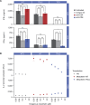
Fig 4. Relative activity of parental and engineered mesothelin HiTs against target cell lines by IFN-γ enzyme-linked immune absorbent spot.
The activity of non-transduced T cells or T cells transduced with the parental HiT or eight engineered HiT mutants (shown in increasing affinity order, from left to right) was assessed against a panel of six target cell lines (shown on key in decreasing order of mesothelin quantitative polymerase chain reaction score from top to bottom). The number of IFN-γ SFU are shown from three replicates for each condition with a representative donor. SFU were limited to a maximum value of 500. Transduction efficiencies for the transduced T cells can be found in S2 Table. HiT, human leukocyte antigen–independent T-cell receptor; IFN-γ, interferon γ; ntd, non-transduced; SFU, spot-forming units; WT, wild type.
Fig 5
Fig 5. Mechanistic investigations of HiT function.
(A) The activity of primary T cells in either the CD4 or CD8 subset were studied for the mesothelin HiT, a mesothelin TRuC, and an HLA-A02*01 displayed MAGE-A4 peptide targeting TCR against a MAGE-A4–positive and mesothelin–over-expressing A375 cell line. Before co-culture with target cells, CD4 or CD8 T cells were incubated with anti-CD4 or anti-CD8 antibodies, respectively; after overnight incubation, IFN-γ release into the supernatants was measured using enzyme-linked immunosorbent assay. Data were analyzed using a two-way analysis of variance n.s. >0.05 * p ≤ 0.05, ** p ≤ 0.01 (GraphPad Prism). (B) Inhibition of the mesothelin-targeting HiT and TRuC by soluble mesothelin was measured using a modified Jurkat cell line expressing luciferase from an IL-2 promoter (TCR/CD3 Effector Cells IL-2, Promega). A mesothelin-positive cell line (Capan-2) and a mesothelin-negative cell line (K562) were incubated with a range of mesothelin concentrations between 0.04 and 40 μM, before being cultured with effector cells. Activation of the IL-2 promoter on effector cells was detected at 6 h. Transduction efficiencies for the transduced T cells can be found in S2 Table. ctrl, control; HiT, human leukocyte antigen–independent T-cell receptor; IFN-γ, interferon γ; IL-2, interkeukin-2; n.s., non-significant; ntd, non-transduced; RLU, relative light units; TCR, T-cell receptor; TRuC, T-cell receptor fusion construct.
Fig 6
Fig 6. In vivo efficacy of parental mesothelin HiT T cells in a mouse xenograft solid tumor model.
Human pancreatic Capan-2 tumor cells were inoculated subcutaneously with either non-transduced or transduced HiT or TRuC T cells (transduced dose as per legend) administered intravenously on day 0. Statistical analysis of these data is shown in S3 Table. HiT, human leukocyte antigen–independent T-cell receptor; SEM, standard error of the mean; TRuC, T-cell receptor fusion construct.

References

    1. Davis MM, Bjorkman PJ. T-cell antigen receptor genes and T-cell recognition. Nature. 1988; 334: 395–402. doi: 10.1038/334395a0 . - DOI - PubMed
    1. Gascoigne NR, Chien Y, Becker DM, Kavaler J, Davis MM. Genomic organization and sequence of T-cell receptor beta-chain constant- and joining-region genes. Nature. 1984; 310: 387–391. doi: 10.1038/310387a0 . - DOI - PubMed
    1. Ganju RK, Smiley ST, Bajorath J, Novotny J, Reinherz EL. Similarity between fluorescein-specific T-cell receptor and antibody in chemical details of antigen recognition. Proc Natl Acad Sci U S A. 1992; 89: 11552–11556. doi: 10.1073/pnas.89.23.11552 . - DOI - PMC - PubMed
    1. Rao A, Ko WW, Faas SJ, Cantor H. Binding of antigen in the absence of histocompatibility proteins by arsonate-reactive T-cell clones. Cell. 1984; 36: 879–888. doi: 10.1016/0092-8674(84)90037-0 . - DOI - PubMed
    1. Moulon C, Wild D, Dormoy A, Weltzien HU. MHC-dependent and -independent activation of human nickel-specific CD8+ cytotoxic T cells from allergic donors. J Invest Dermatol. 1998; 111: 360–366. doi: 10.1046/j.1523-1747.1998.00306.x . - DOI - PubMed

MeSH terms