. 2024 Apr 5;384(6691):106-112.
doi: 10.1126/science.adl5364. Epub 2024 Apr 4.

De novo design of drug-binding proteins with predictable binding energy and specificity

Affiliations

De novo design of drug-binding proteins with predictable binding energy and specificity

Lei Lu et al. Science. .

Abstract

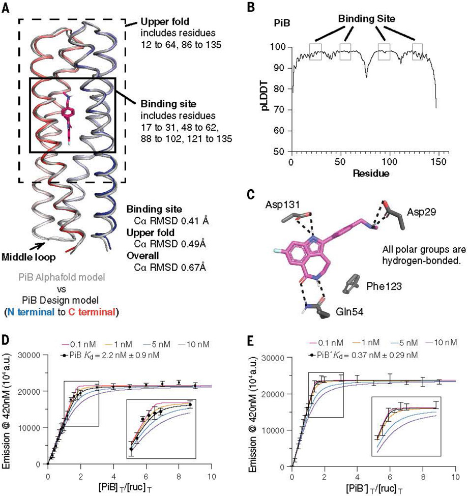
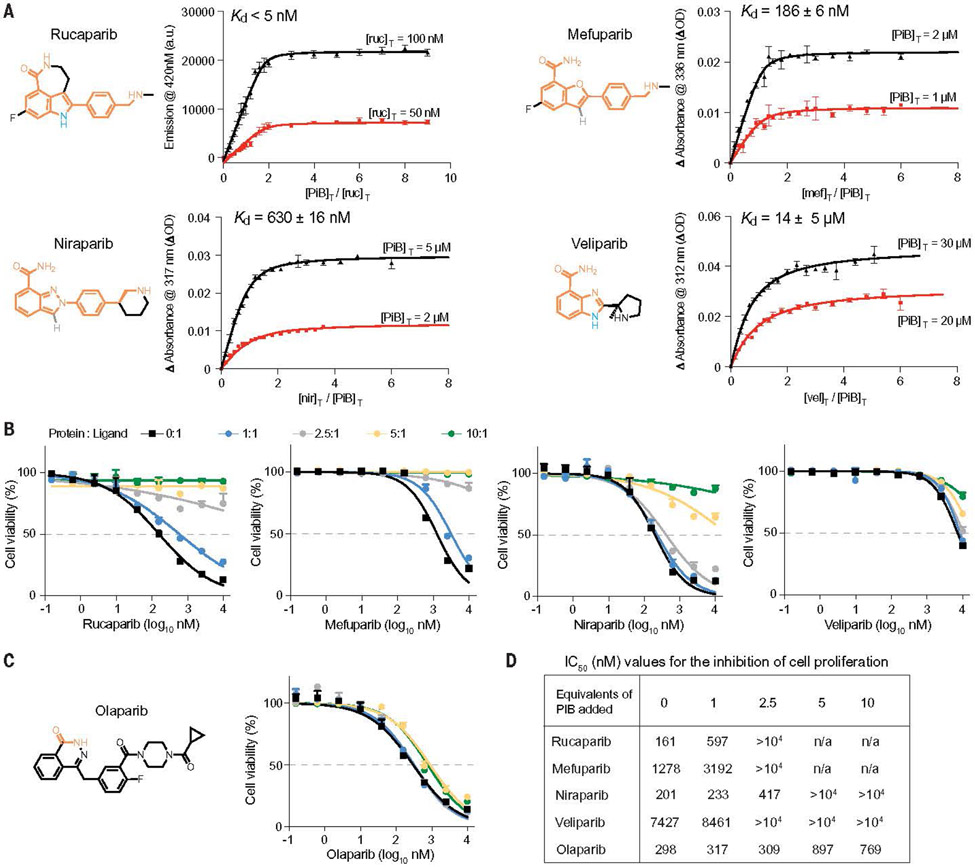
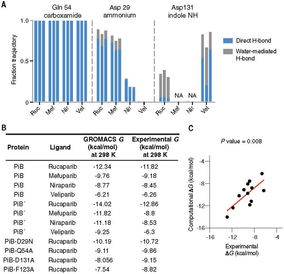
The de novo design of small molecule-binding proteins has seen exciting recent progress; however, high-affinity binding and tunable specificity typically require laborious screening and optimization after computational design. We developed a computational procedure to design a protein that recognizes a common pharmacophore in a series of poly(ADP-ribose) polymerase-1 inhibitors. One of three designed proteins bound different inhibitors with affinities ranging from <5 nM to low micromolar. X-ray crystal structures confirmed the accuracy of the designed protein-drug interactions. Molecular dynamics simulations informed the role of water in binding. Binding free energy calculations performed directly on the designed models were in excellent agreement with the experimentally measured affinities. We conclude that de novo design of high-affinity small molecule-binding proteins with tuned interaction energies is feasible entirely from computation.


Figures

Fig. 1.
Fig. 1.
Computational design of PARPi protein binders. (A) PARPi analogs. The shared chemical features are marked in orange. Olaparib is used as a negative control in the design and binding assay. (B to E) The overall design strategy. [(B) and (C)] We first defined the pharmacophore and used COMBS to sample vdMs on the selected protein backbones. At the outset of the design, we chose chemical groups that should form hydrogen bonds when the drug is bound to the binding site. These groups included rucaparib’s indole NH and carboxamide groups (marked in orange). The carboxamide group is present in our vdM library. However, there were relatively few examples of indole NH vdMs in the database, so we used imidazole as a proxy for the indole’s pyrole ring (in the vdMs, the oxygen is marked in red, and nitrogens are marked in blue). We then used COMBS to discover sidechains at different positions of a four-helix bundle template that could simultaneously form hydrogen bonds to the indole and carboxamide chemical groups of the drug. (The COMBS algorithm samples vdMs on a protein backbone and then performs superpositions of a ligand onto the chemical groups of the vdMs; next, COMBS finds all the vdMs with nearby chemical groups to each superposed ligand; and finally, COMBS computes a specific combination of vdMs for each ligand that optimizes a score, such as the vdM prevalence or cluster score.) We discovered a solution in which the carboxamide formed bidentate hydrogen bonds with sidechain of Q54, and the drug’s indole NH interacted with the N131 [in (C), carbon atoms of protein, green; those of rucaparib, orange]. A second-shell interaction to Q54 that was discovered by COMBS was D58 (carbon atoms, brown). (D) We applied flexible backbone sequence design with a custom Rosetta script while fixing the interactions selected from COMBS (rucaparib, purple). (E) We searched for vdMs again based on the design output from the previous sequence step (D). The slightly different (~ 1-Å Ca RMSD) backbone now preferred different vdMs at some locations (higher cluster scores), and these mutations were made. Three residues at 29, 90, and 131 (deep blue) were changed based on COMBS results. Single-letter abbreviations for the amino acid residues referenced throughout the paper are as follows: L, Leu; N, Asn; D, Asp; Q, Gln; W, Trp; F, Phe.
Fig. 2.
Fig. 2.
Assessing the computational model and experimental binding of PiB to rucaparib. (A) The AlphaFold2 model agreed with the designed PiB, with the binding site Ca RMSD of 0.41 Å, the upper fold Ca RMSD of 0.49 Å, and overall Ca RMSD of 0.67 Å. (B) The predicted local distance difference test scores (pLDDTs) concurred with the trend of RMSD difference of the design model. For example, the N terminal, C terminal, and the middle loop with low pLDDTs (<90) showed higher Ca RMSD. (C) The design model showing that the polar groups of rucaparib are all hydrogen bonded. (D and E) A fluorescence titration showed that PiB and PiB′ bind rucaparib with Kd < 5 nM. The fluorescence emission intensity at 420 nm of rucaparib (excitation wavelength, 355 nm) was measured after titrating aliquots of PiB (D) or PiB′ (E) to a final concentration indicated in the abscissa, in which [PIB]T/[ruc]T or [PIB′]T/[ruc]T refers to the ratio of the molar concentrations of PiB or PiB′ to rucaparib, respectively. The data are well described by a single-site protein-ligand binding model, and a nonlinear least-squares fit to the data returned Kd values of 2.2 ± 0.9 nM for PIB and 0.37 ± 0.29 nM for PiB′. Although the fitting error was relatively small, a sensitivity analysis in which the value of Kd was held constant at various values showed that the data for both proteins were fit within experimental error so long as the Kd was <5 nM. Therefore, although the most probable binding constants were 2 and 0.4 nM, respectively, we can confidently conclude that the values for PiB and PiB′ are <5 nM. The titration was carried out in buffer containing 50-mM Tris and 100-mM NaCl (pH 7.4). a.u., arbitrary units.
Fig. 3.
Fig. 3.
Spectral titrations and cell viability assay of PiB with PARPi. (A) The Kd values of various drugs for PiB as obtained from a global fit of a single-site binding model to the fluorescence or absorbance changes as a function of the concentration of PiB or drugs. The indicated wavelengths for the titration were chosen to maximize the difference in absorption for the free versus bound drug. (B) Growth assays in DLD-1 BRCA2-mutated cells showed that PiB alleviates the effects of rucaparib, mefuparib, niraparib, and veliparib toxicity in a dose-dependent manner. The PARP inhibitors were preincubated with PiB in media at room temperature for 5 min at multiple concentration ratios (protein:ligand) of 0:1, 1:1, 2.5:1, 5:1, and 10:1. (C) Cell viability assay as in Fig. 3B showing that PiB had no effect on the olaparib dose response. (D) Table showing IC50 values for the inhibition of cell proliferation by PARPi drugs in the presence of increasing mole ratios of added PiB protein.
Fig. 4.
Fig. 4.
The structure of drug-bound PiB′ agrees with the design. (A) The design model agreed well with the rucaparib-bound PiB′ crystal structure, with the binding site Ca RMSD range between 0.38 and 0.46 Å for the three monomers in the asymmetric unit. (B) The binding site of PiB′. A 2mFo-DFc composite omit map contoured at 1.6 s. The map was generated from a model that omitted coordinates of rucaparib. Overlay of the design (gray) and the structure (protein, orange; rucaparib, pink). The sidechains of the binding pocket in rucaparib-bound PiB′ agreed with the design. Asp131 interacts with the indole NH through a bridging water as in MD simulations. (C) The structure of drug-free PiB′ shows a preorganized open pocket filled with multiple waters, which are displaced in the drug-bound conformation. (D) Reversal of the three designed substitutions from the vdM optimization procedure led to lower binding affinity (higher Kd) for rucaparib by fluorescence emission titrations. (E) Alanine mutations of the direct binding residues decreased binding affinity confirmed by fluorescence emission titrations
Fig. 5.
Fig. 5.
The MD simulations of PiB, PiB′, and mutants. (A) By using unbiased MD simulations in Amber, we calculated (in triplicate) the frequency with which the intermolecular hydrogen bonds formed between the protein scaffold and the bound drug molecule. PiB was found to form a hydrogen bond between Gln54 and the targeted drug carboxamide in 100% of all simulations for each drug complex. The charged ammonium groups of rucaparib and mefuparib interacted with Asp29 through a combination of direct and water-mediated hydrogen bonds, totaling to more than half of the full simulation time, which contrasts niraparib and veliparib’s inabilities to form equivalent hydrogen bonds (owing to changes in chemical structure around the ammonium tail of the ligand). In a small fraction of each rucaparib and veliparib trajectory, Asp131 engaged in water-mediated hydrogen bonds to the drugs. (B) By using biased simulations in GROMACS, we calculated the binding free energy (DG) for each ligand and found that ranked affinity for each drug was consistent with experimental results. (C) Comparison of DG from the GROMACS calculation with the experimental value from spectral titrations

Update of

References

    1. Schreier B, Stumpp C, Wiesner S, Höcker B, Proc. Natl. Acad. Sci. U.S.A 106, 18491–18496 (2009). - PMC - PubMed
    1. Yang W, Lai L, Curr. Opin. Struct. Biol 45, 67–73 (2017). - PubMed
    1. Polizzi NF, DeGrado WF, Science 369, 1227–1233 (2020). - PMC - PubMed
    1. Thomas F et al., ACS Synth. Biol 7, 1808–1816 (2018). - PubMed
    1. Tinberg CE et al., Nature 501, 212–216 (2013). - PMC - PubMed

Publication types