. 2024 Apr 4;15(4):244.
doi: 10.1038/s41419-024-06589-7.

The GFPT2-O-GlcNAcylation-YBX1 axis promotes IL-18 secretion to regulate the tumor immune microenvironment in pancreatic cancer

Affiliations

The GFPT2-O-GlcNAcylation-YBX1 axis promotes IL-18 secretion to regulate the tumor immune microenvironment in pancreatic cancer

Hui-Ru Zhang et al. Cell Death Dis. .

Abstract

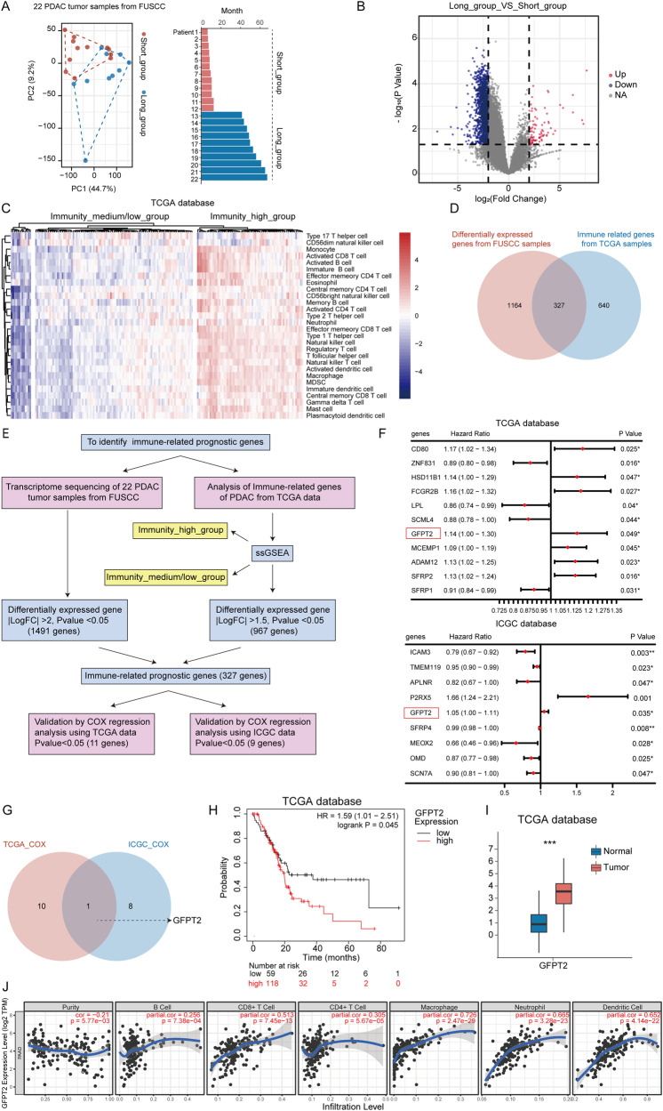
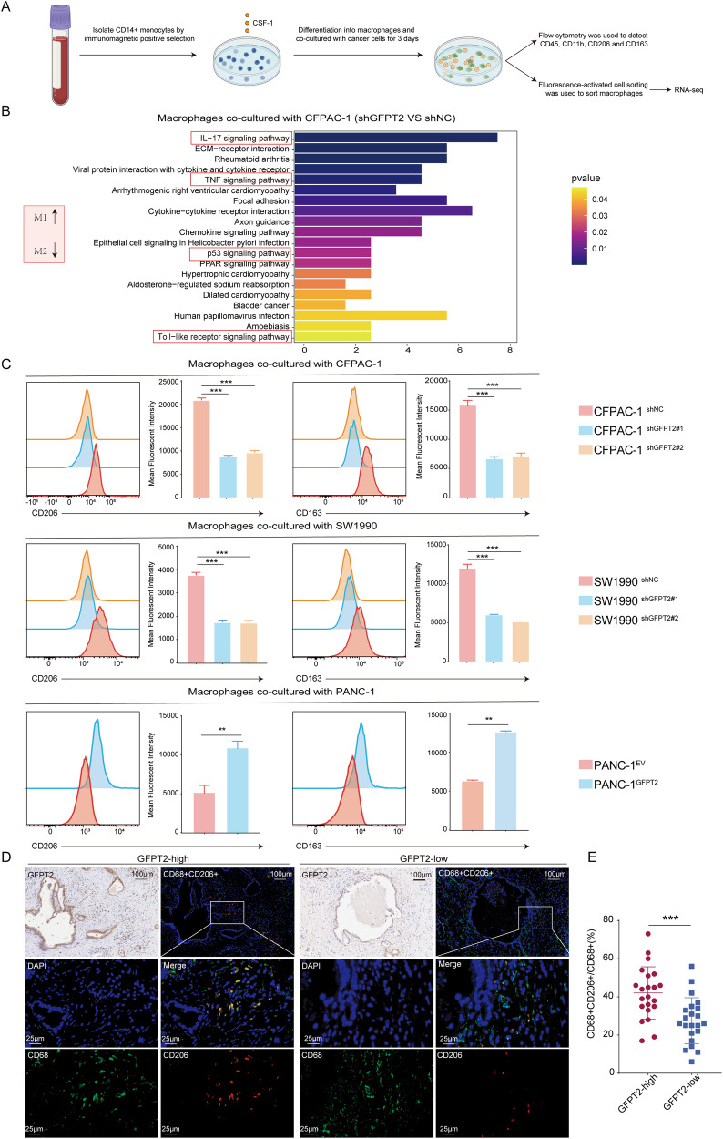
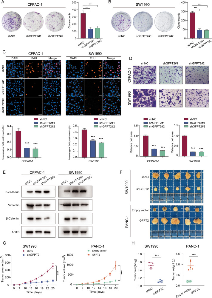
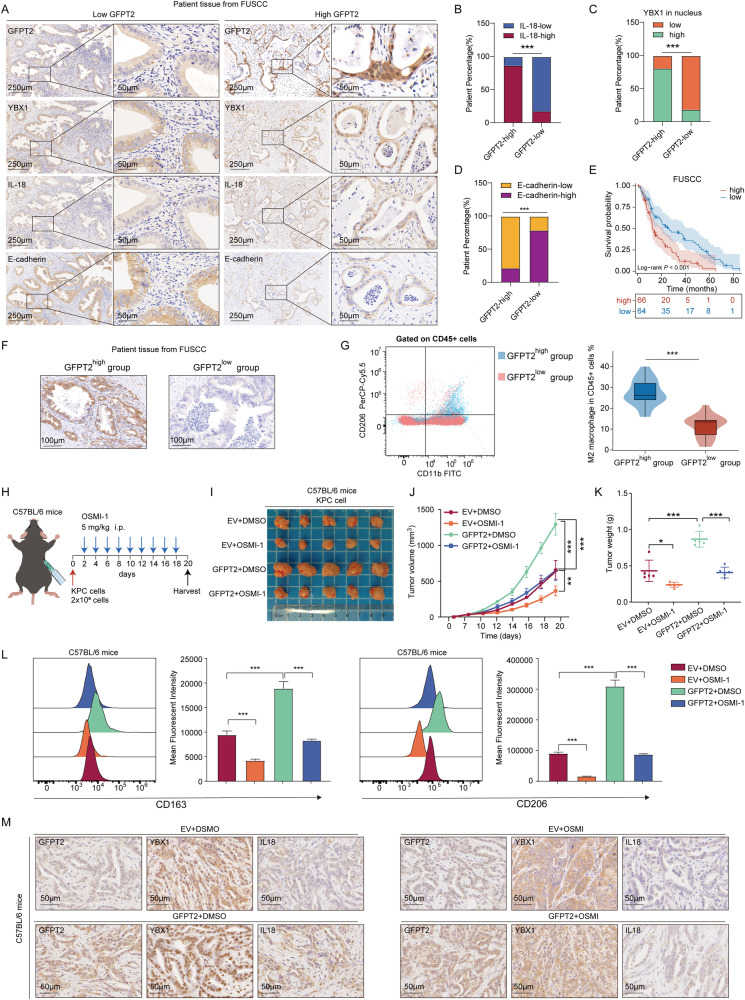
The immunosuppressive microenvironment caused by several intrinsic and extrinsic mechanism has brought great challenges to the immunotherapy of pancreatic cancer. We identified GFPT2, the key enzyme in hexosamine biosynthesis pathway (HBP), as an immune-related prognostic gene in pancreatic cancer using transcriptome sequencing and further confirmed that GFPT2 promoted macrophage M2 polarization and malignant phenotype of pancreatic cancer. HBP is a glucose metabolism pathway leading to the generation of uridine diphosphate N-acetylglucosamine (UDP-GlcNAc), which is further utilized for protein O-GlcNAcylation. We confirmed GFPT2-mediated O-GlcNAcylation played an important role in regulating immune microenvironment. Through cellular proteomics, we identified IL-18 as a key downstream of GFPT2 in regulating the immune microenvironment. Through CO-IP and protein mass spectrum, we confirmed that YBX1 was O-GlcNAcylated and nuclear translocated by GFPT2-mediated O-GlcNAcylation. Then, YBX1 functioned as a transcription factor to promote IL-18 transcription. Our study elucidated the relationship between the metabolic pathway of HBP in cancer cells and the immune microenvironment, which might provide some insights into the combination therapy of HBP vulnerability and immunotherapy in pancreatic cancer.


Conflict of interest statement

The authors declare no competing interests.

Figures

Fig. 1
Fig. 1. GFPT2 was an immune-related prognostic gene in pancreatic cancer.
A Principal component analysis showed transcriptome sequencing sample features of 22 pancreatic ductal adenocarcinoma (PDAC) patients from FUSCC, and the bar chart showed the survival time of the patients. B Volcano map showing differences in gene expression between groups with an overall survival of less than 1 year and those with an overall survival of more than 3 years (fold change > 4 or < -4). C Heatmap showing the results of ssGSEA in 177 PDAC transcriptome datasets ranked according to the degree of immune cell infiltration. D Venn diagram showing the intersection of prognostic genes from FUSCC transcriptome sequencing and immune-related genes from TCGA database. E The flow chart showing our analysis process. F Univariate cox hazard analysis of these 327 genes in the data of TCGA and ICGC, and genes with P < 0.05 were shown in the table. G Venn diagram showed the intersection of two sets of data. H Kaplan-Meier survival curve of GFPT2 expression in tumor tissues from TCGA database was showed. I The expression of GFPT2 in tumor tissues and normal adjacent pancreas tissues from TCGA database was showed. J The correlation between the expression of GFPT2 and the infiltration of immune cells (B cells, CD4 + T cells, CD8 + T cells, neutrophils, macrophages and dendritic cells) was analyzed by Tumor Immune Estimation Resource (TIMER).
Fig. 2
Fig. 2. GFPT2 overexpressing pancreatic cancer cells promoted macrophage M2 polarization.
A The pattern diagram showed the coculture system of pancreatic cancer cells and macrophages which was derived from CD14+ monocytes. B KEGG analysis of transcriptome sequencing data on macrophages cocultured with GFPT2-knockdown CFPAC-1 cells. C CFPAC-1, SW1990 and PANC-1 stable cell lines were cocultured with macrophages for 3 days and then M2 macrophage were analyzed by flow cytometry with CD11b, CD45, CD163 and CD206 antibodies. D, E Multiplex immunohistochemistry assay showing the macrophage M2 polarization by CD206 and CD68 antibodies and the proportion of M2 in all macrophages was calculated.
Fig. 3
Fig. 3. GFPT2 promoted the proliferation and migration of pancreatic cancer cells.
A, B Colony formation assay was performed with CFPAC-1 and SW1990 stable cell lines. C 5-Ethynyl -2′- deoxy uridine (EdU) was used to detect the proliferation of CFPAC-1 and SW1990 stable cell lines. Scale bars, 50 μm. D Transwell assays were used to detect the migration abilities of CFPAC-1 and SW1990 stable cell lines. Scale bars, 200 μm. E The protein levels of E-cadherin, vimentin and β-catenin were measured by western blotting in CFPAC-1, SW1990 and PANC-1 stable cell lines. F The image displayed the subcutaneous implanted tumor of GFPT2-knockdown SW1990 and GFPT2-overexpressed PANC-1 cell lines in nude mice. G The effect of GFPT2-knockdown or -overexpression on the growth trends of subcutaneous implanted tumors were shown. H The weight of tumors was weighed.
Fig. 4
Fig. 4. GFPT2 promoted O-GlcNAcylation in pancreatic cancer.
A Western bolt analysis of GFPT2, IL-18 and overall levels of O-GlcNAcylation in 12 tumor tissues, which are derived from Fig. 1. B The overall level of O-GlcNAcylation was measured in pancreatic cancer cell lines (OSMI-1 (40 μm) for 48 h). C GFPT2 overexpressing PANC-1 cells were pretreated with OSMI-1 (40 μm) and then were cocultured with macrophages for 3 days and then M2 macrophage were analyzed by flow cytometry with CD45, CD11b, CD163 and CD206 antibodies. D EdU was used to detect the proliferation of GFPT2 overexpressing PANC-1 cells pretreated with OSMI-1 (40 μm). Scale bars, 50 μm. E Colony formation assay was performed with GFPT2 overexpressing PANC-1 cells pretreated with OSMI-1 (40 μm). F Transwell assays were used to detect the migration abilities of GFPT2 overexpressing PANC-1 cells pretreated with OSMI-1 (40 μm). Scale bars, 200 μm.
Fig. 5
Fig. 5. GFPT2 promoted the synthesis and secretion of IL-18 in pancreatic cancer.
A The diagram shows the proteome analysis in GFPT2 knockdown and control SW1990 cells. B, C Volcano map and heatmap showing the changed proteins in the proteome analysis. D Venn diagram and heatmap showing the changed secreted proteins and membrane proteins in the proteome analysis. E Protein levels of IL-18 in pancreatic cell lines were measured by western blotting. F Protein levels of IL-18 in SW1990 and CFPAC-1 cells were measured by western blotting. G Protein levels of IL-18 in PANC-1 cells pretreated with OSMI-1 (40 μm) were detected by western blotting. H Secretion of IL-18 wad detected by an ELISA kit. I Protein levels of IL-18 in implanted tumors of nude were measured by western blotting. J Macrophages were cocultured with pancreatic cancer cells for 3 days and then M2 macrophage were analyzed by flow cytometry with CD45, CD11b, CD206 and CD163 antibodies. K EdU was used to detect the proliferation of GFPT2 overexpressing PANC-1 cells pretreated with siRNAs targeting IL-18. Scale bars, 50 μm. L Transwell assay was used to detect the migration abilities of GFPT2 overexpressing PANC-1 cells pretreated with siRNAs targeting IL-18. Scale bars, 200 μm. M The O-GlcNAcylation and phosphorylation site of IL-18 were predicted in the O-GlcNAc Database.
Fig. 6
Fig. 6. GFPT2 promoted the O-GlcNAcylation and nuclear translocation of YBX1.
A The diagram showing the immunoprecipitation assay in SW1990 cells. B Silver staining of the proteins pulled down by IgG and O-GlcNAcylation antibodies. C Representative tandem MS spectrum of the RPQYSNPPVQGEVMEGADNQGAGEQGRPVR peptide from YBX1 as determined by IP-Mass Spec. D The O-GlcNAcylation and phosphorylation site of YBX1 were predicted in the O-GlcNAc Database. E The O-GlcNAcylation and phosphorylation site of YBX1 were predicted in YinOYang-1.2 database. F Immunoprecipitation was performed with IgG and O-GlcNAcylation antibodies and YBX1 and OGT proteins were detected by western blotting. G YBX1 was detected by western blotting in GFPT2-knockdown SW1990 and CFPAC-1 cells or GFPT2-overexpressing PANC-1 cells. H Immunoprecipitation was performed with a YBX1 antibody and YBX1, OGT and O-GlcNAcylation were detected by western blotting in CFPAC-1 and SW1990 cells with GFPT2 changes. I Immunoprecipitation was performed with YBX1 antibody and YBX1, OGT, and O-GlcNAcylation were detected by western blotting in PANC-1 cells with GFPT2-changed and OSMI-1 (40 μm) pretreated. J Western blotting to detect the change in cytoplasmic and nuclear YBX1 in SW1990 and CFPAC-1 cells with GFPT2 changes. K Western blotting to detected the change of cytoplasmic and nuclear YBX1 in PANC-1 cells with GFPT2 changes and OSMI-1 (40 μm) pretreated. L Immunofluorescence to detected the change of cytoplasmic and nuclear YBX1 in SW1990 and CFPAC-1 cells with GFPT2 changes. Scale bars, 20 μm. M Immunofluorescence to detected the change of cytoplasmic and nuclear YBX1 in PANC-1 cells with GFPT2 changes and OSMI-1 (40 μm) pretreated.
Fig. 7
Fig. 7. YBX1 nuclear localization promoted IL-18 transcription.
A IL-18 mRNA was detected by qRT-PCR. B IL-18 protein was detected by western blotting. C The secretion of IL-18 was detected. D The binding site of YBX1 in the JASPAR database was showed and the binding sites in IL-18 promoter was showed. P1-6 showed 6 paired primers that covered the 8 binding sites in IL-18 promoter. E ChIP assay was performed with IgG and YBX1 antibodies in PANC-1 cells and qRT-PCR was performed using the designed 6 paired primers and the proportion of YBX1 binding sites was calculated. F Dual-luciferase reporter gene system was performed using full length or deleted (Δ) promoters of IL-18 in 293 T cells. G Dual-luciferase reporter gene system was performed using the wild type site or the mutated site of YBX1 on IL-18 promoter in 293 T cells. H ChIP assay was performed with IgG and YBX1 antibodies in GFPT2-altered PANC-1 cells pretreated with OSMI-1 and qRT-PCR was performed using the primer 4 and the proportion of YBX1 binding sites were calculated. I Macrophage was cocultured with GFPT2 and YBX1-changed PANC-1 cells for 3 days and then M2 macrophage were analyzed by flow cytometry with CD11b, CD163 and CD206 antibodies. J EdU was used to detect the proliferation of GFPT2 overexpressing PANC-1 cells pretreated with siRNAs targeting YBX1. Scale bars, 50 μm. K Transwell assays were used to detect the migration abilities of GFPT2 overexpressing PANC-1 cells pretreated with siRNAs targeting YBX1. Scale bars, 200 μm.
Fig. 8
Fig. 8. The GFPT2-YBX1-IL-18 signaling was further confirmed in clinical specimens and in vivo experiments.
A Immunohistochemical detected GFPT2, YBX1, IL-18, and E-cadherin expression in human pancreatic cancer tissues. BD Chi-square test was performed to analyzed the relationship of GFPT2 with YBX1, IL-18 or E-cadherin in human pancreatic cancer tissues. E Kaplan-Meier survival curve of GFPT2 expression in tumor tissues from our cohort was shown. F 40 cases of pancreatic cancer tissue samples were divided into high and low groups according to the expression level of GFPT2. G Flow cytometric analysis macrophages M2 polarization in the 40 fresh human pancreatic cancer tissues. HK The subcutaneous implant tumor model of KPC cells in C57BL/6 mice. OSMI-1 or DMSO was intraperitoneally injected after cancer cells subcutaneously implanting. L Flow cytometric analysis macrophages M2 polarization in fresh subcutaneous implant tumors. M Immunohistochemical detected GFPT2, YBX1 and IL-18 expression in subcutaneous implant tumors of C57BL/6 mice.

References

    1. Halbrook CJ, Lyssiotis CA, Pasca di Magliano M, Maitra A. Pancreatic cancer: advances and challenges. Cell. 2023;186:1729–54. doi: 10.1016/j.cell.2023.02.014. - DOI - PMC - PubMed
    1. Kleeff J, Korc M, Apte M, La Vecchia C, Johnson CD, Biankin AV, et al. Pancreatic cancer. Nat Rev Dis Primers. 2016;2:16022. doi: 10.1038/nrdp.2016.22. - DOI - PubMed
    1. Siegel RL, Miller KD, Fuchs HE, Jemal A. Cancer statistics, 2022. CA Cancer J Clin. 2022;72:7–33. doi: 10.3322/caac.21708. - DOI - PubMed
    1. Brahmer JR, Drake CG, Wollner I, Powderly JD, Picus J, Sharfman WH, et al. Phase I study of single-agent anti-programmed death-1 (MDX-1106) in refractory solid tumors: safety, clinical activity, pharmacodynamics, and immunologic correlates. J Clin Oncol. 2010;28:3167–75. doi: 10.1200/JCO.2009.26.7609. - DOI - PMC - PubMed
    1. Topalian SL, Sznol M, McDermott DF, Kluger HM, Carvajal RD, Sharfman WH, et al. Survival, durable tumor remission, and long-term safety in patients with advanced melanoma receiving nivolumab. J Clin Oncol. 2014;32:1020–30. doi: 10.1200/JCO.2013.53.0105. - DOI - PMC - PubMed

Publication types

MeSH terms