. 2024 Apr 4;14(1):7962.
doi: 10.1038/s41598-024-58033-7.

Microneedle combined with iontophoresis and electroporation for assisted transdermal delivery of goniothalamus macrophyllus for enhancement sonophotodynamic activated cancer therapy

Affiliations

Microneedle combined with iontophoresis and electroporation for assisted transdermal delivery of goniothalamus macrophyllus for enhancement sonophotodynamic activated cancer therapy

Samir Ali Abd El-Kaream et al. Sci Rep. .

Abstract

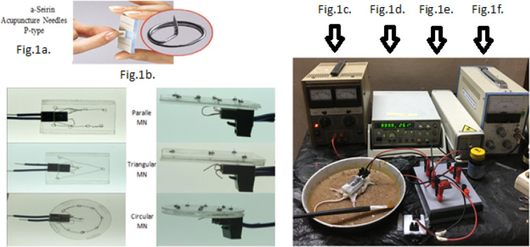
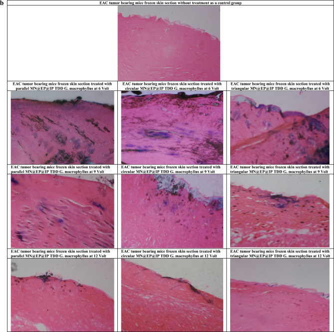
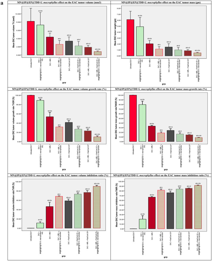
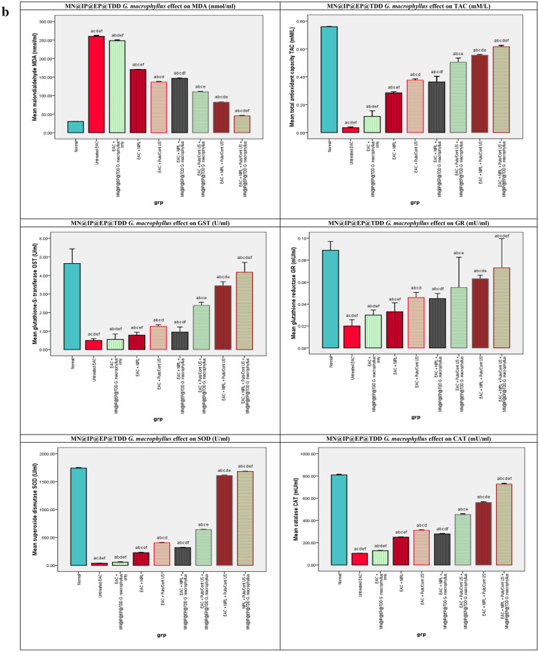
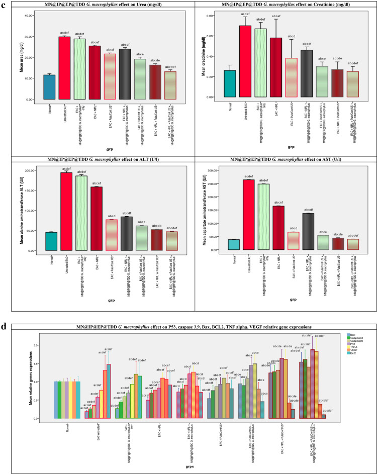
The underlying study was carried out aiming at transdermal drug delivery (TDD) of Goniothalamus macrophyllus as sono-photo-sensitizer (SPS) using microneedle (MN) arrays with iontophoresis (MN-IP), electroporation (MN-EP) in conjunction with applying photodynamic therapy (PDT), sonodynamic therapy (SDT) and sono-photodynamic therapy (SPDT) as an up-to-date activated cancer treatment modality. Study was conducted on 120 male Swiss Albino mice, inoculated with Ehrlich ascites carcinoma (EAC) divided into 9 groups. We employed three different arrays of MN electrodes were used (parallel, triangular, and circular), EP, IP with different volts (6, 9, 12 V), an infrared laser and an ultrasound (pulsed and continuous wave) as our two energy sources. Results revealed that parallel 6 V TDD@MN@IP@EP can be used as effective delivery system for G. macrophyllus from skin directly to target EAC cells. In addition MN@IP@EP@TDD G. macrophyllus is a potential SPS for SPDT treatment of EAC. With respect to normal control mice and as opposed to the EAC untreated control mice, MN@EP@IP TDD G. macrophyllus in the laser, ultrasound, and combination activated groups showed a significant increase in the antioxidant markers TAC level and the GST, GR, Catalase, and SOD activities, while decrease in lipid peroxidation oxidative stress parameter MDA levels. In addition significantly increased apoptotic genes expressions (p53, caspase (3, 9), Bax, and TNF alpha) and on the other hand decreased anti- apoptotic (Bcl-2) and angiogenic (VEGF) genes expressions. Moreover significantly ameliorate liver and kidney function decreasing ALT, AST, urea and creatinine respectively. Furthermore MN@IP@EP@TDD G. macrophyllus combined with SPDT was very effective at reducing the growth of tumors and even causing cell death according to microscopic H&E stain results. This process may be related to a sono- and/or photochemical activation mechanism. According to the findings, MN@IP@EP@TDD G. macrophyllus has a lot of potential as a novel, efficient delivery method that in combination with infrared laser and ultrasound activation SPDT demonstrated promising anticancer impact for treating cancer.


Conflict of interest statement

The authors declare no competing interests.

Figures

Figure 1
Figure 1
Microneedle, electroporation, iontophoresis assisted transdermal delivery of G. macrophyllus [MN@IP@EP@TDD G. macrophyllus] (a,b). (Serine type- parallel, circular and triangular) Microneedle [MN], (c). Electroporation [EP], (d). Iontophoresis [IP], (e). Laser [INRL], (f). Ultrasound [US].
Figure 2
Figure 2
(a) MN@IP@EP@TDD G. macrophyllus pore diameter penetrated through (µm) F(p) = 237.212(0.001*), transported (µg/cm2) F(p) = 1.921E3(0.001*), absorbed (µg/mg tissue) F(p) = 856.956(0.001*), flux rate (µg/cm2/5min) F(p) = 272.534(0.001*) and penetration depth (µm) F(p) = 213.125(0.001*) at 5 s electroporation EP for 5s followed by microneedle (parallel, circular and triangular) assisted DC-Iontophoresis (Square wave, 0.5 mA for 5min) at different volts (6, 9 and 12 V for 5min). (b) Frozen skin cryo-sections illustrating microneedle assisted electroporation-iontophoresis transdermal delivery of Goniothalmus macrophyllus (MN@EP@IP TDD G. macrophyllus) and penetration depth using different MN arrays and IP volts 2; parallel MN array@EP@IP (6V) TDD, 3; circular MN array@EP@IP (6V) TDD, 4; triangular MN array@EP@IP (6V) TDD, 5; parallel MN array@EP@IP (9V) TDD, 6; circular MN array@EP@IP (9V) TDD, 7; triangular MN array@EP@IP (9V) TDD, 8; parallel MN array@EP@IP (12V) TDD, 9; circular MN array@EP@IP (12V) TDD, 10; triangular MN array@EP@IP (12V) TDD. [1; untreated EAC implanted group skin section without any treatment,].
Figure 2
Figure 2
(a) MN@IP@EP@TDD G. macrophyllus pore diameter penetrated through (µm) F(p) = 237.212(0.001*), transported (µg/cm2) F(p) = 1.921E3(0.001*), absorbed (µg/mg tissue) F(p) = 856.956(0.001*), flux rate (µg/cm2/5min) F(p) = 272.534(0.001*) and penetration depth (µm) F(p) = 213.125(0.001*) at 5 s electroporation EP for 5s followed by microneedle (parallel, circular and triangular) assisted DC-Iontophoresis (Square wave, 0.5 mA for 5min) at different volts (6, 9 and 12 V for 5min). (b) Frozen skin cryo-sections illustrating microneedle assisted electroporation-iontophoresis transdermal delivery of Goniothalmus macrophyllus (MN@EP@IP TDD G. macrophyllus) and penetration depth using different MN arrays and IP volts 2; parallel MN array@EP@IP (6V) TDD, 3; circular MN array@EP@IP (6V) TDD, 4; triangular MN array@EP@IP (6V) TDD, 5; parallel MN array@EP@IP (9V) TDD, 6; circular MN array@EP@IP (9V) TDD, 7; triangular MN array@EP@IP (9V) TDD, 8; parallel MN array@EP@IP (12V) TDD, 9; circular MN array@EP@IP (12V) TDD, 10; triangular MN array@EP@IP (12V) TDD. [1; untreated EAC implanted group skin section without any treatment,].
Figure 3
Figure 3
(a) The effect of laser, ultrasound continuous/pulsed and Combined modalities on the EAC tumor volume (mm3 ) F(p) = 44.073(0.001*), tumor volume growth rate (%)F(p) = 1.128E3(0.001*), tumor volume inhibition ratio (%)F(p) = 1.128E3(0.001*), tumor mass (gm) F(p) = 25.731(0.001*), tumor mass growth rate (%)F(p) = 449.995(0.001*), tumor mass inhibition ratio (%)F(p) = 449.995(0.001*), of untreated and treated groups. (b) The effect of laser, ultrasound continuous/pulsed and Combined modalities on antioxidants activities, capacities and MDA, of untreated and treated groups. F: value for ANOVA test MDA (nmol/ml): 2.149E4 p < 0.001*, TAC (mM/L): 440.982 p < 0.001*, GST (U/ml): 80.472 p < 0.001*, GR (mU/ml): 10.170 p < 0.001*, CAT (mU/ml): 1.695E4 p < 0.001*, SOD (U/ml): 4.113E5 p < 0.001*. (c) The effect of laser, ultrasound continuous/pulsed and Combined modalities on renal and hepatic biomarkers, of untreated and treated groups. Urea (mg/dl): 310.998 p < 0.001*, Creatinine (mg/dl): 12.490 p < 0.001*, ALT (U/l): 4.140E3p < 0.001*, AST (U/l): 5.461E4 p < 0.001*. (d) p53, Bax, Caspase (9,3), TNF alpha, VEGF, Bcl-2 qRT-PCR relative gene expression of all study groups p53 F(P): F = 67.190 p < 0.001*, Bax F(P): F = 106.888 p < 0.001*, Caspase 9 F(P): F = 66.025 p < 0.001*, Caspase 3 F(P): F = 100.944 p < 0.001*, TNFalpha F(P): F = 33.830 p < 0.001*, VEGF F(P): F = 19.537 p < 0.001*, Bcl-2 F(P): F = 34.116 p < 0.001*.
Figure 3
Figure 3
(a) The effect of laser, ultrasound continuous/pulsed and Combined modalities on the EAC tumor volume (mm3 ) F(p) = 44.073(0.001*), tumor volume growth rate (%)F(p) = 1.128E3(0.001*), tumor volume inhibition ratio (%)F(p) = 1.128E3(0.001*), tumor mass (gm) F(p) = 25.731(0.001*), tumor mass growth rate (%)F(p) = 449.995(0.001*), tumor mass inhibition ratio (%)F(p) = 449.995(0.001*), of untreated and treated groups. (b) The effect of laser, ultrasound continuous/pulsed and Combined modalities on antioxidants activities, capacities and MDA, of untreated and treated groups. F: value for ANOVA test MDA (nmol/ml): 2.149E4 p < 0.001*, TAC (mM/L): 440.982 p < 0.001*, GST (U/ml): 80.472 p < 0.001*, GR (mU/ml): 10.170 p < 0.001*, CAT (mU/ml): 1.695E4 p < 0.001*, SOD (U/ml): 4.113E5 p < 0.001*. (c) The effect of laser, ultrasound continuous/pulsed and Combined modalities on renal and hepatic biomarkers, of untreated and treated groups. Urea (mg/dl): 310.998 p < 0.001*, Creatinine (mg/dl): 12.490 p < 0.001*, ALT (U/l): 4.140E3p < 0.001*, AST (U/l): 5.461E4 p < 0.001*. (d) p53, Bax, Caspase (9,3), TNF alpha, VEGF, Bcl-2 qRT-PCR relative gene expression of all study groups p53 F(P): F = 67.190 p < 0.001*, Bax F(P): F = 106.888 p < 0.001*, Caspase 9 F(P): F = 66.025 p < 0.001*, Caspase 3 F(P): F = 100.944 p < 0.001*, TNFalpha F(P): F = 33.830 p < 0.001*, VEGF F(P): F = 19.537 p < 0.001*, Bcl-2 F(P): F = 34.116 p < 0.001*.
Figure 4
Figure 4
The effect of 3; NIRL only, 4; NIRL in presence of MN@EP@IP TDD G. macrophyllus, 5; pulsed/continuous US in absence only, 6; pulsed/continuous US in presence of MN@EP@IP TDD G. macrophyllus, 7; Combined modalities NIRL/US only and 8; Combined modalities NIRL/US in presence of MN@EP@IP TDD G. macrophyllus on cellular level. [1; untreated EAC implanted group without any treatment, 2; MN@EP@IP TDD G. macrophyllus treated group without activation].

References

    1. Cagan R, Meyer P. Rethinking cancer: Current challenges and opportunities in cancer research. Dis. Model Mech. 2017;10:349–352. doi: 10.1242/dmm.030007. - DOI - PMC - PubMed
    1. Kebebe D, Liu Y, Wu Y, Vilakhamxay M, Liu Z, Li J. Tumor-targeting delivery of herb-based drugs with cell-penetrating/tumor-targeting peptide-modified nanocarriers. Int. J. Nanomed. 2018;13:1425–1442. doi: 10.2147/IJN.S156616. - DOI - PMC - PubMed
    1. Brodin NP, Guha C, Tomé WA. Photodynamic therapy and its role in combined modality anticancer treatment. Technol. Cancer Res. Treat. 2015;14:355–368. doi: 10.1177/1533034614556192. - DOI - PubMed
    1. Liu Y, Wang P, Liu Q, Wang X. Sinoporphyrin sodium triggered sono-photodynamic effects on breast cancer both in vitro and in vivo. Ultrason. Sonochem. 2016;31:437–448. doi: 10.1016/j.ultsonch.2016.01.038. - DOI - PubMed
    1. Liu Q, Sun S, Xiao Y. Synergistic anti-tumor effect of ultrasound and hematoporphyrin on sarcoma180 cells with special reference to the changes of morphology and cytochrome oxidase activity of tumor cells. J. Exp. Clin. Cancer Res. 2004;23:333–3341. - PubMed