. 2024 Apr;13(4):e12429.
doi: 10.1002/jev2.12429.

Synthetic biology-based bacterial extracellular vesicles displaying BMP-2 and CXCR4 to ameliorate osteoporosis

Affiliations

Synthetic biology-based bacterial extracellular vesicles displaying BMP-2 and CXCR4 to ameliorate osteoporosis

Han Liu et al. J Extracell Vesicles. 2024 Apr.

Erratum in

Abstract

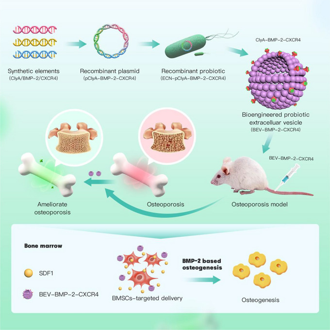
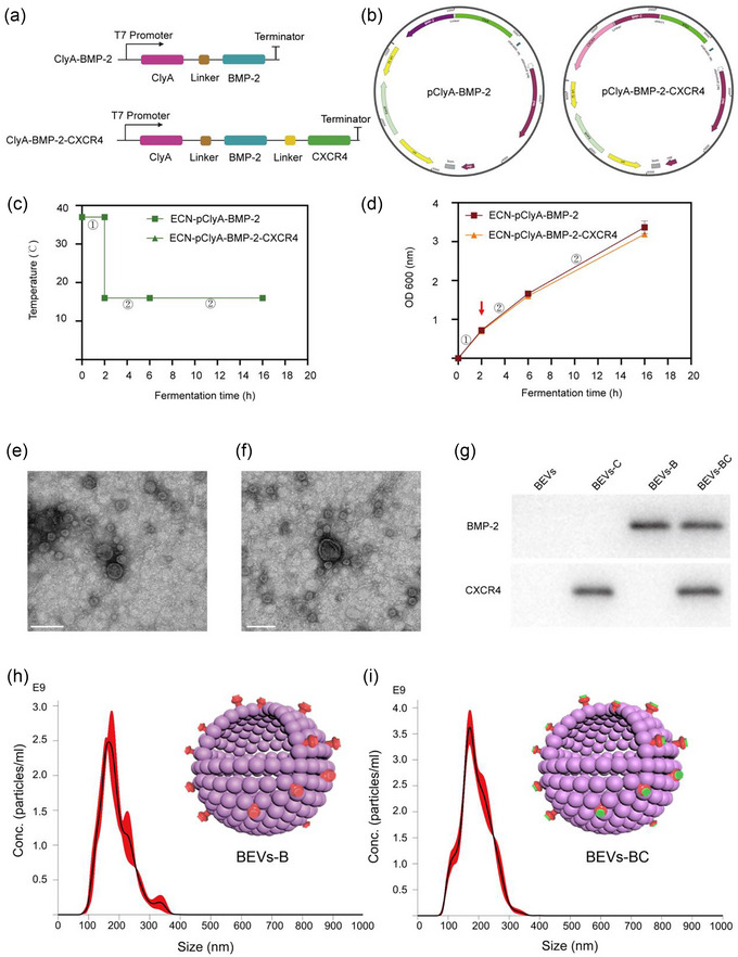
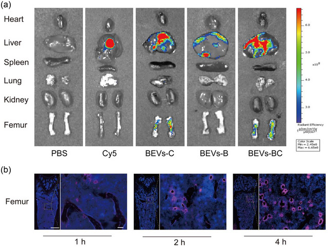
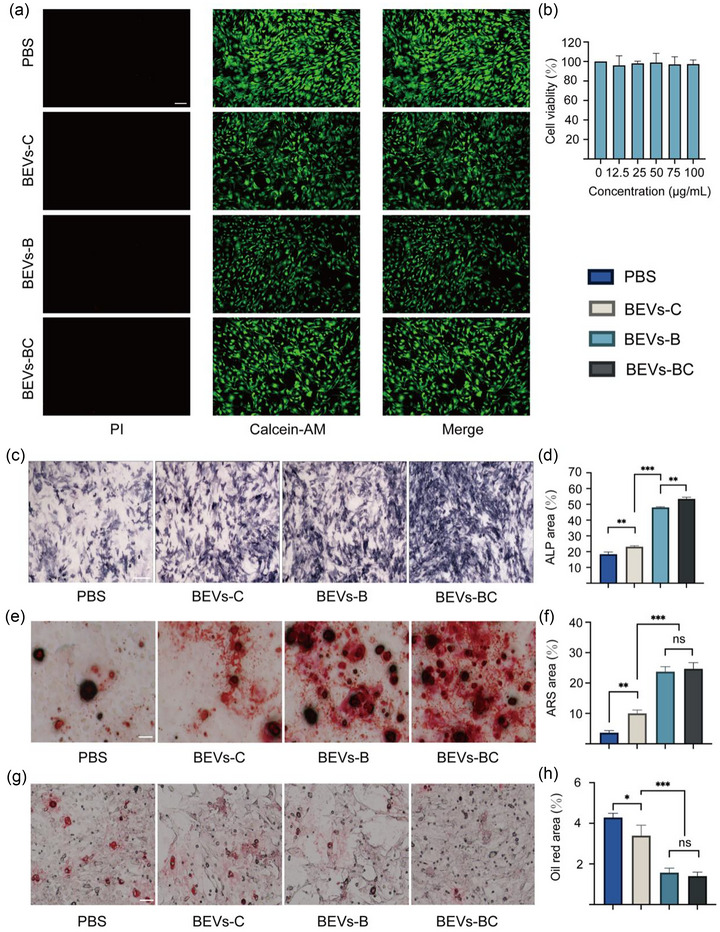
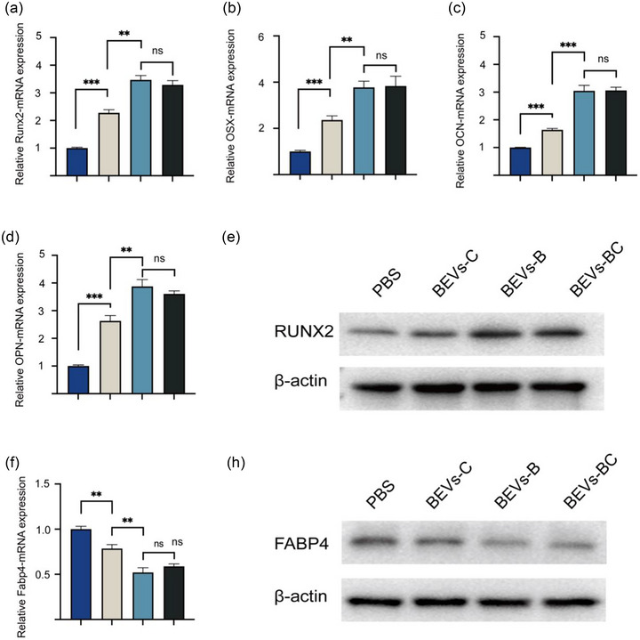
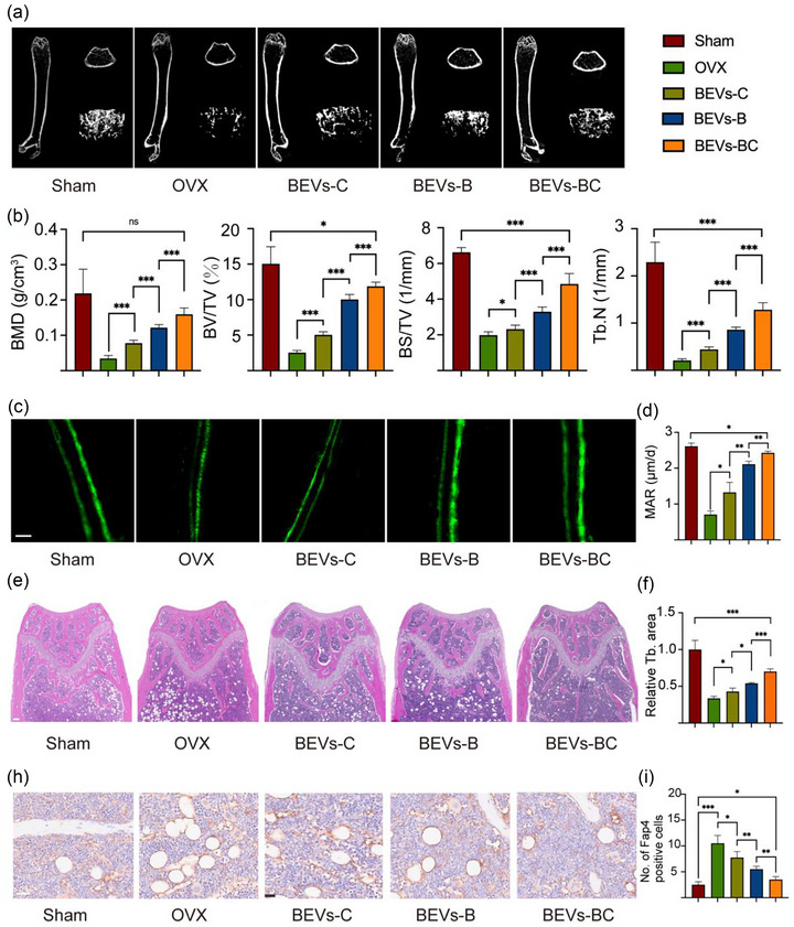
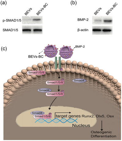
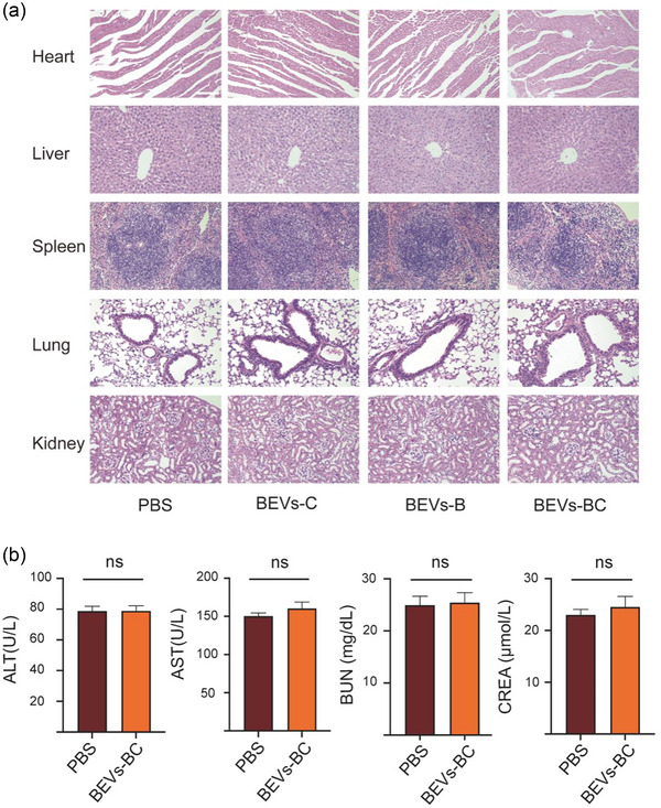
Osteoporosis (OP) is a systematic bone disease characterized by low bone mass and fragile bone microarchitecture. Conventional treatment for OP has limited efficacy and long-term toxicity. Synthetic biology makes bacterial extracellular vesicle (BEVs)-based therapeutic strategies a promising alternative for the treatment of OP. Here, we constructed a recombinant probiotics Escherichia coli Nissle 1917-pET28a-ClyA-BMP-2-CXCR4 (ECN-pClyA-BMP-2-CXCR4), in which BMP-2 and CXCR4 were overexpressed in fusion with BEVs surface protein ClyA. Subsequently, we isolated engineered BEVs-BMP-2-CXCR4 (BEVs-BC) for OP therapy. The engineered BEVs-BC exhibited great bone targeting in vivo. In addition, BEVs-BC had good biocompatibility and remarkable ability to promote osteogenic differentiation of BMSCs. Finally, the synthetic biology-based BEVs-BC significantly prevented the OP in an ovariectomized (OVX) mouse model. In conclusion, we constructed BEVs-BC with both bone-targeting and bone-forming in one-step using synthetic biology, which provides an effective strategy for OP and has great potential for industrialization.

Keywords: bacterial extracellular vesicle; bone targeting; osteogenic differentiation; osteoporosis; synthetic biology.


Conflict of interest statement

The authors declare they have no conflict of interest.

Figures

FIGURE 1
FIGURE 1
The construction and anti‐osteoporosis mechanism of bioengineered BEVs. The elements of BMP‐2 and CXCR4 were synthesized to construct the recombinant plasmid pClyA‐BMP‐2‐CXCR4, thereby constructing the recombinant probiotic ECN‐pClyA‐BMP‐2‐CXCR4. Subsequently, recombinant ECN‐pClyA‐BMP‐2‐CXCR4 were cultured to obtain bioengineered BEVs‐BC, which could target BMSCs and induce osteogenic differentiation of BMSCs, ultimately improving OP.
FIGURE 2
FIGURE 2
Design and characterization of bioengineered BEVs. (a) Schematic illustration of the construct used to express ClyA‐BMP‐2 and ClyA‐BMP‐2‐CXCR4. (b) Schematic illustration of the pClyA‐BMP‐2 and pClyA‐BMP‐2‐CXCR4 construct. (c) Schematic diagram of the two‐stage temperature culture for ECN‐pClyA‐BMP‐2 and ECN‐pClyA‐BMP‐2‐CXCR4. (d) The growth of ECN‐pClyA‐BMP‐2 and ECN‐pClyA‐BMP‐2‐CXCR4. (e) TEM images of BEVs‐B. Scale bars represent 100 µm. (f) TEM images of BEVs‐BC. Scale bars represent 100 µm. (g) Western blot analysis of the expression of BMP‐2 in BEVs‐B and the expressions of BMP‐2 and CXCR4 in BEVs‐BC. (h) NTA results of BEVs‐B. (I) NTA results of BEVs‐BC, the red colour represents BMP‐2 and green colour represents CXCR4.
FIGURE 3
FIGURE 3
in vivo distribution of BEVs‐BC. (a) in vivo biodistribution of Cy5‐labelled BEVs‐C, BEVs‐B, and BEVs‐BC 4 h after injection, n = 3. (b) Representative fluorescence microscopic images of the femur in 1, 2, and 4 h after injection of Cy5‐labeled BEVs‐CSs, n = 3. Scale bar on femur represent 500 µm (left) and 50 µm (right).
FIGURE 4
FIGURE 4
BEVs‐BC promote osteogenic differentiation and inhibit adipogenic differentiation of BMSCs in vitro. (a) Representative D/L staining of BMSCs with PBS, BEVs‐C, BEVs‐B, and BEVs‐BC, n = 3. (b) The potential cytoxicity of BEVs‐CSs in vitro, n = 6. (c) Representative ALP staining of BMSCs after treating with PBS, BEVs‐C, BEVs‐B, and BEVs‐BC, n = 3. Scale bars represent 250 µm. (d) Quantification of ALP staining. (e) Representative ARS staining of BMSCs after treating with PBS, BEVs‐C, BEVs‐B, and BEVs‐BC, n = 3. Scale bars represent 100 µm. (f) Quantitative analysis of ARS staining. (g) Representative Oil Red O staining of BMSCs after treating with PBS, BEVs‐C, BEVs‐B, and BEVs‐BC, n = 3. Scale bars represent 100 µm. (h) Quantitative analysis of Oil Red O staining. *P < 0.05, **P < 0.01, ***P < 0.001.
FIGURE 5
FIGURE 5
qRT‐PCR and WB were used for assessing the expression of osteogenic‐ and adipogenic related gene. (a) The relative mRNA expression of Runx2, n = 3. (b) The relative mRNA expression of OSX, n = 3. (c) The relative mRNA expression of OCN, n = 3. (d) The relative mRNA expression of OPN, n = 3. (e) WB assay of Runx2. (f) The relative mRNA expression of Fabp4, n = 3. (g) WB assay of FABP4. *P < 0.05, **P < 0.01, ***P < 0.001.
FIGURE 6
FIGURE 6
BEVs‐BC ameliorate osteoporosis in vivo. (a) Representative Micro‐CT images of the distal femur. (b) Quantitative analysis of BMD, BV/TV, BS/TV, and Tb. N, respectively, n = 6. (c) Representative calcein double‐labeling assay. scale bar = 20 µm. (d) Quantitative analysis of MAR, n = 6. (e) Representative H&E staining of distal femur sections, n = 6. Scale bar = 200 µm. (f) Quantitative analysis of trabecular bone area. (h) Representative immunohistochemistry staining of FABP4 of the distal femora, n = 6. scale bar = 50 µm. (i) Quantification of FABP4 positive cells. *P < 0.05, **P < 0.01, ***P < 0.001.
FIGURE 7
FIGURE 7
BEVs‐BC promote chondrogenic differentiation by activating SMAD5 signalling. (a) The expression of SMAD5 and p‐SMAD5 proteins after BEVs and BEVs‐BC treatment. (b) The expression of BMP‐2 proteins after BEVs and BEVs‐BC treatment. (c) Schematic diagram of the signalling pathways. *P < 0.05, **P < 0.01, ***P < 0.001.
FIGURE 8
FIGURE 8
in vivo toxicity assay of bioengineered BEVs. (a) H&E staining of the heart, liver, spleen, lung, and kidney after administration of PBS, BEVs‐C, BEVs‐B, and BEVs‐BC, n = 3. Scale bar = 100 µm. (b) Toxicity test of alanine transaminase (ALT), aspartate aminotransferase (AST), blood urea nitrogen (BUN), and creatinine (CREA), n = 3. *P < 0.05, **P < 0.01, ***P < 0.001.

Similar articles

Cited by

References

    1. Kim, H. Y. , Song, M. K. , Gho, Y. S. , Kim, H. H. , & Choi, B. K. (2021). Extracellular vesicles derived from the periodontal pathogen Filifactor alocis induce systemic bone loss through Toll‐like receptor 2. Journal of Extracellular Vesicles, 10(12), e12157. 10.1002/jev2.12157 - DOI - PMC - PubMed
    1. Lee, K. S. , Lee, J. , Kim, H. K. , Yeom, S. H. , Woo, C. H. , Jung, Y. J. , Yun, Y. E. , Park, S. Y. , Han, J. , Kim, E. , Sul, J. H. , Jung, J. M. , Park, J. H. , Choi, J. S. , Cho, Y. W. , & Jo, D.‐G. (2021). Extracellular vesicles from adipose tissue‐derived stem cells alleviate osteoporosis through osteoprotegerin and miR‐21‐5p. Journal of Extracellular Vesicles, 10(12), e12152. 10.1002/jev2.12152 - DOI - PMC - PubMed
    1. Reid, I. R. , & Billington, E. O. (2022). Drug therapy for osteoporosis in older adults. Lancet, 399(10329), 1080–1092. 10.1016/s0140-6736(21)02646-5 - DOI - PubMed
    1. Lei, C. , Song, J. H. , Li, S. , Zhu, Y. N. , Liu, M. Y. , Wan, M. C. , Mu, Z. , Tay, F. R. , & Niu, L. N. (2023). Advances in materials‐based therapeutic strategies against osteoporosis. Biomaterials, 296, 122066. 10.1016/j.biomaterials.2023.122066 - DOI - PubMed
    1. Song, S. , Guo, Y. , Yang, Y. , & Fu, D. (2022). Advances in pathogenesis and therapeutic strategies for osteoporosis. Pharmacology & Therapeutics, 237, 108168. 10.1016/j.pharmthera.2022.108168 - DOI - PubMed