

A study on the ultimate mechanical properties of middle-aged and elderly human aorta based on uniaxial tensile test
- PMID: 38576445
- PMCID: PMC10991712
- DOI: 10.3389/fbioe.2024.1357056
A study on the ultimate mechanical properties of middle-aged and elderly human aorta based on uniaxial tensile test
Abstract
Background: The mechanical properties of the aorta are particularly important in clinical medicine and forensic science, serving as basic data for further exploration of aortic disease or injury mechanisms.
Objective: To study the influence of various factors (age, gender, test direction, anatomical location, and pathological characteristics) on the mechanical properties and thickness of the aorta.
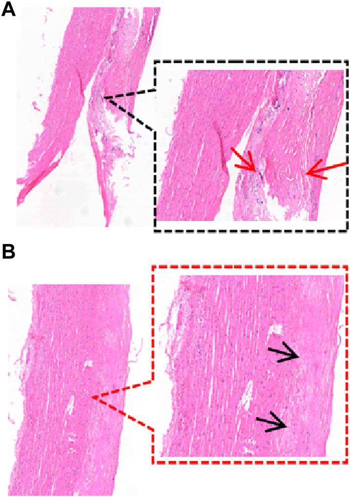
Methods: In this study, a total of 24 aortas (age range: 54-88 years old) were collected, one hundred and seventy-four dog-bone-shaped samples were made, and then the uniaxial tensile test was run, finally, pathological grouping was performed through histological staining.
Results: Atherosclerotic plaques were mainly distributed near the openings of blood vessel branches. The distribution was most severe in the abdominal aorta, followed by the aortic arch. Aortic atherosclerosis was a more severe trend in the male group. In the comparison of thickness, there were no significant differences in age (over 50 years) and test direction, the average thickness of the aorta was greater in the male group than the female group and decreased progressively from the ascending aorta to the abdominal aorta. Comparing the mechanical parameters, various parameters are mainly negatively correlated with age, especially in the circumferential ascending aorta (εp "Y = -0.01402*X + 1.762, R2 = 0.6882", εt "Y = -0.01062*X + 1.250, R2 = 0.6772"); the parameters of males in the healthy group were larger, while the parameters of females were larger in atherosclerosis group; the aorta has anisotropy, the parameters in the circumferential direction were greater than those in the axial direction; the parameters of the ascending aorta were the largest in the circumferential direction, the ultimate stress [σp "1.69 (1.08,2.32)"] and ultimate elastic modulus [E2"8.28 (6.67,10.25)"] of the abdominal aorta were significantly larger in the axial direction; In the circumferential direction, the stress [σp "2.2 (1.31,3.98)", σt "0.13 (0.09,0.31)"] and ultimate elastic modulus (E2 "14.10 ± 7.21") of adaptive intimal thickening were greater than those of other groups, the strain (εp "0.82 ± 0.17", εt "0.53 ± 0.14") of pathological intimal thickening was the largest in the pathological group.
Conclusion: The present study systematically analyzed the influence of age, sex, test direction, anatomical site, and pathological characteristics on the biomechanical properties of the aorta, described the distribution of aortic atherosclerosis, and illustrated the characteristics of aortic thickness changes. At the same time, new insights into the grouping of pathological features were presented.
Keywords: atherosclerosis; human aorta; material properties; pathology; uniaxial tensile test.
Copyright © 2024 Chen, Zhao, Li, Wang, Xing, Bian and Li.
Conflict of interest statement
The authors declare that the research was conducted in the absence of any commercial or financial relationships that could be construed as a potential conflict of interest.
Figures


Similar articles
-
Human dilated ascending aorta: Mechanical characterization via uniaxial tensile tests.J Mech Behav Biomed Mater. 2016 Jan;53:257-271. doi: 10.1016/j.jmbbm.2015.08.021. Epub 2015 Aug 20. J Mech Behav Biomed Mater. 2016. PMID: 26356765
-
A study to characterize the mechanical properties and material constitution of adult descending thoracic aorta based on uniaxial tensile test and digital image correlation.Front Bioeng Biotechnol. 2023 Jun 14;11:1178199. doi: 10.3389/fbioe.2023.1178199. eCollection 2023. Front Bioeng Biotechnol. 2023. PMID: 37388776 Free PMC article.
-
Re-examination of the mechanical anisotropy of porcine thoracic aorta by uniaxial tensile tests.Biomed Eng Online. 2016 Dec 28;15(Suppl 2):167. doi: 10.1186/s12938-016-0279-6. Biomed Eng Online. 2016. PMID: 28155705 Free PMC article.
-
Effects of clinico-pathological risk factors on in-vitro mechanical properties of human dilated ascending aorta.J Mech Behav Biomed Mater. 2018 Jan;77:1-11. doi: 10.1016/j.jmbbm.2017.08.032. Epub 2017 Aug 31. J Mech Behav Biomed Mater. 2018. PMID: 28886508
-
Biaxial tensile testing system for measuring mechanical properties of both sides of biological tissues.J Mech Behav Biomed Mater. 2023 Oct;146:106028. doi: 10.1016/j.jmbbm.2023.106028. Epub 2023 Jul 15. J Mech Behav Biomed Mater. 2023. PMID: 37531771
Cited by
-
From Biomechanical Properties to Morphological Variations: Exploring the Interplay between Aortic Valve Cuspidity and Ascending Aortic Aneurysm.J Clin Med. 2024 Jul 19;13(14):4225. doi: 10.3390/jcm13144225. J Clin Med. 2024. PMID: 39064264 Free PMC article.
References
-
- Astrand H., Stalhand J., Karlsson J., Karlsson M., Sonesson B., Länne T. (2011). In vivo estimation of the contribution of elastin and collagen to the mechanical properties in the human abdominal aorta: effect of age and sex. J. Appl. physiology 110 (1), 176–187. 10.1152/japplphysiol.00579.2010 - DOI - PubMed
-
- Bel-Brunon A., Kehl S., Martin C., Uhlig S., Wall W. A. (2014). Numerical identification method for the non-linear viscoelastic compressible behavior of soft tissue using uniaxial tensile tests and image registration - application to rat lung parenchyma. J. Mech. Behav. Biomed. Mater 29, 360–374. 10.1016/j.jmbbm.2013.09.018 - DOI - PubMed
LinkOut - more resources
Full Text Sources