

A rare case of pancreatic adenocarcinoma accompanied by venous thrombosis, pleural and pericardial effusions
- PMID: 38576945
- PMCID: PMC10990354
- DOI: 10.1097/MS9.0000000000001870
A rare case of pancreatic adenocarcinoma accompanied by venous thrombosis, pleural and pericardial effusions
Abstract
Introduction: Pancreatic cancer is a deadly type of cancer with few symptoms until metastasis. It poses a high risk of cancer-associated thrombosis.
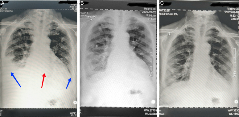
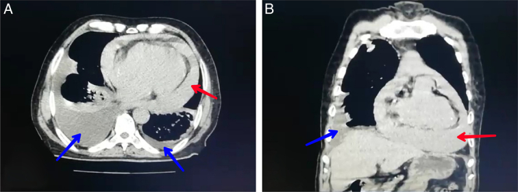
Case presentation: A 73-year-old male presented with fatigue, shortness of breath, weight loss since 9 months, and blood clots recently in his legs. Chest radiography revealed fluid accumulation in pleural and pericardial cavities. Later, a fluid examination revealed the presence of malignant cells in the pericardial fluid. After immunological tests and an upper gastrointestinal endoscopy were performed, a pancreatic tumour was suspected. The patient was administered anticoagulant treatment and palliative care, which resulted in improvement after one month.
Discussion: Pancreatic adenocarcinoma is a highly aggressive cancer with a strong tendency to metastasize, leading to pericardial and pleural effusion, thrombophlebitis, and poor prognosis.
Conclusion: This case indicates that venous thrombosis, pleural and pericardial effusions could be symptoms related to a pancreatic tumour.
Keywords: case report; pancreatic adenocarcinoma; pericardial effusions; pleural effusions; venous thrombosis.
Copyright © 2024 The Author(s). Published by Wolters Kluwer Health, Inc.
Conflict of interest statement
The authors declare no conflicts of interest.Sponsorships or competing interests that may be relevant to content are disclosed at the end of this article.
Figures
Similar articles
-
Malignant Pericardial Effusion Presenting as a Sequela of Lung Adenocarcinoma.Cureus. 2024 Mar 30;16(3):e57287. doi: 10.7759/cureus.57287. eCollection 2024 Mar. Cureus. 2024. PMID: 38690490 Free PMC article.
-
A case of Meigs' syndrome with preceding pericardial effusion in advance of pleural effusion.BMC Pulm Med. 2016 May 10;16(1):71. doi: 10.1186/s12890-016-0241-1. BMC Pulm Med. 2016. PMID: 27160723 Free PMC article.
-
Early chest tube removal following cardiac surgery is associated with pleural and/or pericardial effusions requiring invasive treatment.Eur J Cardiothorac Surg. 2016 Jan;49(1):288-92. doi: 10.1093/ejcts/ezv005. Epub 2015 Feb 7. Eur J Cardiothorac Surg. 2016. PMID: 25661079
-
An Unusual Presentation of Adult-Onset Still's Disease in a Patient with Recurrent Pleural and Pericardial Effusions.Am J Med Sci. 2021 May;361(5):655-658. doi: 10.1016/j.amjms.2020.06.024. Epub 2020 Jun 27. Am J Med Sci. 2021. PMID: 34024355 Review.
-
[Pancreatic pleural effusion accompanied by bronchopleural fistula].Nihon Kokyuki Gakkai Zasshi. 1999 Aug;37(8):662-6. Nihon Kokyuki Gakkai Zasshi. 1999. PMID: 10496109 Review. Japanese.
References
-
- Tam THC, Lui RN. Pancreatic cancer-associated thrombosis. Hong Kong Med J Xianggang yi xue za zhi 2023;29:378–379. - PubMed
Publication types
LinkOut - more resources
Full Text Sources