. 2024 Mar 20;27(4):109541.
doi: 10.1016/j.isci.2024.109541. eCollection 2024 Apr 19.

TRPA5 encodes a thermosensitive ankyrin ion channel receptor in a triatomine insect

Affiliations

TRPA5 encodes a thermosensitive ankyrin ion channel receptor in a triatomine insect

Marjorie A Liénard et al. iScience. .

Abstract

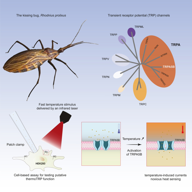
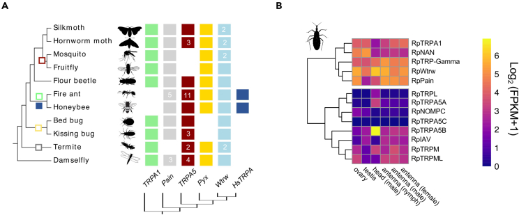
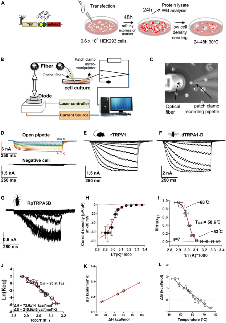
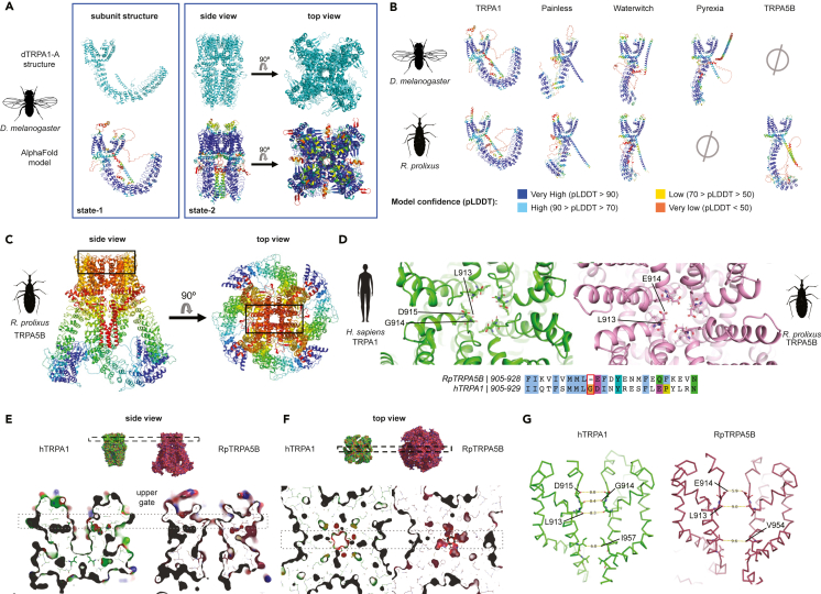
As ectotherms, insects need heat-sensitive receptors to monitor environmental temperatures and facilitate thermoregulation. We show that TRPA5, a class of ankyrin transient receptor potential (TRP) channels absent in dipteran genomes, may function as insect heat receptors. In the triatomine bug Rhodnius prolixus (order: Hemiptera), a vector of Chagas disease, the channel RpTRPA5B displays a uniquely high thermosensitivity, with biophysical determinants including a large channel activation enthalpy change (72 kcal/mol), a high temperature coefficient (Q10 = 25), and in vitro temperature-induced currents from 53°C to 68°C (T0.5 = 58.6°C), similar to noxious TRPV receptors in mammals. Monomeric and tetrameric ion channel structure predictions show reliable parallels with fruit fly dTRPA1, with structural uniqueness in ankyrin repeat domains, the channel selectivity filter, and potential TRP functional modulator regions. Overall, the finding of a member of TRPA5 as a temperature-activated receptor illustrates the diversity of insect molecular heat detectors.

Keywords: Biological sciences; Entomology; Molecular biology.


Conflict of interest statement

The authors declare no competing interests.

Figures

None
Graphical abstract
Figure 1
Figure 1
Phylogeny and expression of Rhodnius prolixus TRPs (A) Phylogenetic reconstruction of the ankyrin TRP (TRPA) channel subfamilies in representative insect species. TRPA5 channels are present across insect orders but absent from dipteran genomes (see also Table S1; Figures S1 and S2). Gene abbreviations: Painless (Pain), Pyrexia (Pyx), Waterwitch (Wtrw), TRPA Hymenoptera-specific (HsTRPA). Silkmoth, Bombyx mori; Hornworm moth, Manduca sexta; Mosquito, Anopheles gambiae; Fruit fly, Drosophila melanogaster; Flour beetle, Tribolium castaneum; Fire ant, Solenopsis invicta; Honeybee, Apis mellifera; Bed bug, Cimex lectularis; Kissing bug, Rhodnius prolixus; Termite, Zootermopsis nevadensis; Bluetail Damselfly, Ischnura elegans. Gene gain: filled square; gene loss: empty square. Numbers within squares indicate gene number when different from 1. (B) TRP genes in R. prolixus and their relative expression levels across tissues in compiled transcriptomic data (seeSTAR Methods). Heat maps compare the expression levels across tissues and developmental stages. Expression levels are represented as Log2 FPKM +1 and depicted with a gradient color scale. Gene models are based on genomic annotations, and de novo transcriptome assembly (see also Tables S2 and S3; Figure S3).
Figure 2
Figure 2
Thermodynamics of RpTRPA5B temperature-activated currents (A–C) Experimental workflow. (A) Each TRP channel subcloned in the pFRT-TO-FLAG-T2A-mRuby2 expression cassette, was transfected in HEK293T cells seeded at low density and incubated at 37°C for 48 h. Cells were then prepared for patch-clamp recording by seeding in a 30-mm2 culture dish overlaid with round glass cover slips and incubated at 30°C. (B) Electrophysiology recordings took place after 24–48 h using an optical fiber-based setup adapted after Yao et al. 2010, designed to couple manual patch-clamp recordings with fiber optics as a way to provide controllable optical and thermal stimulations to individual cells expressing candidate thermosensitive receptor proteins. The setup consists of a fiber launch system combining a high-power optical fiber tuned to near-infrared wavelengths (λc = 1,460 nm (+/−20 nm), Po = 4.8 W), a visible alignment laser (red), and a laser diode controller, forming a PID control loop using the patch-clamp current as the feedback signal. (C) During the experiment, a laser spot is aligned with one single patched cell (see Figure S6) stably expressing the membrane receptor protein of interest in the coverslip placed in the recording chamber. (D) Upper panel, current traces through the open patch-clamp pipette in response to temperature calibration steps from room temperature up to 71°C elicited by increments in the IR laser voltage input (see STAR Methods). Each 700 ms voltage pulse is represented in different colors for the different temperatures calculated from the open pipette currents. Lower panel, representative recording of non-transfected cells; these cells did not show robust temperature-elicited currents, like negative cells on the recording plate. (E) Whole-cell currents evoked by temperature steps from HEK293T cells expressing rat TRPV1 (heat-activated mammalian vanilloid thermoTRP); cells were held at −30 mV during the recording. (F) Whole-cell currents evoked by temperature steps from HEK293T cells expressing dTRPA1-D (holding potential of −30 mV). The sinusoidal pattern observed within the current curves is inherent to the cyclic modulation of the laser’s rapid “on-off” cycles. (G) Whole-cell currents evoked by temperature steps in HEK293T cells expressing RpTRPA5B; cells were held at −30 mV. (H) Current-temperature relationship for RpTRPA5B whole-cell current was normalized by cell membrane capacitance (current density); the red line corresponds to a modified Boltzmann function that includes the leak and unitary current temperature dependence (see STAR Methods). (I) Fraction of RpTRPA5B channels in the open state (open probability, Po) as a function of the temperature. The Po vs. 1/T was fitted to a Boltzman function with the midpoint of activation (T0.5) reached at 58.6°C. (J) van’t Hoff plot estimates of RpTRPA5B with an activation enthalpy of the endothermic transition at 92 kcal/mol and an entropic change associated with the temperature activation process at 274 cal/mol∗K at −30 mV. (K) Coupling between enthalpic (ΔH) and entropic (ΔS) changes for each one of the experiments recorded. (L) Free energy (ΔG) associated with the activation process as a function of temperature for RpTRP5AB channels. The receptor activation is associated with small free energy changes, as reported before for other families of mammalian thermoTRP receptors. ΔG was calculated as -RT∗ln(Keq). Data are represented as mean ± standard error.
Figure 3
Figure 3
Monomeric and tetrameric assemblies of RpTRPA5B channels modeled using AlphaFold, after validation with dTRPA1 structure (A–D) (A) (left panel) Upper row, cartoon representation of chain A in tetramer of dTRPA1 in state 1 (PDB: 7YKR). The fold of a monomer in the experimentally determined structure of the dTRPA1 tetramer is very similar to the fold of an AlphaFold model of a single dTRPA1 monomer (bottom row). AlphaFold model colored from red to blue according to pLDDT confidence scores as shown in (B). The low-confidence regions (red) are not resolved in the reported structure and are likely to be intrinsically disordered. (Right panel) Upper row, experimentally determined structure of dTRPA1 in state 2 (PDB: 7YKS). Bottom row, tetrameric AlphaFold model of dTRPA1 depicted as cartoons colored from red to blue according to confidence scores as in (B). The N- and C-terminal regions, which are not resolved in 7YKS, were excluded in the prediction. Only the last five of the 17 ankyrin repeats (AR12-16) are visible in the structure and overall regions with low confidence in the model (red-yellow) are not resolved in the structure. (B) Monomers of Drosophila and Rhodnius TRPAs colored by pLDDT score from the AlphaFold modeling. (C) Tetrameric model of RpTRPA5B, colored as chain bows (N terminus, blue; C terminus, red). The black box indicates the location of the pore and selectivity filter shown in (D). (D) Top view of the selectivity filter of the pore of hTRPA1 (left: human TRPA1, PDB: 6V9Y) and RpTRPA5B (right). Three important residues – L913, G914, and D915, are marked in hTRPA1. The equivalent residues L913 and E914 are marked in RpTRPA5B, and G914 absent in RpTRPA5B is highlighted in the sequence alignment, together with additional residue changes adjacent to the selectivity filter. (E and F) Comparison of the pore in hTRPA1 and model of RpTRPA5B indicates a closed upper gate and an open lower gate in the RpTRPA5B model. E. (upper row) Surface representation of hTRPA1 (green; PDB: 6V9Y) and RpTRPA5B (pink), side view. The dashed box indicates the location of the upper gate toward the outside of the cell. (lower row) Slab along the pore through the transmembrane domain of the hTRPA1 structure and RpTRPA5B model. F. (upper row) Top view of hTRPA1 and RpTRPA5B shown in E. (lower row) The slab is perpendicular to the pore at the level of the upper gate shown in E. The dashed box indicates the location of slab in (E). (G) Distances between corresponding residues in the upper and lower gate in structures of hTRPA1 and the model of RpTRPA5B, shown as sticks.

Similar articles

Cited by

  • Temperature-sensing riboceptors.
    Anbalagan S. Anbalagan S. RNA Biol. 2024 Jan;21(1):1-6. doi: 10.1080/15476286.2024.2379118. Epub 2024 Jul 17. RNA Biol. 2024. PMID: 39016038 Free PMC article.
  • Hot breath, quick exit: aphids flee mammalian heat via TRPA1.
    Wang B, Yang L, Wei J, Huang T, Zhang B, Bohbot J, Wang G. Wang B, et al. Sci China Life Sci. 2025 Jun 19. doi: 10.1007/s11427-025-2925-9. Online ahead of print. Sci China Life Sci. 2025. PMID: 40550996
  • Evolution of Sensory Receptors.
    Valencia-Montoya WA, Pierce NE, Bellono NW. Valencia-Montoya WA, et al. Annu Rev Cell Dev Biol. 2024 Oct;40(1):353-379. doi: 10.1146/annurev-cellbio-120123-112853. Epub 2024 Sep 21. Annu Rev Cell Dev Biol. 2024. PMID: 38985841 Free PMC article. Review.

References

    1. McKemy D.D. Temperature sensing across species. Pflugers Arch. 2007;454:777–791. doi: 10.1007/s00424-006-0199-6. - DOI - PMC - PubMed
    1. Vriens J., Nilius B., Voets T. Peripheral thermosensation in mammals. Nat. Rev. Neurosci. 2014;15:573–589. doi: 10.1038/nrn3784. - DOI - PubMed
    1. Xiao R., Xu X.Z.S. Temperature sensation: from molecular themosensors to neural circuits and coding principles. Annu. Rev. Physiol. 2021;83:205–230. - PMC - PubMed
    1. Castillo K., Diaz-Franulic I., Canan J., Gonzalez-Nilo F., Latorre R. Thermally activated TRP channels: molecular sensors for temperature detection. Phys. Biol. 2018;15 doi: 10.1088/1478-3975/aa9a6f. - DOI - PubMed
    1. Garrity P.A., Goodman M.B., Samuel A.D., Sengupta P. Running hot and cold: behavioral strategies, neural circuits, and the molecular machinery for thermotaxis in C. elegans and Drosophila. Genes Dev. 2010;24:2365–2382. - PMC - PubMed