

Evaluation of a 3D-Printed Cleft Palate Obturator Using a Low-Dose Cone Beam Computed Tomography Acquisition Protocol: A Proof-of-Concept Study
- PMID: 38577166
- PMCID: PMC10994164
- DOI: 10.7759/cureus.57602
Evaluation of a 3D-Printed Cleft Palate Obturator Using a Low-Dose Cone Beam Computed Tomography Acquisition Protocol: A Proof-of-Concept Study
Abstract
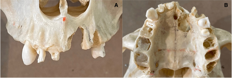
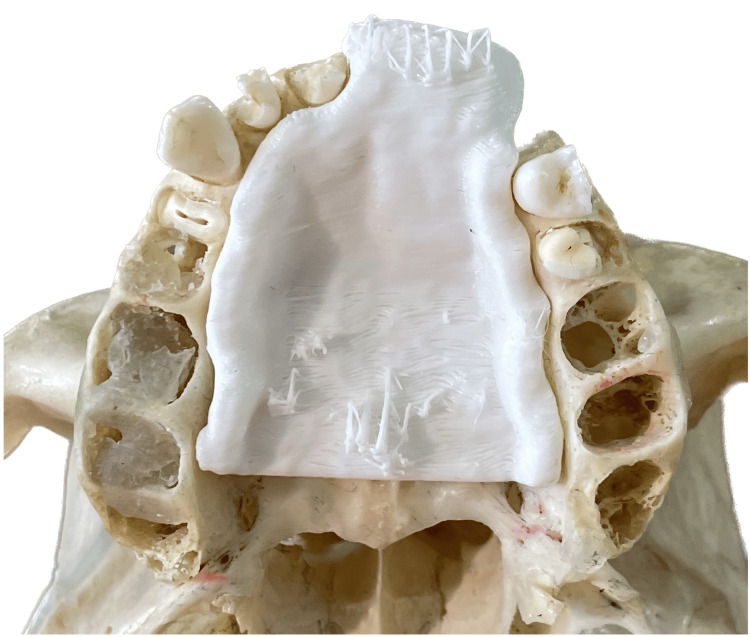
Cone beam computed tomography (CBCT) technology is increasingly utilized in the head and neck region and is valuable in treatment planning for cleft palate patients, potentially enabling the creation of 3D-printed obturators to assist with feeding and speech. This technical report investigates the feasibility of using data from a 360-degree CBCT scan to accurately produce a cleft palate obturator and assesses whether a lower-dose 180-degree CBCT scan can achieve a comparable result. A simulated cleft palate was crafted on a dehydrated human skull, which was then scanned using both 360-degree and 180-degree CBCT scanning protocols. Two obturators were digitally designed based on the segmented images from each scan and subsequently 3D printed. Evaluation of the segmented images and 3D-printed obturators from both protocols demonstrated clear visualization of anatomical landmarks and identical scores across all parameters, suggesting that the 180-degree CBCT scan can produce an obturator of comparable quality to that of the 360-degree scan, with the added benefit of reduced radiation exposure.
Keywords: 3d digital models; 3d image reconstruction; cleft lip & palate; cone-beam computed tomography; maxillary obturator.
Copyright © 2024, Nelson et al.
Conflict of interest statement
The authors have declared that no competing interests exist.
Figures


References
-
- Updated national birth prevalence estimates for selected birth defects in the United States, 2004-2006. Parker SE, Mai CT, Canfield MA, et al. Birth Defects Res A Clin Mol Teratol. 2010;88:1008–1016. - PubMed
-
- A report on the hazards encountered when taking neonatal cleft palate impressions (1983-1992) Chate RA. Br J Orthod. 1995;22:299–307. - PubMed
-
- Review of the role of potential teratogens in the origin of human nonsyndromic oral clefts. Wyszynski DF, Beaty TH. Teratology. 1996;53:309–317. - PubMed
LinkOut - more resources
Full Text Sources