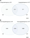
Comparative analysis of basal and etoposide-induced alterations in gene expression by DNA-PKcs kinase activity
- PMID: 38577247
- PMCID: PMC10991847
- DOI: 10.3389/fgene.2024.1276365
Comparative analysis of basal and etoposide-induced alterations in gene expression by DNA-PKcs kinase activity
Abstract
Background: Maintenance of the genome is essential for cell survival, and impairment of the DNA damage response is associated with multiple pathologies including cancer and neurological abnormalities. DNA-PKcs is a DNA repair protein and a core component of the classical nonhomologous end-joining pathway, but it also has roles in modulating gene expression and thus, the overall cellular response to DNA damage. Methods: Using cells producing either wild-type (WT) or kinase-inactive (KR) DNA-PKcs, we assessed global alterations in gene expression in the absence or presence of DNA damage. We evaluated differential gene expression in untreated cells and observed differences in genes associated with cellular adhesion, cell cycle regulation, and inflammation-related pathways. Following exposure to etoposide, we compared how KR versus WT cells responded transcriptionally to DNA damage. Results: Downregulated genes were mostly involved in protein, sugar, and nucleic acid biosynthesis pathways in both genotypes, but enriched biological pathways were divergent, again with KR cells manifesting a more robust inflammatory response compared to WT cells. To determine what major transcriptional regulators are controlling the differences in gene expression noted, we used pathway analysis and found that many master regulators of histone modifications, proinflammatory pathways, cell cycle regulation, Wnt/β-catenin signaling, and cellular development and differentiation were impacted by DNA-PKcs status. Finally, we have used qPCR to validate selected genes among the differentially regulated pathways to validate RNA sequence data. Conclusion: Overall, our results indicate that DNA-PKcs, in a kinase-dependent fashion, decreases proinflammatory signaling following genotoxic insult. As multiple DNA-PK kinase inhibitors are in clinical trials as cancer therapeutics utilized in combination with DNA damaging agents, understanding the transcriptional response when DNA-PKcs cannot phosphorylate downstream targets will inform the overall patient response to combined treatment.
Keywords: DNA damage repair; DNA-PKcs; etoposide; gene expression; non-homologous end-joining; transcriptome.
Copyright © 2024 Ali, Najaf-Panah, Pyper, Lujan, Sena and Ashley.
Conflict of interest statement
Author JS was employed by the National Center for Genome Resources. The remaining authors declare that the research was conducted in the absence of any commercial or financial relationships that could be construed as a potential conflict of interest.
Figures


Similar articles
-
Inactivation of DNA-dependent protein kinase promotes heat-induced apoptosis independently of heat-shock protein induction in human cancer cell lines.PLoS One. 2013;8(3):e58325. doi: 10.1371/journal.pone.0058325. Epub 2013 Mar 11. PLoS One. 2013. PMID: 23505488 Free PMC article.
-
The role of insulin-like growth factor binding protein-3 in the breast cancer cell response to DNA-damaging agents.Oncogene. 2014 Jan 2;33(1):85-96. doi: 10.1038/onc.2012.538. Epub 2012 Nov 26. Oncogene. 2014. PMID: 23178489
-
Progression of chromosomal damage induced by etoposide in G2 phase in a DNA-PKcs-deficient context.Chromosome Res. 2015 Dec;23(4):719-32. doi: 10.1007/s10577-015-9478-4. Epub 2015 Jul 8. Chromosome Res. 2015. PMID: 26152239
-
DNA-PKcs: A Multi-Faceted Player in DNA Damage Response.Front Genet. 2020 Dec 23;11:607428. doi: 10.3389/fgene.2020.607428. eCollection 2020. Front Genet. 2020. PMID: 33424929 Free PMC article. Review.
-
The multifaceted functions of DNA-PKcs: implications for the therapy of human diseases.MedComm (2020). 2024 Jun 19;5(7):e613. doi: 10.1002/mco2.613. eCollection 2024 Jul. MedComm (2020). 2024. PMID: 38898995 Free PMC article. Review.
Cited by
-
Metabolic changes in the one-carbon metabolism-related amino acids during etoposide-induced cellular senescence of neuronal cells.Cytotechnology. 2025 Aug;77(4):131. doi: 10.1007/s10616-025-00803-w. Epub 2025 Jun 30. Cytotechnology. 2025. PMID: 40607102
References
-
- Ashley A. K., Shrivastav M., Nie J., Amerin C., Troksa K., Glanzer J. G., et al. (2014). DNA-PK phosphorylation of RPA32 Ser4/Ser8 regulates replication stress checkpoint activation, fork restart, homologous recombination and mitotic catastrophe. DNA Repair (Amst) 21, 131–139. 10.1016/j.dnarep.2014.04.008 - DOI - PMC - PubMed
Associated data
Grants and funding
LinkOut - more resources
Full Text Sources
Research Materials

