. 2024 Mar 27;3(4):pgae124.
doi: 10.1093/pnasnexus/pgae124. eCollection 2024 Apr.

Structural constraints of pyocin S2 import through the ferripyoverdine receptor FpvAI

Affiliations

Structural constraints of pyocin S2 import through the ferripyoverdine receptor FpvAI

Jonathan D Goult et al. PNAS Nexus. .

Abstract

TonB-dependent transporters (TBDTs) mediate energized transport of essential nutrients into gram-negative bacteria. TBDTs are increasingly being exploited for the delivery of antibiotics to drug-resistant bacteria. While much is known about ground state complexes of TBDTs, few details have emerged about the transport process itself. In this study, we exploit bacteriocin parasitization of a TBDT to probe the mechanics of transport. Previous work has shown that the N-terminal domain of Pseudomonas aeruginosa-specific bacteriocin pyocin S2 (PyoS2NTD) is imported through the pyoverdine receptor FpvAI. PyoS2NTD transport follows the opening of a proton-motive force-dependent pore through FpvAI and the delivery of its own TonB box that engages TonB. We use molecular models and simulations to formulate a complete translocation pathway for PyoS2NTD that we validate using protein engineering and cytotoxicity measurements. We show that following partial removal of the FpvAI plug domain which occludes the channel, the pyocin's N-terminus enters the channel by electrostatic steering and ratchets to the periplasm. Application of force, mimicking that exerted by TonB, leads to unraveling of PyoS2NTD as it squeezes through the channel. Remarkably, while some parts of PyoS2NTD must unfold, complete unfolding is not required for transport, a result we confirmed by disulfide bond engineering. Moreover, the section of the FpvAI plug that remains embedded in the channel appears to serve as a buttress against which PyoS2NTD is pushed to destabilize the domain. Our study reveals the limits of structural deformation that accompanies import through a TBDT and the role the TBDT itself plays in accommodating transport.


Figures

Fig. 1.
Fig. 1.
Application of force on the FpvAI N-terminus of the FpvAI–PyoS2NTD complex partially unfolds the plug domain but does not stimulate PyoS2 translocation in silico. A) Snapshot of the structure of PyoS2NTD (blue) in complex with FpvAI (gray) during simulation of force-dependent unfolding of the FpvAI plug domain, highlighting the labile (red) and nonlabile (green) subdomains of the plug domain (from the FpvAI–PyoS2NTD crystal structure [PDB: 5ODW]). The PyoS2 TonB1 box is located within a β-hairpin (yellow) that associates with the N-terminal kTHB. B) Retraction of Asp126 in the −z direction (perpendicularly to the membrane toward the periplasm) led to unfolding of the labile plug subdomain (red). The nonlabile plug subdomain (green) starts unfolding when the labile plug is completely extended (∼300 Å for the 90 residues that constitute the labile plug). Unfolding of the labile plug subdomain starts at low forces, while the initiation of the unfolding of the nonlabile plug subdomain corresponds to a peak in the force (shown for three different trajectories at different pulling speeds: magenta, cyan, and yellow). Removal of plug domain does not affect the structure of the FpvAI β-barrel (black), or cause remodeling of PyoS2NTD (blue), suggesting that unplugging of the transporter and translocation of pyocin do not occur concomitantly.
Fig. 2.
Fig. 2.
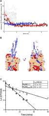
The unstructured N-terminus of PyoS2 is required for optimal import. A) Distance between the (modeled) main chain N of Met1 of PyoS2 from the FpvAI pore entrance demonstrates the disordered N-terminal end of PyoS2 can spontaneously diffuse to the entry of the pore vacated by the unfolded labile plug. Running simulations from random conformations of the N-terminal unstructured residues of PyoS2 reveal a small number of cases in which the N-terminus is observed diffusing toward the pore (identified as the center of mass of residues Val229, Trp707, and Thr797). B) Transverse of FpvAI receptor (surface) with PyoS2NTD bound (blue). Removal of the labile plug domain leaves a pore spanning the membrane through the β-barrel. The pore lining is composed of alternating charges and the unstructured PyoS2 N-terminus (dashed oval) is positioned near the pore entrance, as described previously. The unfolded labile plug domain was removed from this analysis and subsequent simulations. C) PyoS2 kinetic time-course cytotoxicity assay demonstrates that truncation of unstructured residues upstream of the TonB1 box decreases the rate of translocation for PyoS2Δ1-9 (○) compared with PyoS2 (●). Colony counts from four independent YHP17 cultures (with error bars representing SD) were fitted to a pseudo-first order reaction model, and translocation half-lives (t1/2) were calculated from the linear plot.
Fig. 3.
Fig. 3.
The N-terminal domain of PyoS2 must unfold in order to translocate through the pore formed by the FpvAI transporter. A) The rmsd of the different structural components (top) and force applied (bottom) during translocation of PyoS2NTD. PyoS2NTD readily unfolds and displaces from its original position upon retraction of Met1 of PyoS2 in the −z direction. FpvAI barrel and nonlabile plug undergo minimal perturbance in response to PyoS2NTD translocation. B) Snapshots of PyoS2 during the forced translocation shown as blue spheres (see Movie S1). Vertical lines in A) (I, II, and III) correspond to the time at which the snapshots were taken.
Fig. 4.
Fig. 4.
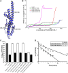
Disulfide bonds impede, but not necessarily prevent, translocation of PyoS2 through the pore formed by the FpvAI transporter. A) Depiction of PyoS2NTD with disulfide positions predicted by Disulfide by Design (disulfide C11–C26 not shown). B) The rmsd of the unplugged transporter FpvAI as a function of the z-coordinate of the first residue of PyoS2. Two variants (C28–C157 and C103–C186) result in a severe structural change in the transporter, while in the case of C11–C26 and C128–C162, the disulfide bond can go through the pore causing only minor structural rearrangements (see Movies S2–S5). C) YHP17 CFU/mL from PyoS2 cytotoxicity assay after 3 h shows disulfide positions C11–C26, C28–C157, and C103–C186 inhibit PyoS2 cytotoxicity when oxidized (white). This effect is reversed upon disulfide bond reduction (black). Disulfide position C128–C162 imparts significantly reduced protection against PyoS2 activity when oxidized. Colony counts from six independent cultures with error bars representing the SD. D) YHP17 CFU/mL from kinetic time-course cytotoxicity assays demonstrate that oxidation of the C128–C162 disulfide bond results in a decrease in the PyoS2 translocation rate (○) compared with that of the reduced form (●). Colony counts from four independent cultures (with error bars representing SD) were fitted to a pseudo-first order reaction model and translocation half-lives (t1/2) were calculated from the linear plot.
Fig. 5.
Fig. 5.
Updated model of PyoS2 import through FpvAI and the structural constraints imposed during translocation. A three-step model for PyoS2NTD import through the FpvAI transporter, supported by in silico, in vitro, and in vivo experimental data presented here. Initial binding of PyoS2NTD to FpvAI mimics Fe3+–PVD binding, activating the transporter for uptake and recruiting TonB1 via its TonB1 box. Step 1 (T1): Force remodeling of the labile portion (red) of the FpvAI plug domain occurs in a PMF-dependent manner via contact to the TonB1–ExbB–ExbD complex in the IM (only the C-terminus of TonB1 is shown for clarity). The N-terminus of PyoS2NTD enters the resultant pore formed, facilitating presentation of its own TonB1 box in the periplasm. This process is blocked by disulfides within the β-hairpin motif. Step 2 (T2): The PyoS2NTD TonB1 box is bound by another copy of TonB1, resulting in the force remodeling of PyoS2NTD itself and driving translocation through the FpvAI lumen. Translocation can be stalled by the presence of intramolecular crosslinks in the form of disulfide bonds (C103–C186). Step 3 (T3): The entire PyoS2NTD is translocated through the FpvAI lumen, presumably followed by the remainder of the protein for the full-length construct. The PyoS2(C128–C162) mutant can successfully translocate across the OM despite the presence of an intramolecular crosslink, however, import as slowed as a result.

Similar articles

References

    1. Lambert PA. 2002. Mechanisms of antibiotic resistance in Pseudomonas aeruginosa. J R Soc Med. 95:22–26. - PMC - PubMed
    1. Nikaido H. 2003. Molecular basis of bacterial outer membrane permeability revisited. Microbiol Mol Biol Rev. 67:593–656. - PMC - PubMed
    1. Hancock REW, Speert DP. 2000. Antibiotic resistance in Pseudomonas aeruginosa: mechanisms and impact on treatment. Drug Resist Updat. 3:247–255. - PubMed
    1. Cascales E, et al. 2007. Colicin biology. Microbiol Mol Biol Rev. 71:158–229. - PMC - PubMed
    1. Kirkup BC, Riley MA. 2004. Antibiotic-mediated antagonism leads to a bacterial game of rock-paper-scissors in vivo. Nature. 428:412–414. - PubMed